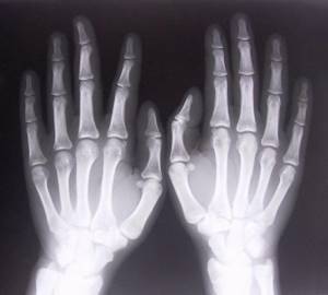
X-ray of the hand and wrist is a way to study their internal structure without invasive intervention. The hand and fingers are complex anatomical parts of the body, consisting of many small bones and performing important tasks in human functioning. This is the ideal and precise tool we use. Every day they are exposed to mechanical stress, their condition is affected by diet, quality of metabolism and other life processes. With the help of radiography, diseases in this area, cracks, dislocations and fractures are detected. Timely diagnosis prevents the development of severe diseases of the hands and wrists.
What does a hand x-ray show?
In everyday life, the human skeleton is subject to constant loads, including the hands. Excessive physical activity, infectious diseases, and mechanical damage change the structure of this part of the body and can sometimes lead to partial or complete loss of functionality.
To diagnose the disease and its cause in a timely manner, radiography is used. This method has been known for a long time and is still popular among traumatologists, surgeons, and therapists.
Content:
- What does a hand x-ray show?
- Indications for radiography
- Features of the study
- Research results
- Contraindications for hand radiography
- Alternative to radiography
- Where to take the study
- The myth about the dangers of research
The technique works on the principle of x-ray radiation. Beams of rays pass through the object and leave a contour mark on the film. Depending on the density of the tissue, the beam passes through it differently. Thus, bone tissue is reflected best, while soft tissue is reflected poorly.
However, based on the shadow in the image, a radiologist or other doctor can see changes in the lymph nodes, tendons, and muscles. The x-ray image shows the entire internal structure of the wrist and hand.
In the picture you can see all parts of the hand: wrist, phalanges of fingers, metacarpus. In turn, each of these sections contains several subgroups of bones, ligaments, joints, and muscles. Damage to even one unit of this system leads to pain, swelling, and deformation of the entire arm or its parts.
An x-ray clearly shows any damage to the bone tissue: thinning, cracks and microcracks, dislocations, inflammatory processes. To accurately diagnose problems with soft tissue, the doctor may prescribe a more informative computed tomography scan, or angiography to examine blood vessels.
Today medicine uses several options for examining the hands and wrist; they are more modern, more accurate, and faster. These include MRI, CT, MSCT and others. However, conventional X-rays continue to be used because they are simpler and more accessible.
Every clinic has such equipment; it costs much less or is completely free. In addition, the radiation dose for all progressive options and radiography is almost the same.
Preparing for an X-ray
To take an x-ray of the right and left hands, no preliminary preparation is needed. Before the procedure, you need to remove rings, bracelets, watches and place your hands (or arm) on the work table of the X-ray machine as directed by a radiologist.
To protect from the influence of ionizing radiation those parts of the patient’s body that are not involved in the study, special lead aprons or vests are used.
Indications for radiography
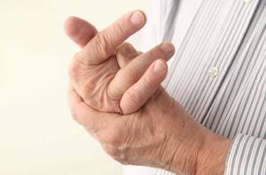
Like every part of the body, the hands and wrists are subject to illness and injury, and the latter happens to them most often.
To accurately establish the diagnosis and future treatment plan, the doctor prescribes an x-ray of the hand and wrist. The images also help to clearly establish the location of the injury, determine its severity and recovery methods.
X-rays of the wrist and hand are prescribed for:
- mechanical damage: bruises, fractures, dislocations;
- arthritis and arthrosis;
- rheumatism;
- swelling and hyperemia of the joints;
- osteomyelitis;
- congenital anomalies in the structure of the hands;
- neoplasms: tumors, cysts, papillomas;
- systemic lupus erythematosus;
- scleroderma.
It is impossible to accurately determine the cause of the disease and its stage by visual examination, so the doctor first prescribes a diagnosis. Any changes in the structure of the hand and wrist skeleton will be visible in the photographs. In addition, radiography is prescribed for children if a lag in physical development and too rapid development is noticed. The patient can undergo this procedure if he experiences pain in any part of the hand, notices curvature of the fingers, numbness, and swelling.
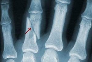
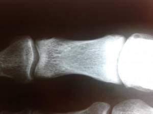
Fracture of the phalanges of the fingers
The cause is a blow with the fingers, an injury while fixing the fingers, or a direct blow to the phalanges. Fractures of the phalanges of the fingers can be:
- intra-articular;
- extra-articular;
- single;
- multiple - within one finger or several;
- combined with dislocations in the metacarpophalangeal or interphalangeal joints.
Symptoms: pain, swelling, hematoma, deformity. The pain intensifies when you try to move your fingers. The diagnosis is established on the basis of complaints, trauma history, objective examination and X-ray results. To treat a fracture of the phalanges of the fingers without displacement, fixation is performed with a plaster cast for 3-4 weeks. In case of fracture-dislocations, joint reduction is performed; in case of displacement of fragments, closed reduction is performed. If it is not possible to compare the fragments using a closed method, skeletal traction or pin osteosynthesis is indicated.
Features of the study
Pictures of the hands are taken quickly and painlessly. No preparation is required for the test; you need to take with you to the session directions from the doctor who prescribed it. X-rays are carried out in a separate room equipped with equipment. Before diagnosis, the patient removes jewelry so that it does not shade the image.
It is worth warning the doctor if there are metal inserts in the palms and wrists. To protect the rest of the body, the technician provides a lead membrane that blocks X-rays.
The installation consists of an X-ray table and a chair. The patient sits at the table and bends his limbs at the elbows, placing his hands on the table. Depending on the desired area of study, the projection of the images is changed. The patient's hands can be placed in different positions to obtain the most informative data.
For wrist x-rays, use:
- Direct projection. A cassette is placed under the palms, on which the photograph will then be reflected. Pictures can be taken from the back or from the palm, sometimes from two projections at once. The patient takes a stationary position, the laboratory assistant takes a photo using a remote button. This way the doctor will be able to examine all segments of the wrist: scaphoid, triquetrum, capitate and others. The phalanges and metacarpal regions are also visible. However, there are several segments that are not visible with this installation.
- Lateral projection. The hand is placed sideways on the cassette, with the thumb protruding slightly forward. This arrangement allows you to view the phalanges of the fingers, carpal and metacarpal bones from a different angle.
- I slant my palm. For this projection, the patient's hand is positioned so that the palm with the cassette creates an angle of 45 degrees. It is important that during the examination the hand does not move or shift; the laboratory assistant will monitor the correct positioning. Images from this position show the trapezium, scaphoid, and trapezius bones.
- I slant the back. In this case, the back of the hand creates a 45-degree angle with the cassette. In this way, the pisiform, hamate and triquetrum bones are best visible.
In addition to these methods, other types of styling can be used to examine the wrists. The radiologist can take targeted pictures of each individual bone. I also have my own radiography methods for reflecting fingers.
Each phalanx is placed in a direct and lateral perspective; the doctor can assess the condition of each of them separately. The accuracy of the results depends on the experience and competence of the doctor; he must set the minimum level of radiation and ensure the correct position of the patient.
For the patient, this procedure takes place without discomfort and lasts 5-15 minutes. If necessary, the doctor can take pictures in all projections for a detailed and complete study of the hands; for example, in case of infectious diseases, it is necessary to establish the entire affected area.
Also, in case of injuries or impaired development, a set of images is needed. No matter how many images need to be taken, the process will be quick and painless.
What other research are we doing?
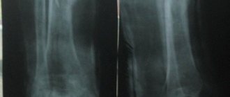
For dislocations, severe bruises, displacement and crushing of bones, radiography of the wrist joint in two projections . The images are taken so that the lower third of the forearm bones, the wrist bones, and the proximal metacarpal bones (the long bones that extend from the wrist in rays) are also visible.
The wrist joint is made up of many fragile bones. The risk of injury increases in old age, with a lack of physical activity, osteoporosis (because with this disease the bones become more fragile and less strong).
Sometimes only radiography of 1 or more fingers is performed in two projections . Depends on what exactly you damaged.
Also, in some cases, we perform x-rays of the scaphoid bone of the hand in three projections . It is one of the largest among its “neighbors” and is quite often susceptible to fractures.
X-ray of the scapula
A fracture of the scapula usually occurs due to direct impact, a direct blow. X-ray of the scapula allows you to detect not only injury, but also detect pathological inflammation, benign and malignant tumors.
Research results
After the images are taken, they must be interpreted and described by a radiologist. This will take 15-40 minutes, depending on the severity of the condition. Based on these data, the attending physician will determine the treatment regimen.
For hand and wrist injuries, therapy is carried out and monitored by a traumatologist; for other diseases, this can be a surgeon or therapist. The patient must take the results of the x-ray to the doctor who referred him for research. The images may reveal mechanical damage or rheumatic diseases.
Rheumatic diseases include scleroderma, joint inflammation, rheumatoid arthritis and other diseases. X-rays can also reveal dangerous joint psoriasis, bone calcification, and cysts. Advanced types of such inflammation lead to erosion of bone tissue and loss of functionality. In severe cases, X-rays indicate surgery, but systematic treatment is usually sufficient.
Wrist fractures
The bones of the wrist, due to their shape, structure and position, are broken quite rarely. The most susceptible bone to fracture is the scaphoid, the large bone at the base of the big toe. Injuries to the lunate and pisiform bones of the wrist also occur. The triquetral bone, as well as the bones of the distal row - polygonal, trapezoid, capitate and hamate - are subject to fractures extremely rarely; usually their fractures are combined with dislocations in the corresponding joints.
Scaphoid fractures
The cause is a fall on a bent hand, a blow with a fist, or a direct injury to the wrist. The following options are possible:
- intra-articular fracture of the scaphoid - the fracture line is located inside the cavity of the wrist joint;
- extra-articular fracture - separation of the tubercle of the scaphoid;
- de Quervain's fracture-dislocation - a simultaneous fracture of the scaphoid and dislocation of its proximal fragment and the lunate from the wrist joint.
Symptoms are pain and swelling at the base of the thumb, inability to move the hand at the wrist joint, or clench the hand into a fist. The diagnosis is established based on the patient’s complaints, data on the nature of the injury, examination and radiography of the hand bones. Sometimes, in the absence of displacement of fragments, the fracture line with all its signs is not determined. In this case, immobilization is still carried out with repeated radiography after 7-10 days, when, due to the activation of regenerative processes, the fracture line becomes clearly visible.
Treatment is immobilization with a plaster cast for a period of 4 weeks, followed by monitoring and prolongation of immobilization in case of insufficient consolidation of the fracture. In case of displacement of fragments and fracture dislocation, closed reduction is ineffective; fixation of fragments of the scaphoid bone with a wire is indicated. Fractures of the scaphoid are often complicated by the development of a false joint or lysis of bone fragments due to damage to the blood vessels supplying them during injury. Therefore, it is important to follow all the doctor’s recommendations and take control photographs in a timely manner to avoid complications and deterioration in the function of the wrist joint. After restoring the integrity of the scaphoid bone, physiotherapeutic treatment and exercise therapy are indicated to restore hand function.
Lunate fractures
The cause is a fall on a bent hand or direct injury, a blow to the wrist. It manifests itself as pain and swelling, intensifying with movements in the third, fourth and fifth fingers and with extension of the hand. The diagnosis is established taking into account complaints, the mechanism of injury, an objective examination of the area of damage and the results of an x-ray examination. To treat a lunate fracture, a plaster cast is applied for 4-8 weeks. Recovery usually proceeds without complications.
Fractures of the pisiform bone
The cause is a blow with the edge of the palm or direct injury. It manifests itself as pain and swelling of the wrist on the little finger side, increasing pain when it moves. The diagnosis is made taking into account complaints, anamnesis of injury, examination of the area of damage and radiography of the bones of the hand. For complete consolidation of a fracture of the pisiform bone, 4-5 weeks of immobilization are sufficient. The injury is rarely complicated.
Contraindications for hand radiography

Radiation examination carries a dose of radiation to the patient, so patients are often afraid of this procedure. However, when examining the hands and wrists, the dose is very small, so there are no categorical contraindications. This procedure is carried out even for small children; for maximum protection, they are completely covered with a lead apron, freeing only the hand.
Pregnant women are still not taken pictures unless absolutely necessary, but in case of fractures, the patient is also covered with a membrane. Breastfeeding mothers also undergo x-rays if necessary.
The session may be complicated if the patient has serious mental problems. To obtain clear images, the patient requires complete temporary immobility, which is not always possible in case of mental disorders.
Also, in a critically ill patient, such a study cannot be performed. If x-rays cannot be taken for some reason, alternative methods of examination are prescribed.
Alternative to radiography
Radiological diagnostic results can be obtained in other ways. The main competitor to radiography is computed tomography. It is based on the same radiation, but at a lower dose.
The CT scan process is faster and the results are more comprehensive. Thus, with a regular x-ray, soft tissues are poorly distinguished, although they are also often susceptible to disease. CT scan reflects vessels, muscles, and ligaments much better. However, the conventional technique is better suited for diagnosing bones.
Also, sometimes the X-ray method is replaced by MRI. This option is considered, like computed tomography, to be very informative. It provides comprehensive information about the condition of bone tissue and its surrounding elements. In this case, the patient does not receive radiation exposure at all, since the process is based on electromagnetic resonance. For a detailed examination of individual systems, such as blood or lymphatic vessels, lymphangiography or angiography is prescribed.
Where to take the study
If a simple x-ray is prescribed, it can be done at any emergency room, clinic, or hospital. The procedure requires conventional equipment, which is available in private and public medical institutions. More advanced techniques such as MRI or CT will have to be sought in private clinics.
The price for a regular photo of one brush in one projection will be 5-10 dollars. The cost may vary depending on the reputation and financial policies of the institution.
Two projections will cost twice as much. The price includes the examination and photographs; interpretation of the results in a public clinic should be free, in a private clinic you will need to pay an additional 5-8 dollars.