- What is upper gastrointestinal radiography?
- In what areas is upper gastrointestinal radiography used?
- How should you prepare for research?
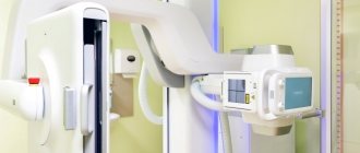
- What does the diagnostic equipment look like?
- What is the basis for the research?
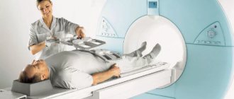
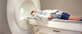
- How is the research conducted?
- What should you expect during and after the procedure?
- Who reviews the research results and where can they be obtained?
- What are the benefits and risks of upper gastrointestinal radiography?
- Limitations of upper gastrointestinal radiography
What is upper gastrointestinal radiography?
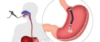
Upper gastrointestinal radiography is an x-ray examination of the pharynx, esophagus, stomach, and the initial portions of the small intestine (duodenum).
The images are obtained using a special type of test called fluoroscopy and an orally administered barium contrast material. X-ray testing is a non-invasive diagnostic technique that helps doctors detect and treat various diseases. In this case, certain parts of the body are exposed to a small dose of ionizing radiation, which allows them to be photographed. X-ray examination is the oldest imaging method and is most often used in diagnosis.
Fluoroscopy allows you to see internal organs in motion. After the barium coats the walls of the gastrointestinal tract, the radiologist can view and evaluate the anatomy and function of the esophagus, stomach, and duodenum.
X-ray examination of only the pharynx and esophagus with a barium suspension is called esophagography or esophagoscopy.
In addition to barium solution, some patients are asked to take crystalline soda orally, which improves the visibility of internal organs. This procedure is called air or double contrast of the upper gastrointestinal tract.
In rare cases, an oral iodinated contrast material in the form of a clear liquid is used instead of barium. For example, an alternative contrast technique may be used in patients who have recently undergone upper gastrointestinal surgery.
Up
In what areas is upper gastrointestinal radiography used?
X-rays of the upper gastrointestinal tract help evaluate the function of the digestive system and are used to diagnose the following conditions:
- Ulcerative defects
- Tumors
- Inflammation of the mucous membrane of the esophagus, stomach and duodenum
- Hiatal hernia
- Scarring
- Impaired patency
- Pathological changes in the muscular wall of the gastrointestinal tract
The test is often used to discover the causes of the following symptoms:
- Difficulty swallowing
- Chest or abdominal pain
- Reflux: backflow of partially digested food and digestive juices
- Unexplained vomiting
- Severe digestive disorders
- Blood in the stool, indicating bleeding from the digestive tract
Up
Pharyngeal-esophageal (Zenker's) diverticulum.
The main role in the pathogenesis of the formation of this type of diverticula is achalasia (inability to relax) of the cricopharyngeal muscles, which results in a violation of the opening of the upper esophageal sphincter in response to swallowing. Excessive intraluminal pressure pushes the submucosal layer into the resulting muscle defect. The diverticulum then descends between the posterior wall of the esophagus and the spine. The inner surface of the diverticulum is covered with a mucous membrane; it may have superficial erosions, foci of inflammation and scars.
How should you prepare for research?
Detailed instructions for preparing for x-rays of the upper gastrointestinal tract are provided to the patient by the attending physician.
The physician should be notified of all medications the patient is taking and of any allergies, especially to barium or iodinated contrast materials. It is also important to tell your doctor about recent and any chronic illnesses.
Women should inform their doctor and radiologist of any possibility of pregnancy. As a rule, X-ray examinations are not performed during pregnancy to avoid exposure of the fetus to radiation. If x-rays are necessary, special precautions should be taken to protect the developing child.
For best image quality, your stomach should be empty. Therefore, the doctor asks the patient to refrain from eating and drinking any liquids, including medications (especially antacids), for 12 hours before the test, and from chewing gum after midnight.
During the examination, you will need to remove some or all of your clothing and wear a special hospital gown. In addition, you should remove all jewelry, glasses, dentures, and any metal or clothing that could interfere with the x-ray image.
Up
Treatment of esophageal diverticula
If the diverticula are small in size, there are no complications, and there are absolute contraindications to surgical treatment, conservative therapy is carried out aimed at preventing the retention of food masses in the diverticulum and reducing the possibility of developing diverticulitis.
Conservative therapy includes adherence to nutrition and diet. Food should be warm, pureed, and not irritate the mucous membrane. It is advisable to eat food in fractional portions, up to 6 times a day. After each meal, the patient drinks up to 100-200 ml of mineral water or other heated liquid and performs postural drainage of the diverticulum (drainage by body position). If esophagitis is present, drug therapy is added.
What is the basis for the research?
X-rays are similar to other forms of radiation such as light or radio waves. It has the ability to pass through most objects, including the human body. When used for diagnostic purposes, an X-ray machine produces a small beam of radiation that passes through the body and creates an image on photographic film or a special matrix for obtaining digital images.
In fluoroscopy, radiation is produced continuously or in pulses, which produces a sequence of images projected on a monitor screen. Using a contrast material that clearly highlights the area being examined, turning it bright white on the screen, helps doctors see joints and internal organs in motion. In addition, you can take a snapshot of the image, which will be stored either on film or in the computer's memory. Until recently, X-ray images were stored as copies on film, similar to photographic negatives. Nowadays, most images are available as digital files that are stored electronically. Such images are readily available and are used for comparison with the results of subsequent examinations to assess the effectiveness of treatment.
Up
Epiphrenic diverticula
Epiphrenic diverticula, usually pulsational, are located along the posterior or right wall of the esophagus 2-11 cm above the diaphragm. Diverticula are spherical or mushroom-shaped. The leading factors in the formation of epiphrenic diverticula are the following factors: weakness of the muscular wall, increased intra-esophageal pressure and pressure of the food bolus on weak areas of the esophageal wall. Muscular weakness can be congenital or acquired. An increase in intra-esophageal pressure occurs due to uncoordinated peristalsis of the esophagus and its lower sphincter.
How is radiography of the upper gastrointestinal tract performed?
The examination is carried out by a radiologist (a doctor who specializes in conducting x-ray examinations and interpreting their results) or an x-ray technician.
While the patient drinks the barium slurry, which is a relatively thick, milky liquid, the radiologist watches the barium pass through the digestive tract on a fluoroscope screen, where the image appears in real time. To maximize the distribution of barium along the walls of the internal organs, the patient's table is tilted at different angles. In addition, the doctor may apply pressure to the patient's abdomen. Once the barium suspension has sufficiently coated the organ walls, images are taken that can be used in the future for further analysis.
Children usually drink barium suspension without resistance. If the child refuses contrast, the radiologist may need to insert a small diameter tube into the stomach to complete the test.
When examining very young children, special rotating platforms are used that provide an inclined position of the body. This allows the doctor to examine the internal organs in detail. During the X-ray, the radiologist asks older children to remain as still as possible and hold their breath for a few seconds.
Older children often undergo a double contrast study. In this case, the patient ingests crystalline soda, which leads to the formation of gas in the stomach, against which additional images are taken.
After the examination is completed, the radiologist asks the patient to wait until the images are analyzed, as additional series of images may be required.
A barium test usually takes about 20 minutes.
Up
What should you expect during and after the study?
In some cases, patients complain of the thick consistency of barium suspension, which makes it difficult to swallow. Liquid barium has a chalky taste that is masked by flavorings such as strawberry or chocolate.
Some patients experience certain discomfort from tilting the table and external pressure on the abdomen. In addition, the study may be accompanied by a feeling of bloating.
When using crystalline soda, the urge to burp often occurs. However, the doctor asks the patient to be patient, swallowing saliva if necessary, since this method increases the clarity of the X-ray images.
Some diagnostic departments use an automatic table tilt system, which reduces patient movement. Tilting the body ensures uniform envelopment of the walls of the organs of the upper gastrointestinal tract with barium. As the x-ray progresses, the doctor may ask the patient to drink more barium. Movements of the equipment during the examination are accompanied by various mechanical sounds.
If there are no contraindications from the doctor, after the x-ray you can return to your usual diet and medication.
Within 48-72 hours after the end of the study, the stool may become grayish or white, which is due to the presence of barium in it. Sometimes barium suspension causes constipation, which can be helped by laxatives. After the study, an extended drinking regimen is recommended for the next few days. If, after a barium x-ray, you do not have independent bowel movements or your bowel habits change significantly, you should contact your doctor.
Up
Surgical treatment of pharyngeal-esophageal (Zenker) diverticula.
Indications for surgical treatment are as follows: complications (perforation, penetration, bleeding, stenosis of the esophagus, cancer, development of fistulas), large diverticula complicated by at least short-term retention of food masses in them, long-term retention of food in the diverticulum, regardless of its size. Ineffectiveness of conservative therapy.
The essence of surgical treatment is complete removal of the diverticulum.
(cutting off from the esophagus, followed by suturing the organ wall) - diverticulectomy: the diverticulum is isolated from the surrounding tissues to the neck, a myotomy is performed, it is excised and the hole in the wall of the esophagus is sutured.
We isolate pharyngoesophageal diverticula from the cervical approach. A cosmetic skin incision is made in the projection of the anterior edge of the left sternocleidomastoid muscle. We mobilize the left lobe of the thyroid gland, retract it medially, and the neurovascular bundle laterally. Before surgery, a thick probe is inserted into the esophagus, which greatly facilitates the surgical treatment of esophageal diverticula. Then we perform extramucosal esophagomyotomy
several centimeters long, making sure that all the fibers of the m. cricopharyngeus (cricopharyngeal myotomy). We isolate the diverticulum up to the neck and stitch its base with an EndoGia-30 endoscopic stapler (Switzerland). The specimen is sent for pathohistological examination. The duration of stay in the clinic is 2-3 days. You can eat after 1-2 days.
Watch a video of operations performed by Professor K.V. Puchkov. You can visit the website “Video of operations of the best surgeons in the world.”
Who reviews the research results and where can they be obtained?
The images are analyzed by a radiologist: a doctor who specializes in performing x-ray examinations and interpreting their results. After examining the images, the radiologist draws up and signs a report, which is sent to the attending physician. In some cases, the report can be collected from the radiology department itself. The results of the study should be discussed with your doctor.
A follow-up x-ray examination is often required, the exact reason for which will be explained to the patient by the attending physician. In some cases, additional examination is carried out when doubtful results are obtained that require clarification during repeated images or the use of special imaging techniques. Dynamic observation allows timely identification of any pathological abnormalities that arise over time. In some situations, repeated examination allows us to talk about the effectiveness of treatment or stabilization of tissue condition over time.
Up
Benefits and risks of upper gastrointestinal radiography
Advantages:
- X-ray examination of the upper gastrointestinal tract is a non-invasive, extremely safe procedure.
- X-ray of the upper gastrointestinal tract allows you to fairly accurately assess the condition of the esophagus, stomach and duodenum.
- Allergic reactions accompany the study extremely rarely, since barium is not absorbed into the blood.
- After completion of the examination, no radiation remains in the patient’s body.
- When used for diagnostic purposes, X-rays do not cause any side effects.
Risks:
- With excessive exposure to X-ray radiation on the body, there is always an extremely small risk of developing malignant tumors. However, the benefits of accurate diagnosis significantly outweigh this risk.
- The effective dose of radiation is different for all patients.
- In rare cases, patients develop allergic reactions to flavorings that are added to some types of barium suspension. Therefore, the radiologist must be informed in advance about the presence of allergies to chocolate, some berries and citrus fruits.
- There is a small chance of barium retention in the intestines, which may cause partial obstruction. Therefore, the study is not suitable for patients who have a known gastrointestinal obstruction for any reason.
- A woman should always tell her doctor or radiologist about the possibility of pregnancy.
A few words about reducing the effects of radiation on the body
During an x-ray examination, the doctor takes special measures to minimize radiation exposure to the body while trying to obtain the best quality image. Experts from international radiological safety councils regularly review radiology standards and produce new technical recommendations for radiologists.
State-of-the-art X-ray machines allow you to control the dose of X-ray radiation and provide filtration, which minimizes beam scattering. In this case, the patient’s organs and systems that are not examined receive a minimal dose of radiation.
Up
What are the limitations of upper gastrointestinal radiography?
The study does not allow diagnosing mild irritation or inflammation of the mucous membrane of the esophagus or stomach, as well as small-diameter ulcers. X-rays are aimed at large ulcers. The study can only suspect the presence of a concomitant bacterial infection Helicobacter pylori (Helicobacter pylori), however, additional non-invasive tests, such as a blood test or a breath test, will be required to accurately confirm the diagnosis. And finally, X-ray examination does not allow a biopsy of suspicious areas of the mucosa.
Up
Clinical presentation and diagnosis of pharyngoesophageal diverticulum.
Small diverticula are manifested by a sore throat, dry cough, foreign body sensation, and increased salivation. When it is filled with food, swallowing problems (dysphagia) may develop. A protrusion may appear in the neck, especially when the head is pulled back. As the size of the diverticulum increases, spontaneous reflux of undigested food appears from the lumen of the diverticulum into the esophagus. As a result of compression of the trachea, difficulty breathing may occur, and when the recurrent nerve is compressed, hoarseness may occur. When food is retained for a long time in the diverticulum, a putrid odor from the mouth appears. All these manifestations lead to eating disorders and weight loss.
Puchkov K.V., Ivanov V.V. and others. Technology of dosed ligating electrothermal effects at the stages of laparoscopic operations: monograph. - M.: ID MEDPRACTIKA, 2005. - 176 p.
Puchkov K.V., Bakov V.S., Ivanov V.V. Simultaneous laparoscopic surgical interventions in surgery and gynecology: Monograph. - M.: ID MEDPRACTIKA, 2005. - 168 p.
Puchkov K.V., Rodichenko D.S. Manual suture in endoscopic surgery: monograph. - M.: MEDPRACTIKA, 2004. - 140 p.
Zenker's diverticula can be complicated by the development of diverticulitis (inflammation of the wall), then phlegmon of the neck, mediastinitis, development of an esophageal fistula, etc. Regurgitation (reflux) and aspiration (entry into the respiratory tract) of the contents of the diverticulum lead to chronic bronchitis, repeated pneumonia, and lung abscesses. Bleeding from the eroded mucous membrane of the diverticulum may occur, as well as the development of polyps and mucosal cancer.