From this article you will learn:
- When is it necessary to do an MRI of three parts of the spine?
- Are there any contraindications to MRI of three parts of the spine?
- What does an MRI of the three parts of the spine show?
- How does MRI of the three parts of the spine differ from CT and myelography?
A magnetic resonance imaging (MRI) procedure of the spine is necessary to examine the skeletal system. In this case, it is possible to identify various pathologies at the beginning of their formation. This method allows you to see the vertebral bodies, intervertebral discs, spinal canal, blood vessels, ligaments, and nerves. Tomography gives a clear picture of pathological changes in the spine, showing neoplasms ranging in size from 0.5 mm.
In this article we will tell you what an MRI of three parts of the spine is, in what cases this procedure is necessary, what contraindications exist, etc.
When is it necessary to do an MRI of three parts of the spine?
A tomographic examination of the entire spine is indicated for severe back pain, deterioration of sensitivity and normal mobility of the limbs, partial loss of vision and hearing, tinnitus, frequent fainting, visible disorders in the spinal region, as well as after injuries, bruises, etc.
What MRI shows in three parts of the spine:
- General condition of the spine and blood supply to this section. The procedure allows you to notice violations in a timely manner and begin their treatment as early as possible.
- Areas of nerve compression.
- Spinal cord compression.
- Infectious lesions, inflammatory processes.
- Disturbances in the structure of the spinal cord membrane.
- Degenerative-dystrophic processes.
- Causes of disturbances in the innervation of the legs, pelvis, abdomen, and chest.
- Condition of blood vessels.
- MRI of three parts of the spine is also performed before surgery - tomography allows you to identify areas of possible complications.
MRI diagnostics is considered an advanced method for analyzing the state of the body. In some cases, tomography is performed with contrast enhancement. This allows you to better assess the body’s disorders due to tumors, poor blood supply, and determine the mechanism and influence of the pathological process on other organs.
Contraindications for MRI of three parts of the spine
Magnetic tomography is completely safe for the human body. However, some patients should still refuse the procedure.
Contraindications for diagnosis:
- overweight, obesity;
- progressive claustrophobia;
- inability to remain motionless and lying down for a long time;
- various metal and electronic devices in the patient’s body;
- mental disorders;
- epilepsy, seizures;
- pregnancy period (it is especially dangerous to carry out the procedure in the first trimester of pregnancy).
If you plan to use a contrast agent during scanning, it is necessary to monitor in advance for possible allergic reactions and other sensitivity to the effects. Contrast scans are also not indicated for people with kidney failure.
Is it possible to do an MRI with a pacemaker?
Just 10 years ago, it was believed that an absolute contraindication to MRI was the presence of pacemakers and insulin pumps. But now there are electronic implants that are compatible with MRI. Models of pacemakers have also been developed to which magnetic resonance imaging cannot cause any harm. Therefore, before the examination, it is best to have a passport of the medical device with you and, when making an appointment for an MRI, consult a doctor. He will be able to determine from the implant data whether it is compatible with MRI.
Preparation and performance of MRI of three parts of the spine
MRI of three parts of the spine and coccyx does not require special preparatory manipulations. There are no specific rules regarding the daily routine and meals. However, as a precaution, it is recommended that you refrain from eating a few hours before therapy. Before the examination, a woman needs to make sure that she is not pregnant.
Loose clothing without metal parts is suitable for diagnostics. Before the procedure, it is necessary to remove all jewelry, watches, prostheses, etc. If a tomography of the spine is planned, a number of additional procedures are required. Usually this means taking tests for possible allergic manifestations.
If the patient has minor problems with the nervous system, he will be offered sedatives. They are also prescribed to children.
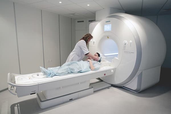
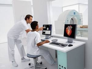
To eliminate the patient’s fear and mistrust, before the session it is explained to him how MRI is performed and how long the diagnosis takes. All actions on the part of the patient are quite simple. He needs to remove all metal objects from his body, then change into special clothes, and then lie down on a moving table for scanning.

The patient's body is fixed with belts, and cushions are placed under certain places. Next, the table moves into the cylindrical tube of the device, and the tomograph is turned on, which scans the patient’s body. If necessary, the patient can contact the doctor through a special microphone.
MRI of three parts of the spine can last different times: from 5 to 30 minutes in normal mode, up to 90 with contrast. After diagnosis, the doctor must collect the results, analyze them and transfer the transcript to the patient. If diagnostics with contrast are necessary, a simple MRI is still performed before it. Next, a scan is performed in three areas with a contrast agent.
If a person does not suffer from allergies and is not observed by a nephrologist, the diagnosis will be carried out without side effects.
Recommended articles on the topic:
- Manual therapy of the cervical spine as a worthy replacement for alternative treatment
- Therapeutic back massage: main types, indications and contraindications
- Myostimulation of body muscles: indications, contraindications and features of the procedure
Contraindications for MRI of the brain
There are a number of contraindications for this type of examination, which are absolute for patients who have:
- Implanted metal-containing pins, crowns and screws in the oral cavity. The ban is due to the fact that the powerful magnetic fields generated by the tomograph have the ability to heat metal objects. Due to the impact on the implants, the latter can shift, expand and cause additional injury to the person.
- CNS stimulants embedded in soft tissues. They can also fail under the influence of the device's fields.
- Metal staples and clamps on the vascular mesh.
- MRI of the brain is not indicated for patients with integrated cochlear implants.
- Foreign metal-containing objects of fragmentation nature in the soft tissues of the organ and the bone shell of the skull.
The following contraindications are conditional for prohibiting the procedure:
- severe post-traumatic condition of the patient, recently undergone surgery;
- decompensation of the heart and other organs;
- taking medications aimed at stimulating the central nervous system;
- implanted fillings containing metal inserts, wearing braces.
What does an MRI of the three parts of the spine show?
The obtained images are analyzed by a radiologist or a functional diagnostics specialist. The current state of the spinal systems is compared with early photos or with a picture of a healthy spine. This is how the doctor identifies various disorders (hernia, osteochondrosis, etc.), their stage and effect on the body. Next, the possibility and type of treatment is determined.
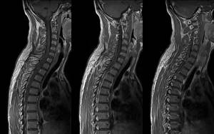
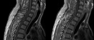
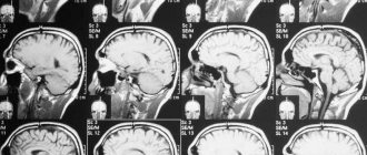
Magnetic tomography of the spine is an effective way to identify the problem and monitor it (for example, before and after surgery). The magnetic scanning method can be used repeatedly to make and clarify the diagnosis. The photographs clearly show the structure of bones, cartilage tissue (colored in a dark color), and the spinal cord (colored in a light color).
MRI of three parts of the spine and coccyx allows:
- see possible disturbances in the development of the spine, determine the extent of the injury;
- identify foci of inflammation, various neoplasms in tissues;
- assess the size of pathologies, as well as the type of disorder;
- analyze the condition of blood vessels, nerves and bone tissue;
- identify osteochondrosis, inflammatory processes in the spinal cord;
- determine the presence of a hernia (in the picture it looks like a protrusion of intervertebral discs, muscles, longitudinal ligaments).
How much does an MRI of three parts of the spine cost?
A magnetic tomography machine is a fairly expensive device, so not all public and private clinics can afford it. Due to the high cost of the tomograph, the price of MRI diagnostics is also high. However, this method gives the clearest picture of disorders and pathologies in the human body. The method allows you to see the problem in the early stages of its manifestation.
In a public clinic you can get a free MRI examination. To do this, you need to take a referral from your attending physician and stand in line for diagnostics. This waiting period can last several months. For many patients, such a loss of time is unacceptable, then you can contact a private clinic.
The approximate cost of a thoracic scan is 4,000 rubles. But for a complete picture, a photo of one department is usually not enough. For a more reliable result, MRI of three or four parts of the spine is used. The price will be quite high, but this study is extremely necessary to identify all pathologies.
Read material on the topic: Manual therapy of the cervical spine - features of the technique
Contraindications for abdominal MRI
The patient is not allowed to undergo the MR procedure if:
- The person's weight exceeds the maximum allowable for using the MR unit. Currently, most devices are manufactured with a patient weight limit of up to 120 kilograms.
- Abdominal girth from seventy centimeters. Tomography results may be incorrect.
- There is an electronic device built into the body, such as a pacemaker. Any electronics fails under the influence of magnetic fields.
- The presence of metal clamps on vessels installed during surgery.
An MRI of the abdominal cavity will be questionable in the following cases:
- if the person previously had an insulin pump implanted;
- if a person is in critical condition and is connected to a life support system;
- if the patient’s skin has tattoos with metallic inclusions.
It is worth noting that the latest generation MR devices are designed in such a way that they do not allow the patient to be disconnected from the ventilator systems. Even with the need for constant monitoring and maintenance of the heartbeat rhythm, it has become possible to diagnose diseases using tomography.
Myelography and its differences from MRI of three parts of the spine
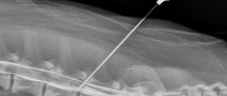
For some problems, conventional tomography is not enough. Then an additional procedure is used - myelography. Its essence is that a contrast agent is injected into the subarachnoid space of the spine, after which a scan is performed.
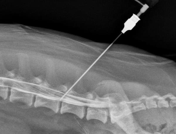
Next, a standard MRI of three parts of the spine is performed, which allows you to see how the contrast agent spreads.
Advantages of myelography:
- the procedure allows you to see the contours and nerve roots (conventional scanning does not provide this information);
- myelography reveals neurological problems, arachnoiditis, damage to the processes of the spinal nerves;
- helps to identify the causes of disturbances in the movement of cerebrospinal fluid;
- makes it possible to see spinal stenosis;
- accurately determines the presence of osteochondrosis, hernia;
- indicates the cause of compression of the spinal canal.
Myelography using Povidone-Iodine or Lipiodol makes it possible to see a three-dimensional image of the spinal cord. The picture displays the smallest parts of tissue structures, thanks to which you can recognize any violation and understand its cause. This procedure is completely harmless to the body. However, the method has contraindications: severe arthritis, previous spinal surgery, disorders in its structure.
Also, myelography followed by MRI of three parts of the spine is contraindicated in case of high body temperature, pregnancy, kidney disease, heart disease in the stage of decompensation, etc. 6 hours before the procedure, you should refuse to eat. If the patient is prescribed a lumbar puncture, a cleansing enema is used before therapy. In some cases, side effects from the administration of a contrast agent are observed. To get rid of its traces in the body, you need to drink as much water as possible.
Simple MRI and tomography with a contrast agent are effective ways to analyze the spinal cord and its pathologies. Scanning is necessary to correctly identify the problem and make a diagnosis. MRI is subsequently used to monitor treatment results. The spine is one of the main structures in the body; the proper functioning of many organs depends on it. If there are abnormalities, it is better to do a tomography as soon as possible in order to identify the pathology as early as possible and begin treatment.
Read material on the topic: Physical therapy for spinal hernia
Contraindications to MRI examination in childhood
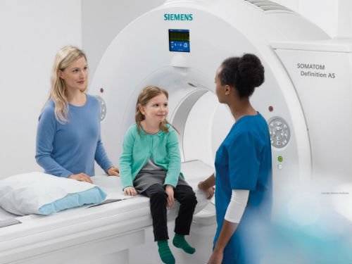
The MRI procedure in children has its own distinctive features. To maintain complete immobility during the session, the child is prescribed a preliminary sedative or general anesthesia.
Absolute contraindications include severe bronchial asthma and allergic reactions to medications.
Computed tomography and MRI of three parts of the spine
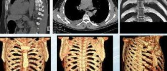
Computed tomography (CT) is a way to get a three-dimensional view of the problem area of the body. This is the main difference from the magnetic resonance method. A three-dimensional image is obtained by taking several photographs at once, taken from different angles of view. The resulting images are processed by a computer program, which creates a picture in 3D format.
In what situations is CT indicated for patients:
- if necessary, examine disorders in bone and joint tissues;
- to identify and analyze traumatic brain injury;
- to determine pathologies of the spinal column (hernias, brittle bones, spinal deformity);
- to analyze the condition of blood vessels (for aneurysm, atherosclerosis);
- if necessary, examination of the chest, abdominal, and pelvic organs.
CT provides a clear image of stones, hollow tumors filled with fluid, and oncological tumors.
The main differences between MRI of the three parts of the spine:
- MRI is commonly used to analyze soft tissue, joints, and blood vessels;
- the procedure is necessary to identify or confirm the presence of tumors in tissues;
- examination of the nerves of the skull and parts of the brain;
- analysis of the condition of the meninges;
- the method is used to examine the brain after cases of circulatory disorders;
- magnetic tomography examines people with neurological disorders;
- using this method, the condition of the connective tissue cords that connect the bones and support the organs in their positions is analyzed, and the muscle tissue is examined;
- identify disorders of the health of joints and bones.
The CT method works better for pathologies of bone tissue and organs. MRI of three parts of the spine is used to identify diseases of muscles, tendons, synovial membranes, to examine connective tissue, blood vessels and nerves.
Computed tomography is a radiation exposure to the human body. However, modern devices make it possible to minimize the negative effects of rays. During diagnostics, the radiation does not last long, and this does not depend on the time of the session. For example, even with hour-long scans, the effect of the rays is minimal. MRI is a painless and safe procedure that, in the absence of contraindications, does not carry any side effects.
Nowadays, you no longer have to spend a lot of time performing complex and unpleasant procedures at home. It is much easier to seek help from real professionals - the Veronika Herba beauty and health center, equipped with effective and modern equipment.
Why clients choose Veronika Herba Beauty and Health Center:
- This is a beauty center where you can take care of yourself at a reasonable cost, while your face and/or body will be treated not by an ordinary cosmetologist, but by one of the best dermatologists in Moscow. This is a completely different, higher level of service!
- You can receive qualified help at any time convenient for you. The beauty center is open from 9:00 to 21:00, seven days a week. The main thing is to agree with your doctor in advance on the date and time of your appointment.
Sign up for a consultation with a specialist by phone +7 (495) 085-15-13
, and you will see for yourself!