- Home /
- Branches /
- Ultrasound diagnostics /
- Triplex scanning of the abdominal aorta and its visceral branches
At the Clinical Hospital on Yauza, triplex scanning of the abdominal aorta and its visceral branches is performed - a highly informative non-invasive study of blood vessels.
The abdominal aorta is its descending part, located in the retroperitoneal space between the abdominal organs and the muscles of the spine. The visceral branches of the abdominal aorta are relatively small arteries extending from it to the internal organs: to the kidneys, to the spleen, to the intestines, etc.
At the Yauza Clinical Hospital, the abdominal aorta and its visceral branches are examined using triplex scanning. The examination is performed by experienced doctors from the ultrasound diagnostics department who have the highest medical category and academic degrees. The department uses modern diagnostic equipment, including Accuvix A30 ultrasound scanners manufactured by Samsung Medisson, South Korea HIVision Prerius - Hitachi, Japan.
Triplex scanning of the abdominal aorta and its visceral branches is a comprehensive expert-level ultrasound examination, including 3 diagnostic techniques.
- ultrasound diagnostics in gray scale scanning mode (B-mode),
- color Doppler mapping (CDC),
- spectral analysis of blood flow based on Doppler ultrasound (USDG).
What is ultrasound of abdominal vessels?
Ultrasound Dopplerography of abdominal vessels (ultrasound Dopplerography, or duplex scanning technique) is a procedure that allows you to study blood flow in the abdominal aorta, celiac trunk, vena cava and its branches, inferior vena cava and its branches, vessels responsible for the blood supply to the digestive organs. The method is based on the Doppler effect - the ability of blood particles to reflect waves emitted by an ultrasound sensor.
This procedure makes it possible to diagnose abdominal vascular diseases, dysfunction of the liver, kidneys, and pancreas at an early stage.
Doppler ultrasound is widely used in medical practice. This procedure has no contraindications or age restrictions. It is painless, safe and is performed without introducing contrast agents into the human body.
Aortic aneurysm
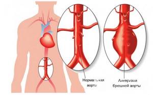
The aorta is the largest, most powerful blood vessel in the human body. Powerful, therefore, it seemed that nothing “took” him. However, aortic aneurysm is the scourge of modern cardiovascular surgery. In normal conditions, in adult women and men, the diameter of the lumen of the ascending aorta is about 3 cm, the descending part is 2.5 cm, the abdominal segment of this large vessel is even smaller - 2 cm. The diagnosis of an aneurysm is announced only if the diameter of the affected aorta increases by 2 or more times compared to the norm.
An aneurysm is an abnormal bulge that occurs in the walls of an artery. The walls of the arteries are quite thick and strong, the muscle fibers of which they are composed allow them to withstand intense blood pressure. However, if there is a weak area in the artery wall, the pressure causes the area to bulge, thereby forming an aneurysm.
An aortic aneurysm can develop in two parts of this artery:
- the abdominal part passing through the lower part of the abdominal cavity is an abdominal aortic aneurysm;
- A thoracic aortic aneurysm developing in the chest cavity. This type of aneurysm is less common, but both types are equally dangerous to human health and life.
Depending on the appearance, the aneurysm can be: 1. fusiform 2. saccular.
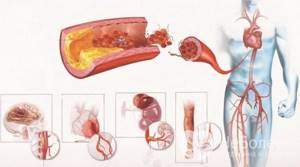
Small aneurysms usually pose no threat. However, they can increase the risk of: the formation of atherosclerotic plaques at the site of the aneurysm, which cause further weakening of the artery walls; formation and separation of a blood clot, therefore increasing the risk of stroke; an increase in the size of the aneurysm, which means compression of nearby organs, which causes pain; aneurysm rupture. The main complication of aneurysms of any location is their dissection followed by possible rupture (mortality rate - 90%).
Causes and risk factors
The main causes of aneurysm are diseases and conditions that reduce the strength and elasticity of the vascular wall:
- atherosclerosis of the aortic wall (according to various sources, from 70 to 90%); inflammation of the aorta (aortitis) of a syphilitic, giant cell, mycotic nature;
- traumatic injury;
- congenital systemic connective tissue diseases (for example, Marfan or Ehlers-Danlos syndrome);
- autoimmune diseases (nonspecific aortoarteritis);
- iatrogenic causes caused by medical manipulations (reconstructive operations on the aorta and its branches, cardiac catheterization, aortography).
Risk factors for the development of atherosclerosis and aneurysm formation:
- male gender (the incidence of aneurysms in men is 2–14 times higher than in women);
- smoking (during screening diagnostics of 455 people aged 50 to 89 years in the Department of Vascular Surgery of the Moscow Regional Research Clinical Institute, it was revealed that 100% of patients with abdominal aortic aneurysms had a smoking history of more than 25 years, and as a result of the Whitehall study it was proven that life-threatening complications of aneurysms occur 4 times more often in smokers than in non-smokers);
- age over 55 years;
- family history;
- long-term arterial hypertension (blood pressure above 140/90 mm Hg);
- physical inactivity;
- excess body weight;
- increased blood cholesterol levels.
They also talk about a dissecting aneurysm, which is formed as a result of rupture of the inner membrane with its subsequent dissection and the formation of a second false channel for blood flow.
Depending on the location and extent of the dissection, 3 types of pathology are distinguished: 1. Dissection begins in the ascending aorta and moves along the arch (50%). 2. Dissection occurs only in the ascending aorta (35%). 3. Dissection begins in the descending aorta and moves down (more often) or up (less often) along the arch (15%). Depending on the duration of the process, a dissecting aneurysm can be: acute (1–2 days from the appearance of the endothelial defect); subacute (2–4 weeks); chronic (4–8 weeks or more, up to several years).
SYMPTOMS OF AN AORTIC ANEURYSM
Aortic aneurysm manifests itself in different ways - it mainly depends on the size of the aneurysmal sac and its location (below is a visual clinical picture using the example of an aneurysm of the sinus of Valsalva). In some cases, no symptoms are observed at all (in particular, before the aneurysm ruptures, but this will be a different diagnosis), which makes early diagnosis difficult. The most common complaints from patients with an aneurysm of the ascending aorta: pain in the chest (in the area of the heart or behind the sternum) - due to the fact that the aneurysmal protrusion presses on nearby organs and tissues, as well as due to the pressure of the blood flow on thinned and weak wall; shortness of breath, increasing over time; feeling of palpitations (“As if something is pounding in the chest” - a comment from patients); dizziness; with large aneurysms, attacks of headaches, swelling of the soft tissues of the face and upper half of the body are disturbing - due to the development of the so-called superior vena cava syndrome (because the aneurysm presses on the superior vena cava).
An aortic arch aneurysm is characterized by:
- difficulty swallowing (due to pressure on the esophagus);
- hoarseness of the voice, sometimes coughing - if the aneurysm puts pressure on the recurrent nerve, which is “responsible” for the voice;
- suddenly increased salivation and slow pulse - if pressure spreads to the vagus nerve, which controls salivation and pulse rate;
- strained breathing, and later shortness of breath if the trachea and bronchi are compressed by a huge aneurysm;
- unilateral pneumonia - if an aneurysm, pressing on the root of the lung, interferes with its normal ventilation, then, as a result, congestion occurs in the lungs, which, when an infection occurs, develops into pneumonia.
With an aneurysm of the descending aorta, the following appear:
- pain in the left hand (sometimes up to the fingers) and shoulder blade;
- with pressure on the intercostal arteries, a lack of oxygen supply to the spinal cord may develop, which is why paresis and paralysis are inevitable;
- in case of constant long-term pressure of a large aneurysm on the vertebrae, they may even be displaced;
- in milder cases, due to pressure on the intercostal nerves and arteries - pain, as with radiculitis or neuralgia.
The most common complaints with an aneurysm of the abdominal aorta:
- a feeling of fullness in the stomach and heaviness in the epigastrium (upper floor of the abdomen), which the patient initially tries to explain by overeating or stomach pathology;
- belching;
- in some cases, reflex vomiting (appears as a reaction to the pressure of the aortic aneurysm on nearby organs and tissues);
- When palpated, a tense, tumor-like pulsating formation is felt. Sometimes patients can independently detect this pulsation.
DIAGNOSIS OF AORTIC ANEURYSMS AND ITS COMPLICATIONS
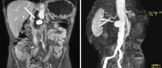
An aortic aneurysm in the period before rupture has rather meager clinical manifestations: murmurs that are heard on auscultation; the doctor listens not only to the chest, but also to the abdominal cavity; a tumor-like pulsating formation, which is found with deep but careful palpation (sometimes it is actually regarded as a tumor, since it is quite dense to the touch); incomprehensible discomfort at the site of aneurysmal protrusion formation. Therefore, to clarify the pathology before it “gives birth” to dangerous complications, instrumental diagnostic methods are used: fluoroscopy and radiography of the chest and abdominal cavity - they visualize a tumor-like formation (its pulsation is visible during fluoroscopy); echocardiography - if an aneurysm of the ascending aorta is suspected; Doppler ultrasound (USDG) - for signs of aneurysm in other parts of the aorta; CT and MRI.
TREATMENT AND SURGERY FOR AORTIC ANEURYSM
If an aneurysm is diagnosed, but its progression is not observed, doctors adopt conservative tactics: further careful observation by a vascular surgeon and cardiologist - monitoring the general condition, blood pressure, pulse, repeated electrocardiography and other more informative methods to monitor the possible progression of the aneurysm and notice in time the prerequisites for complications of an aneurysm; antihypertensive therapy - in order to reduce blood pressure on the thinned wall of the aneurysm; anticoagulant treatment - to prevent the formation of blood clots and possible subsequent thromboembolism of medium and small vessels; reducing the amount of cholesterol in the blood (using both drug therapy and diet). Surgical intervention is resorted to in the following cases: large aneurysms (at least 4 cm in diameter) or with a rapid increase in size (by half a centimeter in six months); complications that threaten the patient’s life - aneurysm rupture and others; complications that, although not critical from the point of view of death, sharply reduce the patient’s quality of life - for example, pressure on nearby organs and tissues, which causes pain, shortness of breath, vomiting, belching and similar symptoms.
PROGNOSIS FOR AORTIC ANEURYSM
Aortic aneurysm is a nosology that should be constantly under close monitoring by doctors. The reason is possible complications, which in most cases threaten human life. Over time, the aneurysm progresses morphologically (the altered wall becomes thinner and thinner, the protrusion increases). The life and health of a patient can be saved only through careful monitoring of the course of the disease and, if necessary, immediate surgical intervention.
PREVENTIVE MEASURES
Prevention, thanks to which it is possible to prevent the occurrence of aortic aneurysm in healthy people, is nonspecific (that is, effective not only in the case of this pathology) and includes: complete cessation of smoking; reducing alcohol standards to the level of “only for holidays”, or better yet, a complete refusal; physical education and sports; elimination of factors that cause a rise in blood pressure (stress, kidney disease); treatment and prevention of pathology that contributes to the formation of aortic aneurysm (atherosclerosis); immediate alertness in the event of a sudden, at first glance inexplicable appearance of interruptions in the functioning of the heart, gastrointestinal tract and respiratory system and immediate examination by specialized specialists to exclude an aortic aneurysm; regular, high-quality, not just for show, medical examinations with a vascular surgeon and cardiologist. If an aortic aneurysm is already present, preventive measures are indicated in order to prevent complications of this disease: well-chosen anticoagulant therapy to prevent the formation of blood clots in the lumen of the aneurysm; a significant reduction in physical activity - otherwise they can cause overstrain of the thinned wall of the aneurysm, which will result in its rupture; sometimes a complete refusal of physical activity is necessary until the doctor clarifies the diagnosis and assesses the risk; antihypertensive treatment - thanks to it, it is possible to avoid an increase in the pressure of the blood flow on the thinned wall of the aneurysm, which can rupture at any moment; careful psychological control - in some patients, even minor stressful situations pushed the aortic aneurysm to rupture.
Indications for use
Having understood what ultrasound of the abdominal aorta is, you need to identify the indications for prescribing this procedure.
Feeling of heaviness
A feeling of heaviness in the abdominal area may not always be associated with overeating, poisoning or eating junk food. Heaviness may be a symptom of kidney or liver failure, impaired blood flow in the vessels of the abdominal cavity that feed the digestive organs. Dopplerography of abdominal vessels (ultrasound of the abdominal cavity) will allow you to accurately determine the quality of blood flow and assess in what quantity and at what speed blood flows to the stomach, liver, kidneys and other organs.
Bloating
Bloating accompanies many diseases associated with the liver, heart, kidneys, stomach and other internal organs. To exclude or confirm the presence of hemodynamic disturbances in the vessels, it is necessary to perform an ultrasound scan of the vessels of the abdominal aorta.
Pain syndrome
Pain in the abdominal cavity may bother patients after eating or be permanent. Pain may be associated with pathology of the internal digestive organs or a disturbance in the functioning of blood vessels. Thus, an abdominal aortic aneurysm may be accompanied by abdominal pain. Other blood flow pathologies accompanied by pain include acute or chronic ischemia of the mesentery, ischemic colitis (when there is insufficient blood flow to the colon), and vasculitis.
Duration of the abdominal aorta ultrasound procedure
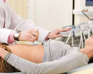
There are no time restrictions when performing ultrasound or color duplex mapping, but according to the standards, 30-40 minutes are allotted for one patient.
This is quite enough for a comprehensive assessment of the abdominal aorta, its branches and internal organs. The diagnostic procedure takes on average about 30 minutes and another 10-15 minutes will be required for the doctor to fill out the research protocol. The time spent on an ultrasound scan of the abdominal aorta depends on the human factor and the location: in private clinics, where the flow of patients is smaller, ultrasound scanning with Doppler sonography is performed longer. The subject should not worry: ultrasound does not imply radiation exposure to the body and can be performed as much as a specific clinical situation requires. In unclear cases, other sonologists are invited to the diagnostic room, and then the presumed diagnosis is established collectively. The final cause of the disease is determined by the attending physician, taking into account all the examination data.
Preparation for ultrasound examination
Ultrasound scanning of abdominal vessels requires special preparation.
Diet
The day before diagnosis, the patient needs to exclude the following foods from the diet: dairy and fermented milk, black bread, soda, sweets and flour, cabbage, legumes. In other words, it is not recommended to consume foods that contribute to excessive gas formation in the intestines.
Body cleansing (one day before the procedure)
A day before the procedure (or two days before, as recommended by the doctor), the patient needs to take 4-5 tablets of activated carbon 3 times a day. You can replace charcoal with any other carminative drug after consulting your doctor. If the patient's digestion is impaired, he is recommended to take the enzyme preparation after meals. The final meal is 5 hours before bedtime. The study is carried out on an empty stomach.
What surgical treatment methods are there?
Currently, there are two methods of surgical treatment:
- open surgery – removal of an enlarged section of the abdominal aorta and replacing it with a synthetic prosthesis through an incision in the abdomen (prosthetics);
- placement of a synthetic prosthesis in the cavity of the aneurysm through small incisions in the groin (endoprosthetics). Unfortunately, this operation has very strict indications and is therefore only available to a small number of patients. In addition, the number of reoperations after such an intervention is significantly higher than the number of complications than after open surgery.
Doppler ultrasound indicators
When analyzing and interpreting the results of Doppler ultrasound of the abdominal vessels, the doctor takes into account the following indicators:
- vascular patency;
- diameter and features of the location of the lumen of the vessel;
- length of visibility of the changed lumen;
- speed of blood flow in arteries and veins.
If blood clots or atherosclerotic plaques are detected (which should not normally be present), the doctor must determine their exact location, analyze the structure and density.
How is the operation performed?
The operation is performed under general anesthesia (anesthesia). You will be informed separately about the details and risks of each anesthesia method.
The purpose of the operation is to exclude the dilated part of the abdominal aorta from the bloodstream and replace it with a synthetic prosthesis. To do this, through an incision along the midline of the abdomen or a lateral incision through the chest and abdomen, the aneurysm is exposed, the aorta above and below the aneurysm is clamped (so that blood does not flow into the abdomen during the operation) and an incision is made along the anterior wall of the aneurysm. A synthetic graft is then sutured between the two relatively intact ends of the abdominal aorta.
If the expansion continues to the iliac arteries or the iliac arteries are narrowed, this expansion is also eliminated, only in this case an already bifurcated bifurcation prosthesis is sewn on (patients also call it “pants”).
If there is damage to the arteries supplying blood to the kidneys (renal) or intestines (mesenteric arteries), then, if necessary, it is also possible to replace them with a vascular prosthesis.