What are the main indications for the procedure? How do you need to prepare to do an ultrasound of the blood vessels of the legs? What can a diagnostic specialist see?
Author:
- Dehissi-Golbrekht Muad Alfredovich
ultrasound diagnostic doctor, phlebologist, surgeon, proctologist
4.92 (Votes: 12)
The lower limbs are an important element of the human musculoskeletal system, which can be susceptible to a number of diseases, pathologies, syndromes and altered conditions. Ultrasound of the vessels and veins of the legs allows us to identify individual or complex disorders of blood flow in the specified location and help in establishing the correct diagnosis.
What are the main indications for the procedure? How do you need to prepare to do an ultrasound of the blood vessels of the legs? What can a diagnostic specialist see? You will read about this and much more in our article.
Types and purposes of ultrasound of the veins of the lower extremities
In recent decades, ultrasound diagnostics has become a simple, inexpensive, and most importantly, accessible research method.
The quality and capabilities of ultrasonic devices are constantly being improved, their classification is constantly expanding. The great advantage of the technique is its non-invasiveness. Vein ultrasound is no exception. Modern ultrasound scanners allow a thorough diagnosis of the venous system.
The main goals of ultrasound of the veins of the lower extremities are to assess the condition of the vessels (their diameter, course, relationship with other structures, wall condition), examine blood flow parameters, and identify blood clots. The article will talk about the types and purposes of ultrasound examination of blood vessels, as well as how ultrasound of the veins of the lower extremities is performed.
Due to the constant improvement of devices, many terms have appeared to denote various components of ultrasound examinations and their combinations.
Let's try to figure out what ultrasound, DS, ultrasound, ultrasound of veins are.
- Doppler ultrasound (Doppler ultrasound) is a method based on the Doppler effect. This effect consists of changing the frequency of an ultrasonic wave when reflected from a moving object. In the lumen of the vessel, such objects are formed elements of blood, for example, red blood cells. Computer processing of reflected signals allows one to obtain a curve of the speed characteristics of blood flow. Currently, ultrasound scanning in its pure form is not used, but is included in the structure of other studies.
- DS (duplex scanning (USD)) - allows you to visually evaluate the vessel - its wall, course, lumen, as well as conduct an ultrasound scan of any part of the vessel - i.e. in addition to examining the vessel, show the characteristics of blood flow at any point. Modern devices also use coloring of blood flows in blue and red colors, based on the Doppler effect (the so-called color Doppler or color Doppler mapping (CDC)). Combining duplex scanning with color flow is also called triplex scanning.
- USAS (ultrasound angioscanning) - a synonym for DS, is the same.
- Ultrasound of veins (ultrasound examination of veins) - this term refers to the entire combination of the above methods. Modern devices provide all the components of the study - ultrasound, DS, and color Doppler mapping (CDC).
Currently, when a doctor prescribes an ultrasound scan of the veins of the lower extremities, an ultrasound scan of the veins of the lower extremities, he means that the patient will undergo an ultrasound scan of the veins according to all possible parameters.
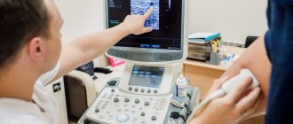
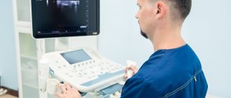
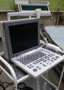
Ultrasound equipment
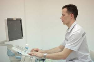
To conduct research, devices are used that are designed for visual diagnostics of the human body. Ultrasound rooms are equipped with multifunctional equipment designed to meet stringent requirements. Modern models of devices have ultra-precise focusing and compact dimensions. They provide high quality images.

To improve performance, the devices are equipped with various modes. For example, two-dimensional visualization, as well as color and power Doppler. Many models are equipped with glare-free monitors that rotate freely around an axis and tilt in the desired direction. It is possible to connect several sensors simultaneously. The received information is recorded on the hard drive and stored in a database.
When developing ultrasound machines, new technologies are being introduced: grain suppression, image detailing, automatic image optimization, wireless information transfer. The scanners are easy to operate. If necessary, a specialist can adjust the settings. The image is instantly transferred to the monitor. The doctor examines the details and assesses the situation.
There are two types of equipment on sale: stationary ultrasound and portable ultrasound. Stationary devices are installed in rooms designed for ultrasound examinations of leg vessels. They are impressive in size and provide reliable diagnostics. Portable devices are portable and small in size. Such devices are designed to conduct research in any place: ward, ambulance, emergency department. They are indispensable in cases where it is necessary to urgently determine a person’s condition and take the right actions.
Who needs to undergo Doppler ultrasound of the extremities?
An ultrasound examination of the veins is usually prescribed by a doctor (surgeon, phlebologist, therapist). Indications for it can be a wide variety of conditions. Most often, the doctor prescribes a study if there are complaints and suspicions of diseases of the venous system. In other cases, ultrasound of the veins is prescribed to exclude hidden pathology of the venous system (for example, before operations, during pregnancy).
The main indications for prescribing ultrasound examination of veins are:
- Swelling of one or both legs
- Leg pain
- Feeling of heaviness and fatigue in the legs
- Night cramps in the legs
- Varicose veins, spider veins
- Before any operations
- Late pregnancy
Differences between duplex and triplex scanning of vessels
In recent years, triplex research, created on the basis of duplex, has become increasingly popular. Triplex vein scanning is not as significant a difference from duplex scanning as it might seem. This is the same set of techniques, supplemented by color coding, thanks to which you can see the direction of blood flow inside the vessels in color. Otherwise, triplex also allows you to assess blood flow, the condition of the vein walls, and the presence or absence of blood clots. Moreover, much more often this type of study is used to diagnose the vessels of the head and neck; for the lower extremities, duplex ultrasound scanning is quite suitable.
How is the examination done?
Many patients are concerned about the question of how ultrasound examination is performed, whether there are any contraindications or painful sensations during the examination.
Ultrasound diagnostics is non-invasive, painless, does not cause any discomfort, and has no contraindications. An ultrasound can be performed either by an ultrasound diagnostics doctor or by a surgeon or phlebologist who knows this method and has undergone appropriate training.
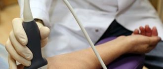
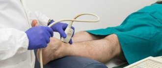
The examination of the veins of the lower extremities is carried out from the inguinal fold to the ankles. The patient completely frees his legs (panties can be left on) and lies down on the couch. The doctor applies a special gel to the lower limbs, along which the sensor will be moved during the examination. The gel is needed to ensure good contact between the skin and the sensor so that there is no obstacle to the passage of ultrasonic waves. The examination can be carried out both lying down and standing. At certain moments, the doctor may ask you to hold your breath, strain, bend your leg, etc.
The study lasts from 10 to 40 minutes (depending on the structural features of the veins and the identified pathology). For example, detected varicose veins increase the examination time, because it is necessary to carry out a number of additional tests. At the end of the study, the doctor gives the patient paper napkins to remove any remaining gel. The gel is absolutely harmless and does not leave stains on clothes.
Then the doctor writes a conclusion and hands it to the patient. It should be remembered that the conclusion itself is not a diagnosis, but only describes the identified ultrasound signs. The diagnosis can only be made by a clinician (surgeon, phlebologist), based on a comparison of the ultrasound picture, examination data and other examinations. In many cases, an ultrasound of the veins is performed not by an ultrasound diagnostics doctor, but by a phlebologist who is proficient in vein ultrasound. Then a clinical diagnosis can be made immediately.
How to decrypt data
Only a doctor can determine what an ultrasound examination of the veins of the lower extremities shows.

The doctor evaluates:
- Vein diameter
- Condition of the vascular wall (irregularities, thickening)
- The condition of the veins of the right and left legs is compared
- Valve condition
- Presence of pathological blood flows (refluxes)
- Vessel lumen (presence of blood clots, their structure, level of extent, apex)
- Presence of varicose veins
- Presence of incompetent perforators (veins connecting superficial and deep veins)

If during the examination the doctor notices changes in other structures that are not related to the veins (for example, pathology of the arteries, Baker's cyst, altered lymph node), he may note this in the protocol or recommend additional research.
Based on the data received, the doctor writes a conclusion; if necessary, photographs are attached to the conclusion.
Example of a conclusion: “Echo signs of varicose veins of the lower extremities. Insufficiency of the great saphenous vein on the left. Incompetent perforator of the left leg." It should be noted that the classification of varicose veins according to this conclusion is impossible - the stage is determined by the attending phlebologist based on a comparison of the ultrasound picture with clinical signs.
Decoding and results
Ultrasound is an affordable imaging method that does not use harmful radiation. During the diagnosis, the doctor determines the thickness of the vascular membranes, pulsation index, and vascular resistance. The minimum and maximum speed of blood movement is also determined.
After conducting the study, the specialist receives accurate data that indicates the condition of the vascular system. It defines a number of important parameters and reflects them in the protocol. Based on such information, the doctor gives an opinion, according to which the vascular surgeon develops treatment tactics.
Using ultrasound examination of the vessels of the lower extremities, various diseases are detected:
- stenosis is a term that refers to the narrowing of blood vessels in the circulatory system. The development of such a pathology leads to undesirable consequences: muscle atrophy, ulcerative formations, pain. Stenosis appears in people who lead a sedentary lifestyle and are overweight. Diabetes mellitus also leads to changes in the structure of blood vessels
- Varicose veins are a serious disease accompanied by impaired blood flow. The trigger for the development of pathology is considered to be disruption of the venous valves with the subsequent occurrence of blood reflux. The main symptom of the disease is dilated saphenous veins. First, a person is bothered by a feeling of heaviness in the legs and a burning sensation, and then swelling appears. Pregnant women and people who spend a lot of time in one position (office workers, salespeople, drivers, bank tellers) are predisposed to varicose veins.
- Phlebitis is a disease in which the vein wall becomes inflamed. Its main symptoms include soreness, a slight increase in temperature, and redness of the skin. Phlebitis can be chronic or acute. The appearance of the disease is provoked by varicose veins, infection, and mechanical damage to the vessel. When diagnosing phlebitis, the doctor prescribes a set of therapeutic agents
- thrombosis - the formation of blood clots inside blood vessels. This is a pathological process because the free flow of liquid connective tissue through the system is limited. The key signs of the disease are severe swelling, pain and induration. Thrombosis may be asymptomatic. In such cases, the disease is detected by ultrasound. This problem cannot be ignored, because it leads to serious consequences.
Ultrasound of the vessels of the lower extremities is a study with which you can detect the disease in time and prescribe effective treatment. To carry it out, equipment with wide functionality is used. This is a guarantee that the results will be reliable.
Where to take the study
Currently in Moscow there are many medical centers that offer ultrasound diagnostics. What should you look for in order to get to a good specialist?
The doctor must have extensive experience, work experience, and have positive reviews from other patients. It is very good to have an ultrasound of the veins done by a doctor who not only knows ultrasound diagnostics, but is also a surgeon or phlebologist. Such a doctor has the most complete understanding of the anatomy and functioning of the venous system, knows well how ultrasound of the veins of the lower extremities is performed, and can also make a clinical diagnosis and prescribe treatment immediately after the ultrasound.

Dr. Elshansky Igor Vitalievich fully meets these requirements, is a phlebologist surgeon, and also has advanced training in ultrasound diagnostics in angiology, has been involved in the diagnosis and treatment of vascular diseases of the lower extremities for many years, Ph.D.