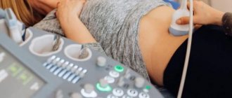
Ultrasound of the retroperitoneal space is a universal, non-invasive, affordable diagnostic method. With its help, a wide range of diseases are detected at an early stage. The study is often carried out in conjunction with an ultrasound of the abdominal organs, since these areas are located nearby, and the doctor can examine both at once. The information content of the method directly depends on the professionalism of the diagnostician and the quality of patient preparation
What organs are located in the retroperitoneal space?
This area is located between the intra-abdominal fascia and the posterior aspect of the parietal peritoneum. Vertically, it fills the space between the diaphragm and the small pelvis. It contains:
- pancreas (partially);
- ureters;
- kidneys and adrenal glands;
- inferior vena cava, abdominal aorta and its branches;
- roots of the azygos and semi-gypsy veins;
- intestines (partially).
The retroperitoneal space contains a large number of nerve endings and lymph nodes.
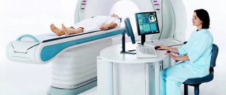
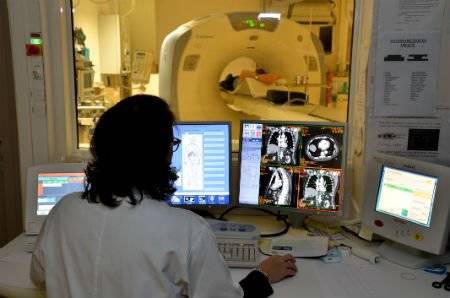
Computed tomography of the retroperitoneum
CT is used to diagnose various pathologies of the urinary system, kidneys, adrenal glands, blood vessels and other organs and tissues. The high information content of CT makes it possible to detect diseases of organs in the retroperitoneal space, predict their course and choose the most effective methods of treatment.
Indications for the study
There are a number of indications for performing a CT scan of the retroperitoneum. Scanning may be required:
- to localize an abscess or phlegmon;
- to study the post-traumatic state of organs;
- if you suspect cancer;
- to clarify diagnostic data before surgery;
- with the formation of stones in the ureter and kidneys, cysts in internal organs;
- to confirm the diagnosis when other examinations were insufficiently informative.
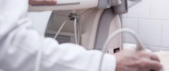
Procedure for magnetic resonance imaging of the abdominal cavity
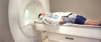
Magnetic resonance imaging allows you to see organs from the inside: the entire structure is scanned layer by layer, the images are transferred to a computer screen, where they are processed by a special program.
Magnets that create electromagnetic waves “shine through” the body. MRI allows you to avoid the use of invasive and laparoscopic diagnostic methods. The procedure is considered absolutely safe, as it does not expose the patient’s body to radiation.
Magnetic resonance imaging is suitable for examining soft tissue; bone structures are best seen on x-rays.
Abdominal examination includes the following:
- inspection of the contours and sizes of organs;
- assessment of blood flow, thickness and integrity of vessel walls;
- searching for the source of pain and the reason for referral for examination;
- examination of the condition of the lymph nodes.
Preparing for the study
CT scanning of the retroperitoneal organs must be performed with the intestine as empty as possible. Therefore, if the procedure is carried out in the morning, it is better to skip breakfast and make dinner light and liquid. If you need to prepare for a CT scan of the retroperitoneum scheduled for lunch, you should limit your breakfast. When performing the procedure in the evening, it is better to skip lunch.
The information content of retroperitoneal CT results may be affected by increased gas formation. Therefore, on the eve of the examination, it is not recommended to drink soda or eat heavy food.
Anatomy
Rice.
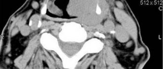
1. Scheme of a parasagittal cut of the lower half of the body through the area of the right kidney and pelvis (the dotted line limits the retroperitoneal space): 1 - diaphragmatic pleura; 2 - diaphragm; 3 - adrenal gland; 4 and 6 - liver; 5 - kidney; 7 - prerenal fascia; 8 - reduced mesentery of the ascending and descending colon (Toldt's fascia); 9— jumpers closing the renal space from below; 10 - ascending colon; 11 - paracolon; 12 - ureter; 13 - periureteral tissue; 14 - fascia surrounding the common iliac artery and vein; 15 - iliacus muscle; 16 - fascia iliaca; 17 - iliac crest; 18 - retrorenal fascia; 19 - fascia of the quadratus lumborum muscle; 20 - quadratus lumborum muscle; 21 - aponeurosis of the transverse abdominal muscle; 22 - XII rib. Rice. 2. Scheme of a horizontal cut of the body through the kidney area (the dotted line limits the retroperitoneal space): 1 - ascending colon; 2 - peritoneum; 3 - retroperitoneal tissue; 4 - transverse abdominal muscle; 5 - internal oblique abdominal muscle; 6 - external oblique abdominal muscle; 7 - right kidney; 8 - quadratus lumborum muscle; 9 - psoas major muscle; 10 - transverse spinalis muscle; 11 - retrorenal fascia; 12 - sacral spinal muscle; 13 - Perinephric fiber; 14 - pericolic tissue; 15 - prerenal fascia; 16 - descending colon; 17 - left kidney; 18— abdominal aorta; 19 - inferior vena cava; 20 - reduced mesentery of the ascending and descending colon. In front, the third section is limited by the posterior (dorsal) part of the parietal peritoneum (peritoneum parietale), in the rear by the posterior wall of the abdominal cavity (see Lumbar region), covered from the inside by intra-abdominal fascia (fascia endoabdominalis), from above by the diaphragm and phrenic fascia; down the 3rd point extends to the promontory of the sacrum (promontorium) and the border line of the pelvis (linea terminalis), where it passes into the subperitoneal space of the small pelvis. 3. p. is made of fatty and loose unformed connective tissue, in which the retroperitoneal organs with their vessels, nerves and nerve plexuses, lymph, and apparatus are located (color. Fig. 1 and 2).
In the 3rd section, three sections are distinguished: a median (according to Pirogov, “mediastinum of the abdomen”) and two lateral, lying on the sides of the spine and limited laterally by the line of transition of the parietal peritoneum from the lateral to the posterior abdominal wall. The middle section of the 3rd section corresponds to the bodies of the lumbar vertebrae and extends from the legs of the diaphragm, through the openings of the cut there is a connection between the mediastinum of the chest and the “mediastinum of the abdomen”, to the promontory of the sacrum. The loose, unformed connective tissue that fills this section of the 3rd section passes into similar tissue between the leaves of the mesenteries of the small and transverse colon. In the middle section of the 3rd section there are the abdominal aorta with its branches, the inferior vena cava, the beginning of the portal vein, part of the head and body of the pancreas, the horizontal part of the duodenum, prevertebral autonomic nerve plexuses, the lumbar section of the sympathetic trunk, numerous lymph nodes and the cistern thoracic lymph duct with its roots. The lateral sections of the 3. p. are also made of adipose and loose connective tissue, which contain the adrenal glands, kidneys, ureters, part of the head of the pancreas, the descending part of the duodenum, the ascending (right) and descending (left) colons, branches of the lumbar nerve plexus. Loose connective and adipose tissue in the lateral sections of the 3. p. are divided into several layers by the retroperitoneal (subperitoneal) fascia (fascia retroperitonealis, s. subperitonealis), the edges originate from the parietal peritoneum at the point of its transition from the side wall of the abdomen to the back.
Within the lateral sections of the 3rd section, 4 cellular spaces are distinguished. 1. The retroperitoneal fiber layer, or the retroperitoneal fiber itself (textus cellulosus retroperitonealis), is located between the intra-abdominal and retroperitoneal fascia. In the external sections, the retroperitoneal tissue itself is more abundant and anteriorly directly passes into the preperitoneal tissue, inwardly into the tissue of the middle section of the 3rd section, below into the pelvic tissue, at the top it reaches the diaphragm. 2. The perinephric cellular space (paranephron, s. capsula adiposa renis) is limited by the layers of the retroperitoneal fascia. Inwardly, this space passes into the tissue surrounding the aorta and the inferior vena cava, outside and above it is closed, and below it passes into the tissue surrounding the ureter. 3. The periureteric tissue space (paraureteron) is located around the ureter between the pre- and retroureteral fascia; downward it connects with the tissue lying behind the bladder. 4. The peri-colic cellular space (paracolon) lies anterior and lateral to the peri-renal space. At the top, the paracolon reaches the right or left flexures of the colon, at the bottom - the cecum (right) or the initial section of the sigmoid colon (left).
The degree of isolation of these spaces varies from person to person, which is associated with different expressions of the retroperitoneal fascia. The possibility of communication of the cellular layers of 3. items is used during diagnostic and treatment. measures (retropneumoperitoneum, lumbar and perinephric blocks, etc.).
Blood supply
organs and fiber 3. p. is carried out by the branches of the abdominal aorta: the celiac trunk, the superior and inferior mesenteric, paired renal, inferior diaphragmatic, middle adrenal and testicular (ovarian), lumbar arteries, between which numerous collateral anastomoses are formed, which play a significant role in disorders of blood circulation in the abdominal cavity. They not only connect all branches of the abdominal aorta, but also connect them with the above and underlying arteries (intercostal, superior epigastric artery - at the top, inferior epigastric, iliopsoas, deep artery, circumflex ilium, artery of the vas deferens, etc. - below ).
A more abundant collateral network in the 3rd section is formed by veins, which belong to three systems: the inferior and superior vena cava and the portal vein. Therefore, among the anastomotic venous networks in the 3rd section there are cavo-caval, porto-portal and portocaval anastomoses. All of the listed groups of anastomoses are of important functional importance in case of blood flow disorders in the inferior vena cava and portal veins. Lymphatic drainage. There are parietal and visceral lymph nodes, 3. p. Parietal lymph nodes include lumbar lymph nodes (nodi lymphatici lumbales), as well as common iliac nodes (nodi lymphatici iliaci communes). Visceral lymph nodes are very numerous and lie near the celiac trunk, along the splenic and gastroduodenal arteries, between the head of the pancreas and the duodenum, etc.
From the left part of the retroperitoneal lymphatic tissue proper, the vessels go to the lumbar nodes located near the abdominal aorta, and from the right part of the same layer - to the same nodes near the inferior vena cava. The lymph vessels of the peri-colic cellular spaces pass respectively to the right and left colic lymph nodes, lymph; the vessels of the peri-renal space reach the renal lymph nodes, and the vessels of the para-ureteral space reach the renal, lumbar and internal iliac lymph nodes.
Innervation
. Afferent innervation of the loose connective and adipose tissue of the third section, the organs located in it, the retroperitoneal and intra-abdominal fascia is carried out by sensitive spinal fibers running as part of the prevertebral autonomic plexuses (celiac, superior and inferior mesenteric, renal, aortic, ureteric, testicular, etc. ), as well as as part of the lumbar nerves.
Methodology
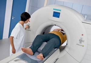
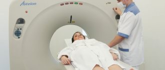
With a CT scan of the retroperitoneum, the patient is placed on a special table around which the tomograph moves. To ensure clear images, the patient should not move during the examination. A retroperitoneal CT scan lasts 15 minutes; with contrast, the examination can take up to half an hour.
Contrast enhancement
A retroperitoneal CT scan with contrast is prescribed when the doctor suspects cancer or in a number of other cases. The method of administering the drug is chosen by a specialist. It depends on which part of the retroperitoneal space CT scan with contrast needs to be performed, and what the examination should show (which diagnosis to confirm or refute).
Contraindications for CT of the retroperitoneum
Retroperitoneal CT is a noninvasive procedure that is considered safe for most patients. Contraindications include pregnancy or breastfeeding, the patient's inability to lie still - hyperkinesis.
For CT of the retroperitoneal space with contrast, relative contraindications will be diseases of the cardiovascular system, liver, kidneys and urinary tract, diseases affecting the condition of blood vessels. A CT scan of the retroperitoneum with contrast is not done in case of an allergy to the drug.