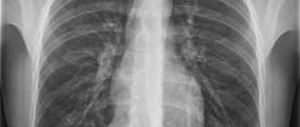
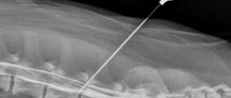
X-ray of the spine with functional tests
The most informative is considered to be radiography of the spine, which is performed in a vertical position.
To obtain maximum information, radiography is performed with a functional load - with bending, maximum flexion or extension in a vertical, sitting or horizontal position. It provides additional information to clarify the diagnosis. Functional tests are strictly individual in each specific case. They allow:
- assess the mobility of the spine: determine the full range of movements, functional blocks (limitations of mobility) in the selected segment;
- more accurately identify the degree and nature of vertebral displacement;
- plan orthopedic surgery.
How is the examination carried out?
Each part of the spine is examined differently and requires a different level of preparation. It is worth considering the conduct for each area separately.
X-ray of the cervical spine
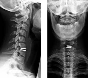
Radiation doses during X-rays of the spine do not exceed the norm, no matter what part we are talking about. To examine the cervical spine
, the patient may be asked to lie down on a special table or remain standing. When you need to get an image of vertebrae 1 through 3, you will need to take a targeted shot through the mouth. The patient is asked to open his mouth so that the shadow of the lower jaw does not cover the necessary vertebrae, after which an x-ray is taken. Functional tests may also be necessary - the patient needs to bend his neck as much as possible and touch his chin to his chest to take the first picture, and also tilt his head back as much as possible to take the second.
X-ray of the thoracic spine
X-rays of this area are not as frequent, which is associated with limitations in the mobility of the thoracic region
. If the procedure is still necessary, it can be performed standing or lying down, and the picture is taken in two projections - lateral and direct. Occasionally, oblique tests may be prescribed, as well as functional tests, which are very rarely necessary due to limited mobility. In the latter case, the doctor himself determines the optimal position and asks the patient to take it.
X-ray of the lumbosacral spine
You need to think not only about how often you can do the X-ray procedure of the spine
, but also how such research is carried out. After careful preparation, the patient is asked to lie on the table. AP and lateral views may be necessary, and the legs may need to be flexed.
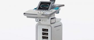
Expert equipment
The study is carried out on the latest generation X-ray system DIGITAL DIAGNOST (PHILIPS, the Netherlands). This is a completely digital x-ray station. Its advantages:
- Maximum data transfer and processing speed
- Obtaining the highest resolution (quality) image possible
- Lowest radiation exposure comparable to a short flight
The results of radiation diagnostics of the Yauza Clinical Hospital are accepted anywhere in the world, which is important for patients planning to undergo certain stages of treatment abroad.
Where can I get an X-ray of the spine?
You can find out how the doctor is treating you and make an appointment at a convenient time by phone or via the Internet at any time from 8 to 21 hours, without days off or breaks.
Make an appointment with a specialist, without queues, at a convenient time
+7
Sign up
The cost of the service is adjusted by the following basic parameters:
- the size of the diagnosed area of the spine;
- type of device, its technical capabilities;
- qualification of a radiologist;
- time: how urgently the research result is needed;
- type of delivery of the result - digital image, film, flash drive, disk;
- the use of additional reagents - MRI of the spine with a contrast agent.
Clinic No. 1 is located within walking distance from Volzhskaya and Maryino metro stations:
Moscow, st. Krasnodarskaya, house. 52, bldg. 2
+7
Alternative research methods
Sometimes patients wonder: what is better – CT, X-ray or MRI of the spine? Each method has its own capabilities, advantages and disadvantages.
- X-ray of the spine is a method that allows for examination with functional tests, as well as standing, with axial load and imaging along the entire length of the spinal column. The radiation dose with it is less than with CT. Limitations of the method - there is no way to assess the structure of the spinal cord and muscle corset.
- MRI is a method that allows you to evaluate the structure of the spinal cord, intervertebral discs and their herniated protrusions, joints, cartilage, nerve roots emerging from the spinal canal, soft tissues, muscle corset and identify inflammatory changes in the vertebral bodies.
- - an informative method for assessing bone structures with a higher resolution, but also with a higher radiation dose than with radiography. CT allows for 3D reconstruction of bones. Indispensable when planning operations. Compared to MRI, CT is significantly inferior in assessing soft tissue and cartilage structures.
Contraindications
Indications
The study is prescribed in traumatology, orthopedics and neurology for the diagnosis of congenital anomalies, pathological deformities and neuropathologies. Indications for this procedure are:
- pain syndrome;
- limitation of motor function;
- suspicions of Schmorl's hernia, protrusion, tumor processes;
- trauma and congenital anomalies;
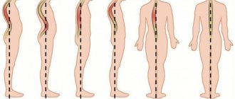
- pathological bending;
- loss of sensation in the lower extremities.
An x-ray allows you to identify the localization of the inflammatory process or traumatic lesion, primary tumors and metastases. Depending on the nature of the pathology, a change in the shape and size of the vertebrae, the stage of development of degenerative changes, and a violation of the integrity of various structures, including intervertebral discs, are revealed.
Identification points of the spine
In the cervical region, the identification point is the superior spinous process of the seventh cervical vertebra, especially clearly visible when the upper limbs are lowered. The line connecting the inner ends of the scapular spines passes through the spinous process of the third thoracic vertebra. The line connecting the angles of the shoulder blades passes through the spinous process of the seventh thoracic vertebra. The line connecting the highest points of the iliac crests passes through the spinous process of the fourth lumbar vertebra. When examining a normal spine, very limited sections are accessible by palpation - only the ends of the spinous processes. When running the palmar surface of the second finger along the spinous processes downwards, starting from the cervical region, you can even detect a slight protrusion of the spinous process posteriorly or to the side.
The localization of painful foci is determined by pressing the distal phalanx of the first finger on the spinous processes of the vertebrae from vertebra to vertebra, from top to bottom. The transverse processes are palpated away from the spinous processes. To determine the localization of the pathological process, tapping on the spine is sometimes used; the shock causes pain in the affected area. The same can be achieved by applying certain pressure on the head or shoulders along the axis of the spinal column. It should be noted that these techniques are quite crude and are not always applicable.
Electromyography (EMG)
With this method, biological muscle potentials are recorded. These data make it possible to assess the condition of the peripheral nerves that work with these muscles, as well as to assess the condition of the muscle tissue itself. If the muscles are damaged due to osteochondrosis, changes in biopotentials will be noticed. Electrodes on the skin are usually used for recording; needle electrodes, which are inserted directly into the muscle, are less commonly used. A variation of the method is stimulation myography, when nerves are stimulated with current through needle electrodes, and then the speed of impulse transmission along the nerve is recorded.
Remember that before using any spinal treatment methods or medications, you should consult with a specialist to know about possible contraindications and the applicability of these treatment methods and medications for you personally!
Preparatory procedures
It is imperative to prepare for an X-ray of the lower back in order to eliminate errors in the image. What should you do first?
- Adjust your diet. A few days before the intended procedure, you should completely avoid foods that can cause gas formation. These include fruits, brown bread, legumes, carbonated drinks, and dairy products.
- Cleanse the intestines. Any accumulated gases will obstruct the X-ray beams, so the image may appear blurry. On the eve of the study, you should do an enema with a decoction of herbs or warm water. An alternative method is to take a laxative.
- Remove all jewelry from yourself. Before irradiation, you will need to remove the piercing and remove all metal objects from clothing. It is advisable not to wear jeans if you are planning an examination, as the inserts on them cannot be removed.
- Sign up for an X-ray room. It is better to do this in the first half of the day so that you come on an empty stomach. In this case, the possibility of gas formation due to the process of digestion of food is eliminated.
Before coming for diagnostics, you do not need to eat, even light foods that may seem at first glance. Three hours is enough for the food to be already in the intestines and cause a decrease in the information content of the image.
No ads 2
Main contraindications
X-ray diagnostics of the cervical, thoracic or lumbar region is carried out for healthy people or those who do not have contraindications to it. Despite the safety of the technique, the dose of radiation can provoke exacerbations. Therefore, it is not recommended to undergo it if you have the following conditions:
- Pregnancy in the first trimester.
- Lactation period.
- Obesity in severe stages.
- Pneumothorax.
- Pathologies of the cardiovascular system.
- Age up to 14 years.
- Mental disorders, panic attacks when standing still.
- Liver and kidney diseases.
In other cases, a doctor’s consultation is required before taking an x-ray. If the use of this technique is prohibited, any similar one is prescribed. Modern diagnostic methods usually use CT or MRI.
X-ray of the spine is a modern method for diagnosing pathologies, fractures, bruises and any degenerative processes in this area. It is prescribed in a variety of situations when determining the condition of the bones is required. Due to the ease of implementation, information content and speed of obtaining results, today the method can be called the best for studying various pathologies.
Ultrasound Dopplerography (USDG)
This method is used to study the blood supply to the brain (for example, to study the patency of the arteries that supply the brain). The basis of the method is the Doppler effect, which consists in the fact that if an object moves, the frequency of the signal from it changes. These objects during ultrasound examination are blood particles. The system sends out an ultrasonic signal that is reflected from these moving particles, and changes in its frequency are the data that are processed by the computer and make it possible to judge the patency of the vessels. The method is absolutely safe and very effective, and therefore is widely used for diagnosing vascular changes that occur due to spinal osteochondrosis.
Rheography
When studying the vessels of the skull and brain, this method is called rheoencephalography (REG); when studying the vessels of the extremities, it is called rheovasography (RVG). To judge the relationship of the identified pathology to cervical osteochondrosis, special tests are performed, for example, rheoencephalographic recording after taking nitroglycerin or when tilting and turning the head. Rheovasography (RVG) is performed using long flat lead electrodes that are wrapped around the limb in the area under study. The entire limb or part of it is examined. The study is carried out at rest and after various tests (vibration stimulation, inducing Lasegue's symptom). With radicular syndromes, spastic phenomena are detected in the vessels of the arms and legs. More pronounced spastic reactions prevail on the side of the radicular syndrome. Rheography is especially informative in vascular syndromes, for example, in vertebral artery syndrome; it also makes it possible to indirectly diagnose the level of damage to the spine and monitor the dynamics (changes) during treatment.
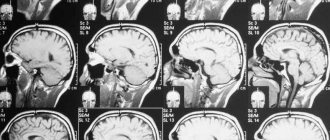
Magnetic resonance imaging (MRI)
This method is used in difficult to diagnose cases. It is mandatory when consulting a neurosurgeon and is always used during surgery. The method is based on the phenomenon of resonance, which is provided by hydrogen atoms in a magnetic field. Our body contains an innumerable number of hydrogen atoms (they are found in molecules of water, proteins, fats and carbohydrates), and as a result, if a magnetic field is applied, a signal is created that is characteristic of a particular tissue of the body. The computer processes these signals and builds an image on the screen. The result is a contrast image of the body in various projections without the use of harmful X-rays. The method allows you to see nerve roots, intervertebral discs, blood vessels, nucleus pulposus, and so on.
Computed tomography (CT)
This method uses a narrow beam of X-ray radiation, which illuminates the desired object and is captured at the output by receiving equipment. All data enters the computer, which processes it, builds an image and displays it on the display. To obtain complete data, the patient is filmed while the system rotates around his body, and recording is carried out at all phases of rotation. This method allows you to see the vertebrae, intervertebral discs, ligaments, blood vessels, and soft tissues. As a result, tomograms can detect deformations of the meninges, ruptures of disc contours, compression of nerve roots, and so on. This method is used to photograph small segments of the body and is usually prescribed after an x-ray to clarify the condition of a specific suspicious intervertebral disc. Let us add that the dose of x-ray radiation during tomography is much higher than when taking a conventional x-ray.