Every pregnant woman registered at the antenatal clinic will have to undergo many examinations over 9 months. A responsible and important procedure for the expectant mother is prenatal screening of pregnant women, which is carried out three times at certain obstetric dates. Some people confuse screening with regular ultrasound, but there are fundamental differences between these terms. What is the difference between ultrasound and prenatal screening?

How much does the first ultrasound and subsequent screenings of pregnant women cost at a medical clinic?
Finding out about pregnancy using an ultrasound will cost from 800 rubles. The price will vary depending on the method of the procedure (transabdominal or transvaginal) and the month of pregnancy.
| Services | Price in rubles |
| Ultrasound of the uterus, appendages (examination with transvaginal and abdominal sensors) | 1 200 |
| Ultrasound of the uterus, appendages (examination with a transvaginal sensor) | 1 200 |
| Ultrasound of the uterus, appendages (examination with an abdominal sensor) | 800 |
| Control of the uterine cavity after m/a | 500 |
What is the difference between a regular ultrasound and a screening ultrasound?
Ultrasound differs from screening in the purposes of the study. In the first case, the doctor will be able to assess the external parameters of the fetus, identify features of intrauterine development and detect growth delays. Screening is a much more detailed examination, which includes a whole range of activities.
Ultrasound is one of the components of screening. An ultrasound examination alone is not enough to fully assess the health of the mother and child. It is important to evaluate all factors together using various techniques. A biochemical blood test should not be neglected, relying only on good ultrasound results. If a high risk of chromosomal abnormalities is detected in the fetus, it is recommended to undergo an expert ultrasound, DOT test, amniocentesis or cordocentesis.
What kind of ultrasound is done during pregnancy?
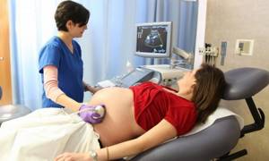
Depending on the stage of pregnancy, ultrasound is performed transvaginally or transabdominally. In the first case, the sensor is inserted into the vagina, and in the second, imaging is carried out through the abdominal wall.
Usually, at any stage, doctors resort to the transabdominal technique so as not to provoke a miscarriage or premature birth. Only the first examination is performed transvaginally, during which the fact of pregnancy is actually established.
It is worth noting that both methods are absolutely safe for the fetus, and if women feel excellent, they can be used without restrictions.

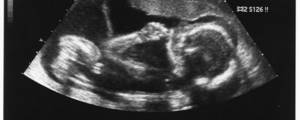
From the point of view of better visualization, doctors distinguish between classic and three-dimensional ultrasound. The latter is also called 3D and even 4D. Three-dimensional images can only be obtained using special ultra-modern equipment. From a medical point of view, in the vast majority of cases there is no need to do a 3D ultrasound, however, it allows you to see the child in the smallest detail, right down to the facial expression in the later stages, which gives future parents a lot of positive emotions.
What is ultrasound?
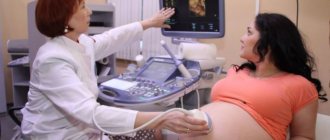
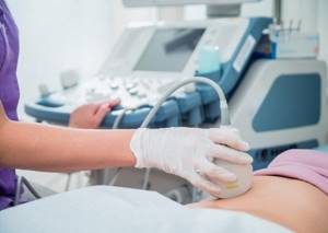
Ultrasound – ultrasound examination of tissues and organs. A sonologist can use a special sensor to see how a particular organ looks and functions. The procedure is a non-invasive technique and does not cause pain or discomfort to the patient.
In obstetrics and gynecology, using ultrasound, the doctor monitors the growth and development of the fetus, determines its heart rate, the amount of amniotic fluid, the condition of the uterus and ovaries. During an ultrasound, on the big screen, the expectant mother can see her baby - how he moves, yawns, and moves his fingers.
Many clinics have modern ultrasound machines with a 3D/4D imaging function, which allows you to see a three-dimensional image of the fetus in real time. It is even possible to record it digitally. Thus, even before the birth of the child, you can visually “get to know” him and look at the baby’s facial features. At the end of the third trimester, the doctor assesses the position of the fetus in the uterus to choose the method of delivery.
Can pregnant women have an ultrasound?
Medicine does not yet have data on the potential harm of ultrasound to the fetus. Moreover, this diagnostic method has been used for several decades, and there is no difference between babies born after ultrasound and babies whose mothers were not examined at all.
However, there are some nuances that should be considered before making an appointment with a doctor. At what week of pregnancy is the first ultrasound done? Usually this procedure is carried out no earlier than the 5th obstetric week, which is approximately the first week of missed menstruation. Before this period, it is almost impossible to see the fertilized egg.
At what stage of pregnancy can a repeat ultrasound be done? Any, if there are medical indications. Almost half of pregnant women undergo 10 or more sonographies over a 9-month period as prescribed by a doctor, and this does not affect the child in any way.
Expert ultrasound examination during pregnancy: luxury or necessity?
Screening ultrasound is a mandatory examination during pregnancy. But often women do not limit themselves to standard services and turn to specialists who perform expert ultrasound. In each trimester of pregnancy, the expectant mother must undergo a screening ultrasound, which provides information about the condition of the fetus and whether the baby’s development is normal. Sometimes, after a screening ultrasound, additional diagnostics may be required. It is not advisable to carry out such diagnostics using invasive procedures: violation of the integrity of the skin can lead to serious problems with the patient’s health, ranging from infection to damage to the embryo. Therefore, invasive procedures are carried out only under strict indications; in all other cases, doctors recommend expert ultrasound examination.
If the expectant mother's pregnancy is progressing normally, there is no need for an expert ultrasound. However, pregnant women often sign up for such a procedure themselves, wanting to see a 3D image of their baby, take a 3D photo as a souvenir, or record a video. But in some cases, expert ultrasound is necessary.
When to perform an expert ultrasound
- If, during a screening ultrasound, the doctor finds signs of hereditary defects or congenital anomalies, an expert ultrasound is necessary.
- If, based on the results of a traditional study, a specialist concludes that the parameters of fetal development do not correspond to the woman’s gestational age, an ultrasound examination is prescribed using expert ultrasound scanners.
- It is also necessary to turn to the possibilities of modern medicine in case of detection of violations of the baby’s vital functions, for example, if during the examination the doctor notes an unusual rate of the fetal heartbeat (slow or, conversely, rapid), if the baby does not move after the eighth week of gestation.
- If the expectant mother has a history of gestosis, miscarriages, or if intrauterine growth retardation was observed in previous pregnancies, it is worth playing it safe and undergoing an expert ultrasound.
- Other reasons for prescribing additional ultrasound on expert scanners include age over 30 years, family history, and the need for the most complete visualization of the fetus.
What is the difference between screening ultrasound and expert ultrasound?
The main difference is the quality of the equipment for the procedure. For expert diagnostics, you need not just an ultrasound scanner, but equipment with increased resolution, which allows you to visualize images in 3D and 4D format. When using the 3D/4D module, the specialist performing the ultrasound can not only show the expectant mother a three-dimensional image of the baby (take photographs, video), but also assess in detail the condition of the internal organs and external anatomical structures. This method provides maximum accuracy in the analysis of the work and condition of temporary organs (placenta, umbilical cord, amniotic fluid), and the vital activity of the fetus.
When performing a three-dimensional ultrasound, the fetometric readings of the fetus, the anatomical structure of the baby’s internal organs, the intrauterine vital activity of the embryo, as well as the condition of the temporary organs of pregnancy and the uterus are assessed.
Expert ultrasound scanners are quite expensive, and district clinics cannot afford them, so if you need to undergo an expert ultrasound scan, you will have to go to private medical clinics or large diagnostic centers.
Based on materials from the site https://www.sonomedica.ru/.
At what stage does an ultrasound show pregnancy?
Most of all, women are interested in the question: will ultrasound determine pregnancy in the early stages? And if so, which ones exactly? Doctors at the Art Medic clinic claim that the fertilized egg is clearly visualized in the uterine cavity already 2.5-3 weeks after conception. Based on a normal 28-day cycle, this is the first week of a missed period. At this stage, it is already possible to determine the week of pregnancy and the very fact of its presence.
Which ultrasound most accurately shows the gestational age? At this stage, doctors resort to the transvaginal method of examination, as it provides better visualization. From about 8-10 weeks, all diagnostics are carried out “in the abdomen”.
Definition of ectopic pregnancy
From the point of view of doctors, it is most advisable to use ultrasound specifically to determine an ectopic pregnancy. This pathology can be detected as early as 5-6 weeks (the first week of delay). An ectopic pregnancy is characterized by the fact that the fertilized egg does not reach the uterus, but implants in the fallopian tube. If it is not detected in a timely manner, then as the fetus grows, it will rupture the tube. Without timely medical care, a woman will face death. Fortunately, modern medicine has all the means to not only save a pregnant woman, but also avoid removal of the fallopian tube, preserving the possibility of childbearing in the future.
Ectopic pregnancy is quite rare and, if detected at 5-6 weeks, can be terminated without irreparable harm to the woman’s fertility.
Definition of frozen pregnancy
A frozen pregnancy is one in which the fetus does not develop. Usually this condition ends in spontaneous miscarriage, but sometimes nature fails. The woman herself practically cannot feel that the pregnancy has stopped, especially if this is the first child.
The fetus can “freeze” at any stage, and the main criterion for this sad fact is the absence of a heartbeat. According to research, the heart of the embryo begins to beat already at the 5th obstetric week (3 weeks after conception), however, this can only be determined using ultrasound starting from the 6th week of pregnancy. Moreover, in the vast majority of cases, doctors recommend waiting until week 7, since imaging errors are possible before this period.
To monitor the situation, especially if there have already been cases of frozen pregnancy in the anamnesis, it is enough to undergo diagnostics at the 7th obstetric week, and then do scheduled screenings on time. An emergency ultrasound is performed according to the doctor’s indications in the presence of threatening symptoms.
What does pregnancy screening include?
Screening includes a set of measures aimed at assessing fetal development, excluding chromosomal abnormalities and intrauterine defects. Prenatal screening for pregnant women includes:
- three times ultrasound at different stages of pregnancy;
- biochemical blood test to determine specific markers;
- Dopplerometry - assessment of blood flow in the vessels of the fetus and placenta;
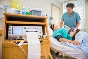
- cardiotocography – assessment of the frequency and rhythm of the fetal heartbeat.
Thus, screening is a broader, more detailed and accurate examination. It is aimed at eliminating all deviations during pregnancy. The examination is recommended for all pregnant women, regardless of age. If desired, a woman can refuse the procedure by signing a written refusal.
Ultrasound examination of the fetus
An ultrasound examination includes three examinations at different stages of pregnancy:
- at 11–14 weeks;
- at 18–21 weeks;
- at 30–34 weeks.
The first ultrasound examination of a pregnant woman is aimed at identifying chromosomal abnormalities of the fetus. Using the device, the doctor takes the necessary measurements, which he describes in detail in the conclusion. During an ultrasound in the 1st trimester, the following parameters are assessed:
- coccygeal-parietal size (CTP);
- biparental fetal head size (BFS);
- collar space thickness (TNT);
- length of the nasal bone;
- heart rate (HR).
These parameters make it possible in the early stages of pregnancy to identify fetal anomalies for trisomy 21, 18 and 13 - Down syndrome, Edwards syndrome and Patau syndrome. At approximately 20 obstetric weeks, a second ultrasound is performed, which either refutes or confirms the data of the first. The transcript of the second survey data contains the following information:
- fetal size and compliance with their standards;
- detailed description of internal organs;
- lung maturity level;
- limb development;
- thickness and structure of the placenta;
- volume of amniotic fluid.
Ultrasound as part of the third screening has others. Unlike the first two, its task is to assess the position of the fetus in the uterine cavity, study the condition of the placenta, and assess the amount and condition of amniotic fluid. After the third examination, the doctor gives the patient recommendations regarding the method of delivery - naturally or by cesarean section.
After the third ultrasound, the patient is sent for Doppler ultrasound and CTG - procedures during which the fetal blood flow and heart rate are assessed. This information will help identify possible disorders in the child’s cardiovascular system.
Biochemical analysis of maternal venous blood
A biochemical blood test is an integral part of the first and second screening of pregnant women. After the first routine ultrasound, the patient donates blood for the following free beta-hCG and pregnancy-associated protein A (PAPP-A).
Based on the size of the fetus according to ultrasound, the results of laboratory tests and the patient’s medical history (age, weight and height, presence of bad habits, endocrine and chronic somatic diseases, abortions, miscarriages, IVF, etc.), the risk of genetic abnormalities is calculated using a computer program . Blood is donated in the morning on an empty stomach or 4 hours after the last snack.
At 18–19 weeks of pregnancy, the patient, if necessary, donates blood a second time - this test is called a “triple test”. When calculating risk, the following data is used:
- results of the 1st ultrasound at 11–14 weeks of pregnancy;
- blood test for hCG;
- blood test for AFP;
- blood test for free estriol.

The second biochemical screening is prescribed to the patient in the following cases:
- with poor results of the first screening;
- in the absence of results of the first screening;
- at the personal request of the pregnant woman.
If the results of the 1st screening are normal, it is enough for the patient to donate blood for AFP to exclude fetal neural tube defects. After passing the second screening, the gynecologist can refer the patient to a geneticist for detailed consultation.
What screenings are prescribed during pregnancy?
Screening of pregnant women is a complex of examinations that can detect the presence of genetic abnormalities and developmental pathologies in the fetus. Screening consists of:
- Ultrasound;
- Blood test.
For unification and ease of interpretation of the results, uniform dates for ultrasound screening and blood donation during pregnancy were adopted. In this case, accuracy down to the day is very important, since computer programs operate in a specific time interval.
Today screening is indicated for certain categories of women:
- If the mother is over 35 years old or the father of the child is over 40.
- There are relatives in the family with genetic disorders.
- There is a history of miscarriages and frozen pregnancies.
- The woman took medications in the first trimester that were potentially dangerous to the fetus.
- A woman suffered from an infectious disease during pregnancy.
- A woman works in hazardous work.
At what stage of pregnancy is 1st ultrasound screening performed, and what is it?
The first screening during pregnancy is done between 11 and 13 weeks. Its decoding suggests the presence of such genetic developmental abnormalities as Down syndrome, Edwards syndrome, Patau syndrome, as well as preeclampsia and fetal death.
At week 12, 2 ultrasounds are performed, during which the following indicators are determined:
- KTP – coccygeal-parietal size (43-65 mm);
- BPR – biparietal size (17-24 mm);
- TVP - thickness of the collar space (1.6-1.7 mm);
- Length of the nasal bone (2-4.2 mm);
- Heart rate – heartbeat (140-160 beats/min), etc.
The next day, a blood test is done, which determines the following hormones:
- B-hCG – human chorionic gonadotropin;
- PAPP-A is plasma protein A.
It is worth noting that screening results only suggest the presence of abnormalities and require further examination.
At what week is the 2nd screening scheduled?
The timing of the second screening varies from 16 to 21 weeks. At 20 weeks of pregnancy, an ultrasound scan can already print out a photo in which the silhouette of the little man can already be guessed.

Ultrasound is used to determine:
- BDP - biparietal size (26–56 mm).
- DBL - length of the femur (13–38 mm).
- KDP - length of the humerus (13–36 mm).
- OG - head circumference (112–186 mm) and a number of other parameters.
A blood test shows the qualitative and quantitative content of the following hormones:
- B-hCG;
- Free estriol;
- AFP.
This examination suggests the presence of Meckel syndrome and pathologies in the development of the neural tube and intestines in the fetus.
Third ultrasound screening during pregnancy
3 Ultrasound screening during pregnancy does not require simultaneous blood donation. It aims to determine the degree of fetal development and helps the doctor decide whether to have a natural birth or whether a caesarean section is necessary.
Using ultrasound, the following parameters are determined:
- BDP - biparietal size (67–91 mm).
- DBC is the length of the femur (47–71 mm).
- KDP - length of the humerus (44–63 mm).
- OG - head circumference (238–336 mm).
The condition of the amniotic fluid, cervix and placenta is also determined. At this stage, they play a decisive role in determining the method of delivery and even the timing of the operation.
Sometimes you can hear the question, at what week is the 4th screening during pregnancy carried out? Screening 4 refers to an ultrasound scan immediately before childbirth, which is done for medical reasons.
Screening ultrasounds: identifying pathologies at an early stage (part 1)
Pregnancy is one of the most amazing periods in a woman’s life. At this time, you are blossoming and preparing to become a Mom. Of course, when you are pregnant, you have to work a lot on yourself in close contact with specialists who sometimes prescribe incomprehensible examinations. For example, most of us do not understand the difference between obstetric ultrasound and screening. What's more important? When to take place? For what?
Today, the Chief Physician of the MC "Samara School of Ultrasound", an ultrasound doctor of the highest category, Elena Vladimirovna Litvinova, will answer your questions and help you understand the studies necessary for every pregnant woman.
1. What are the differences between screening and obstetric ultrasound?
The main difference between screening and obstetric ultrasound is that screening studies are carried out at specific stages of pregnancy. These dates are determined by order of the Ministry of Health with an exact list of those aspects of ultrasound examination that should be reflected at this stage of pregnancy.
2. What is screening during pregnancy? What is screening #1?
As I have already noted, screening is a mandatory study carried out by order of the Ministry of Health at certain stages of pregnancy.
Screening 1 - also called “genetic” - is carried out in the period 11-13 weeks and 6 days (the remaining periods of the first trimester are not considered screening), the best time for examination is 12 weeks. During this period, the mandatory parameters for the study are measuring the coccygeal-parietal size of the fetus (CTP), counting the heart rate, measuring the thickness of the nuchal space and the length of the nasal bones.
3. Is it true that screening No. 1 can detect Down syndrome in a child?
I am often asked about the possibility of accurately diagnosing chromosomal abnormalities (such as Down syndrome, Edwards syndrome, Patau syndrome) based on the results of ultrasound examinations. Unfortunately, ultrasound method itself chromosomes. True, in a certain percentage of cases, ultrasound can identify the signs that may occur with this pathology. After the examination, the expectant mother donates blood for biochemical screening. Ultrasound and biochemical examination data are processed by a mathematical program that determines the risk of chromosomal abnormalities.
4. What to do if the risk of chromosomal abnormalities is high or moderate?
If the risk of chromosomal abnormalities is high , then the pregnant woman is sent for consultation with a geneticist and to resolve the issue of counting the number of chromosomes by taking the necessary material.
If the risk of chromosomal abnormalities is low , the woman continues to be monitored by her obstetrician-gynecologist according to the usual program.
If the risk is average , the pregnant woman is observed and undergoes additional research. In addition to these data, which are determined by the order for the first screening, many other parameters are assessed - very important for further pregnancy management tactics. Establishing the exact period of fetal development during a multiple pregnancy makes it possible to establish chorionicity, which is the most important factor determining the outcome of a multiple pregnancy. Helps identify women at high risk for complicated pregnancies.
Determining the sex of a child by ultrasound
Theoretically, a doctor can tell the gender by ultrasound starting from the 15th week of pregnancy, however, in practice, patients will have to wait until 22-23 weeks. In the early stages, identification is difficult for objective reasons. It often happens that the child does not turn his “face” to the scanner, so his gender remains a mystery until the very birth.

If we take into account the timing from which the gender is visible on an ultrasound, then most often parents find out it during the second screening, which is very convenient, since it does not require an additional visit to the doctor.
Is it possible to go through an entire pregnancy without an ultrasound?
There are often cases when a woman never visits an ultrasound diagnostic room during the entire pregnancy. Most often this is due to religious or other beliefs.
Doctors strongly recommend doing an ultrasound at least after pregnancy in order to prevent possible complications (bleeding, infection, etc.) in time. The examination is also indicated after an abortion or miscarriage. It is carried out three times - the first with an interval of 10 days, and then another after 2 months.
Reviews from patients about ultrasound in medical
Our patients often leave reviews about pelvic ultrasound during pregnancy, which they underwent at the Art Medic clinic. You can view them in a special section of the medical center’s website.
Congratulations to the team on March 8th. My husband and I express our deep gratitude to all the doctors and personally to the director T.P. Kryachek. for good organization. A highly professional team has been selected here and every problem is treated with understanding. The legs carry themselves to such doctors. Happiness and prosperity!
05 March 2012