the whole list
As a rule, the magnetic resonance diagnostic method is quite informative. However, sometimes the patient is prescribed a contrast-enhanced MRI. This is a detailed study that allows you to evaluate the structure of tissues and cells of the body.
This diagnostic method is most often prescribed to cancer patients for a detailed study of the tumor and the extent of its spread. In oncology, MRI with contrast can significantly increase the information content of the procedure. A contrast agent injected into a vein during MRI makes it possible to detect a tumor at the very initial stage of its development, clearly determine the boundaries of the malignant tumor, its structure and consistency.
Substances for research
The substance used for contrast-enhanced MRI is significantly different from the drugs used for conventional magnetic resonance imaging. For a detailed MRI with contrast, the patient is intravenously injected with one of the drugs based on the metal gadolinium:
- Magnevist;
- Omniscan;
- Gadovist;
- Let's do it.
The contrast agent is administered intravenously, using a syringe or injector, carefully calculating the dose and rate of administration of the drug. The supply of contrast agent is synchronized with the progress of the MRI scan.
Contrast MRI
Often, to obtain a more complete picture of the patient's condition, contrast-enhanced MRI is prescribed. With this method of conducting the study, a dye is injected into the patient’s vein, which provides clear images. To study the alimentary tract, the dye is administered orally. The dye illuminates the tissue and highlights the clear boundaries of the internal organs. Affected tissues under the influence of the dye can change their color, thanks to which the doctor can determine the structure, size and boundaries of the tumor in cancer. In this way, the presence of metastases and their location can be detected.
Many patients are concerned about the safety of this diagnostic method. But they don’t have to worry, because the procedure is absolutely safe and the substance injected into the vein is a common dye. Its use does not pose a threat to humans because it:
- does not cause allergic reactions, with the exception of patients who are allergic to some component of the dye;
- is eliminated from the body quickly.
When conducting MRI with contrast, several cases of side effects were recorded: mild dizziness, headache and skin rash. Such symptoms are observed with individual sensitivity to the dye. To examine different parts of the body, certain coloring compounds are used. Diagnosis of multiple sclerosis is carried out using a composition containing iron oxide, because it will allow you to obtain the most accurate images of blood vessels. To examine the organs of the gastrointestinal tract, compositions based on gadolinium and manganese salts are used.
Regular food products (blueberry or green tea) can be used as a contrast agent. Contrast-enhanced MRI accounts for 20% of all magnetic resonance imaging scans. In most cases, such an examination is prescribed for studying the brain, pathologies of the digestive tract, or suspected cancer.
When is it necessary to perform an MRI with contrast?
Indications:
- The rehabilitation period after removal of herniated intervertebral discs is for differential diagnosis of scar tissue from relapse of the disease.
- There is a suspicion of a benign formation in the pituitary gland, whereas conventional magnetic resonance imaging does not detect a tumor.
- Using MRI with contrast, you can determine the degree of activity of multiple sclerosis, as well as judge the effectiveness of the therapy.
- For malignant and benign neoplasms in the brain and spinal cord. Sometimes native MRI does not provide enough information about the boundaries and consistency of the tumor. As a result, the doctor cannot make a diagnosis and prescribe the correct treatment to the patient. In this case, an MRI with contrast is prescribed. Thanks to an extensive and detailed examination, the doctor can determine the size of the tumor, its location, the boundaries of the tumor and its consistency.
- MRI using a contrast agent allows you to assess the condition of the structures of the brain and spinal cord after surgery. During the study, the doctor notes whether the patient has a relapse of the disease.
- To detect metastases in the spinal cord and brain. Due to the small size of metastases, it is difficult for the doctor to identify them, and the MRI method with the introduction of a contrast agent makes it possible to detect metastases at an early stage of their formation.
What is a contrast agent?
The diagnostic method is based on the phenomenon of magnetic nuclear resonance. The device scans the main organogen element - hydrogen. Half of all atoms in our body are hydrogen; it is part of absolutely all compounds. But the natural contrast of some tissues is not enough to obtain accurate scanning results. Therefore, in some cases, for tomographic examination, a water-soluble contrast agent is injected intravenously, which is then excreted from the body along with urine.
MRI uses paramagnetic rather than x-ray contrasts, which change the magnetic susceptibility of the cell and reduce relaxation time. The substance enters the intercellular and intravascular space with the blood, is distributed in it and allows more accurate visualization of the condition of the tissues.
With contrast, you can more accurately diagnose:
- neoplasms and tumors of any size;
- pathologies of central nervous system tissues;
- foci of infection;
- necrotic formations, etc.
If we try to explain the effect of contrast in simple language, we can say that the injected substance amplifies the signal from the nucleus and allows us to accurately determine the composition of the cell, the degree of its changes, and dynamically track the processes occurring in the tissues. Moreover, there are specific substances that accumulate selectively in tumor tissue or the muscle layer, for example, the myocardium. The introduction of contrast makes it possible to differentiate healthy tissues from pathologically altered ones, to establish the exact location of the lesion and its size.
Possibility of allergic reactions
Patients who are allergic to medications often worry about possible complications after the administration of a contrast agent. Due to the fact that the preparations used for contrast do not contain iodine, allergic reactions are practically not observed. The above drugs for MRI with contrast are non-toxic, therefore they are well tolerated by patients, even those with allergies, and do not cause side effects after administration to the body.
Prices for MRI examination →
Book an MRI online →
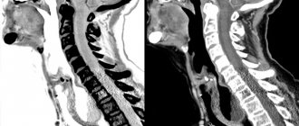
Brain MRI results
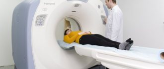
The MRI procedure of the brain does not cause any pain. The patient takes a horizontal position on a special table, which slides into the tunnel, which is the inside of the magnetic coil. Radio waves affect the magnetic position of the atoms of the body, which are recorded by a powerful receiver, which transmits the received information to a computer. Thanks to this, highly accurate black and white images appear on the computer. If necessary, they can be converted into 3D photographs of the scanned area. As a result, the doctor gets the opportunity to see all the problems both in the brain itself and in its stem cells.
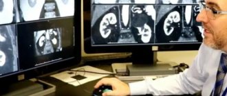
Using MRI of the brain, it is possible to determine and evaluate in detail the location, size and other features of the tumor. In addition, MRI can reveal the degree of hemorrhage resulting from any damage to brain structures. If an MRI of brain vessels is performed using a contrast agent, the doctor receives confirmation of the malignancy of the oncological process. The analysis of images obtained during an MRI of the brain is carried out by a qualified specialist, who draws up his conclusion and sends the results of the examination to the attending physician. In some cases, the patient receives MRI images on film or digitally.
It doesn't take long to wait for MRI results. As a rule, within a few days after the tomography, the doctor can tell the patient in detail what the MRI shows. This is very convenient when a person needs to quickly find out why he has headaches that arise for no apparent reason, or find out the true reason for the unexpected appearance of severe weakness in the limbs, accompanied by numbness. By undergoing an MRI of the brain, you can determine the presence of the disease at a very early stage. In the future, this will help to promptly take all the necessary steps for medical intervention and a speedy recovery.
What is MRI with contrast?
In some cases, to obtain a more accurate clinical picture, the doctor may prescribe an MRI using a contrast agent. Thanks to contrast, doctors have the opportunity to visualize the organ being examined with increased accuracy. The contrast agent is usually administered to the patient intravenously, with the exception of rare cases when the contrast can be administered orally (as a rule, this method of examination is intended for diagnosing pathologies of the gastrointestinal tract). Next, we will consider what advantages this method has and in what cases MRI with contrast is prescribed.
The main function of the contrast agent is to illuminate the tissues during the examination, which is recorded by the tomograph. The powerful magnetic field of the tomograph, when exposed to a contrast agent, makes it possible to obtain the most accurate and clear image of the organ being studied. The speed at which the contrast agent spreads through the vessels directly depends on the intensity of blood flow at the study site.
The principle of operation of the contrast agent is as follows:
- When contrast is administered intravenously, the drug spreads through the vessels along with the blood flow.
- Since the affected tissues are capable of changing their color under the influence of a contrast agent, the doctor is able to accurately determine the boundaries, size and structure of pathologies or all kinds of neoplasms. And what is important is to determine whether there are metastases and visualize their locations.
Features to consider when performing MRI with contrast:
- The patient has an allergic reaction to the contrast agent. If such a reaction exists, then MRI with contrast is not performed. Instead, a conventional magnetic resonance imaging will be prescribed
- Pregnancy in the first trimester. MRI is not prescribed in the early stages of pregnancy, since the effect of the magnetic field on the fetus has not been fully studied
- The presence of any metal objects and implants in the patient’s body. This limitation lies in the fact that a high-power magnetic field can attract any metal and move it from its place, and this can become a threat to the life and health of the patient
- The presence of a pacemaker or other electronic devices in the patient's body. The magnetic field can cause them to malfunction
- Claustrophobia or all kinds of mental disorders in the patient that do not allow him to remain motionless for a long time. In such cases, the doctor may offer the patient a sedative, which can have a calming effect and allow the patient to remain calmly still during the MRI.
What is the main difference between MRI with and without contrast?
Next, let's look at what is the difference between the two types of MRI under consideration:
- MRI with contrast. In this type of study, a contrast agent is intravenously injected into the body, made, as we discussed above, based on gadolinium salts or ions. And then an MRI scan is performed - scanning the organ or body area being examined. Conventional MRI is performed without the introduction of any additional substances.
- MRI with contrast requires simple preparation:
- Before diagnosis, the patient must refuse food and drink 2–5 hours.
- To conduct an MRI of the pelvic organs, the patient will need to drink about one liter of clean water 1 hour before the diagnosis.
- When performing conventional tomography without contrast, no preliminary preparation is required at all.
- An MRI with contrast will require a longer period of time than a diagnosis without it.
- Contrast-enhanced magnetic resonance imaging is not recommended for pregnant women because the contrast agent can easily cross the placental barrier and affect the fetus.
- The cost of MRI with contrast injection is an order of magnitude higher than the price of conventional magnetic resonance imaging.
Briefly about MRI
Unlike computed tomography, fluorography and radiography, MRI scanning does not involve exposure to radiation on the body. The doctor receives images thanks to the phenomenon of magnetic resonance, and in several projections at once, which makes it possible to identify various types of pathologies:
- benign and malignant tumors;
- inflammatory processes;
- damaged muscles, bones and blood vessels;
- functional failures in the functioning of various organs and systems;
- anomalies of congenital etiology;
- foreign objects;
- traces of previous surgical interventions;
- age-related changes and consequences of injuries.
Indications and contraindications
Indications for MRI with contrast:
- If there is a suspicion of malignant formations or the possibility of the development of various pathological processes in the patient’s body
- MRI is used to monitor the patient’s condition before and after surgery
- The need to visualize blood vessels and their condition
- The need to study the musculoskeletal system
- Receiving work-related or sports injuries
- Suspicion of various infectious processes that may occur in the patient’s body.
Next, we will look at what contraindications there are for MRI. They, in turn, are divided into absolute and relative. Absolute contraindications include:
- Presence of electronic devices and metal implants in the patient’s body
- Heart and kidney failure
- Pregnancy in the first trimester
- Lactation period
- Individual intolerance to contrast agent components
- The patient's weight is more than 120 kg (depending on the tomograph apparatus).
Relative contraindications include:
- Claustrophobia and various mental disorders
- The presence of tattoos on the body, the composition of which contains metallic elements
- Bronchial asthma
- Myeloma
- Pregnancy in the 2nd and 3rd trimesters
- Some diseases of the cardiovascular system.
MRI 3 Tesla
Possibilities of MRI of the knee joint
MRI allows you to diagnose the most common diseases of the knee joint and is often used to make an accurate diagnosis or when receiving questionable X-ray results.
During magnetic resonance imaging, it is possible to record even the most minor changes in the knee joint. All structures are visualized, down to the smallest structures. The most informative study is for identifying pathologies of the meniscus and other soft tissues of the joint, which are quite difficult to determine using other diagnostic methods. Using MRI, you can detect a lot of disorders: degenerative changes, hemorrhages, hematomas, the formation of coarse scar tissue, tumors and cancer processes.
MRI indications of the knee joint:
- falling on the knee, accompanied by swelling, pain and limited mobility;
- sports injuries;
- stiffness of movements;
- contraindications to X-ray diagnostics;
- tumors of the knee joint and surrounding tissues;
- pinched nerves;
- rupture of the meniscus, joint capsule;
- severe swelling of tissues, accumulation of fluid in the joint cavity;
- arthritis, arthrosis;
- dislocations, joint instability;
- osteochondropathy;
- development of inflammatory processes;
- preparation for surgical operations;
- monitoring the effectiveness of treatment.
Contraindications for MRI of the knee joint
The list of contraindications depends on whether contrast enhancement is used in MRI or not. The contrast clearly visualizes tissues with increased blood circulation - tumors, inflamed areas. It allows you to deal with complex, complex damage. The procedure is carried out with contrast on an empty stomach.
But for patients with kidney pathologies and decompensated diabetes mellitus, contrast MRI is contraindicated, since diagnostic drugs for these diseases can be removed from the body with a delay, causing a toxic effect. Contrast enhancement is also contraindicated for pregnant women and nursing mothers.
MRI without contrast has no strict contraindications. The technique is safe and is used in various branches of medicine. There is a limitation for patients with metal elements in the body - a pacemaker, pump, staples, etc. For claustrophobia, an open MRI is used. If there is no equipment of this type in a medical institution, then it is possible to conduct the study in a state of medicated sleep.