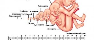
Many “advanced first-time parents” will be able to show you not only photos and videos of their child from the first days of life, but also a video about the unborn child. On the screen you can see how the baby moves his legs, squints his eyes, covers himself with his hands, etc. Some parents even have a video collection at various stages of their child’s development. Pregnant women begin to feel their connection with their baby early and their desire to have a video as a keepsake is understandable. However, few of them think about why fetal ultrasound is performed during pregnancy.
From a medical point of view, ultrasound scanning of the fetus is today considered a safe study, provided that it is carried out for medical reasons. Ultrasound is the most common method of prenatal diagnosis and carried out at the right time (20-24th week of pregnancy) and at the proper level, it can detect up to 70% of visible malformations. Down's disease, like other chromosomal diseases, is not detected by ultrasound.
If diagnostics, including ultrasound, reveal a hereditary disease or malformation in the fetus, parents must carefully weigh the possibilities for treatment and rehabilitation of the child and decide whether to continue carrying the pregnancy or not.
When do you need to take the Prenatal screening test in the 2nd trimester (14-20 weeks)?
1. Age over 35 years; 2. The presence of hereditary diseases in close relatives; 3. Down's disease or other chromosomal abnormalities of the fetus in previous pregnancies; 4. Exposure to radiation, harmful factors, or taking medications with a teratogenic effect by one of the spouses on the eve of conception; 5. with borderline results of the calculated risk of chromosomal pathology during screening of the first trimester, as well as if the screening examination of the first trimester was not carried out on time.
Prenatal screening for fetal malformations
Screening methods - completely safe studies conducted at certain stages of pregnancy - make it possible with a very high degree of probability to identify groups of women at risk of developing Down syndrome in the fetus.
There are risk groups for the development of miscarriage, gestosis (late toxicosis), various complications during childbirth, etc. If a woman, as a result of an examination, is found to be at risk for a particular pathology, this only means that this patient has a certain type of pathology may be more likely to occur than in other women.
What is a risk group?
Risk groups are groups of patients among whom the likelihood of detecting a particular pregnancy pathology is higher than among all women in a given region. Detection of an increased risk of developing fetal malformations using prenatal screening methods is not a diagnosis.
The diagnosis can be made or rejected with additional tests. Thus, the risk group is not identical to the diagnosis.
Why are risk groups needed? Knowing that the patient is in one or another risk group helps the doctor correctly plan the management of pregnancy and childbirth.
Identification of risk groups allows us to protect patients from unnecessary medical interventions, and allows us to justify the appointment of certain procedures or studies to patients.
Detailed description of the study
Prenatal screening in the second trimester:
- It is carried out during 14-20 weeks of pregnancy;
- Biochemical (serum) markers: alpha-fetoprotein (AFP), human chorionic gonadotropin (hCG), free estriol, free β-hCG;
- Ultrasound data of the second trimester indicating the date of execution: biparietal fetal size (BFS) to calculate the gestational age;
- Personal details of the pregnant woman: age, weight, race, bad habits (smoking);
- History of the pregnant woman: number of previous pregnancies, presence of multiple pregnancies, in vitro fertilization, presence of diseases (insulin-dependent diabetes mellitus).
To correctly calculate the risk of developing chromosomal abnormalities, the laboratory must have accurate data on the gestational age, ultrasound data (CTR, TVP and visualization of the nasal bone for the first trimester) and complete information about all factors necessary for screening (indicated in the referral form). The gestational age can be calculated by the date of the last menstruation (LMP), the date of conception (estimated period). For prenatal screening, it is recommended to use a more accurate and informative method - calculating the gestational age according to ultrasound data (CTR and BPR). Due to the existence of several statistical methods for determining the gestational age based on fetal ultrasound data, the calculation results performed using the PRISCA software may differ slightly from the gestational age determined by the ultrasound physician (range up to 7 days in the second trimester).
The computer program for calculating the analysis results PRISCA allows you to:
- calculate the probability of various risks of fetal pathology
- take into account the patient’s individual data
- consider factors influencing the detection of abnormal levels of biochemical markers.
Screening parameters in the 2nd trimester (14-20 weeks):
- Ultrasound measurement of BDP (Biparetal size) 26-56 mm. (limitation period 1-3 days)
- Immunochemical determination of 1) beta-hCG - free and total, 2) AFP, 3) free estriol.
Features of risk calculation:
- Risk calculation depends on the accuracy of the data provided for analysis
- Risk calculation is the result of statistical data processing
- Results must be confirmed (or excluded) by cytogenetic studies.
The result is presented in the form of MoM - this is the magnitude of the deviation of the indicators of a given pregnant woman from the average norm. Beta-hCG is produced in the fetal membrane of the human embryo. It is an important indicator of the development of pregnancy and its deviations. The level of beta-hCG reaches its maximum in the 10-11th week, and then gradually decreases. An increase in the concentration of beta-hCG in pregnant women in the second trimester of pregnancy may indicate a risk of developing trisomy 21 chromosome (Down syndrome) in the fetus. A decrease in hormone concentration may indicate the development of other chromosomal abnormalities in the fetus, in particular Edwards syndrome, trisomy 18.
Alpha fetoprotein (AFP) is produced in the embryonic yolk sac, liver and intestinal epithelium of the fetus, its level depends on the state of the gastrointestinal tract, fetal kidneys and placental barrier. In the mother's blood, its concentration gradually increases from the 10th week of pregnancy and reaches a maximum at 30-32 weeks. A decrease in concentration is observed in Down syndrome. With a neural tube defect, AFP levels increase.
Free estriol is produced by the placenta, and then by the fetal liver. It stimulates the synthesis of vasodilating prostaglandins in endometrial cells and enhances uteroplacental blood flow. In addition, it increases the receptor-mediated uptake of low-density lipoprotein cholesterol for subsequent synthesis of progesterone by the placenta, and also stimulates mammary gland growth. In Down syndrome and Edwards syndrome, the concentration of free estriol decreases. Also, the level of the hormone may decrease due to taking medications: dexamethasone, prednisolone, metipred, antibacterials. Low concentrations of free estriol in combination with high levels of beta-hCG and AFP are associated with an increased risk of intrauterine growth restriction and complications of the third trimester of pregnancy (premature placental abruption and preeclampsia).
In accordance with the order of the Ministry of Health of the Russian Federation dated November 1, 2012 No. 572n. The procedure for providing medical care in the field of “obstetrics and gynecology (except for the use of assisted reproductive technologies)”: it is necessary to conduct prenatal screening in the second trimester at 18-20+6 weeks of pregnancy.
If for some reason an ultrasound was not performed in the second trimester, you can provide data (CTR, TVP, visualization of the nasal bone) from an ultrasound of the first trimester, indicating the date of performance. Calculating the risk of developing chromosomal abnormalities in the second trimester using ultrasound data from the first trimester has lower accuracy and greater error compared to using ultrasound data from the second trimester.
Algorithm for prenatal screening and the second trimester of pregnancy:
1. Calculate the gestational age based on the date of the last menstruation or the day of conception and determine the date of the ultrasound and blood donation for biochemical markers. 2. Perform an ultrasound. If ultrasound data are not suitable for risk calculation (CTE<38mm), repeat ultrasound only on the recommendation of a doctor after a certain time. 3. Knowing the exact gestational age, calculated by ultrasound, take a blood sample to conduct studies of biochemical markers. The time interval between the ultrasound and blood donation should be minimal (no more than 2-3 days).
Attention! Taking medications can affect the results of prenatal screening!
What kind of screening is performed?
Prenatal screening is a complex of mass diagnostic measures in pregnant women to search for gross developmental anomalies and indirect signs of fetal pathology. Studies conducted as part of prenatal screening are safe, do not have a negative impact on the course of pregnancy and fetal development, and therefore can and should be used on a mass scale for all pregnant women.
Types of research:
- Biochemical screening: blood test for various indicators
- Ultrasound screening: identifying signs of developmental anomalies using ultrasound diagnostics.
- Combined screening: a combination of biochemical and ultrasound screening.
The general direction in the development of prenatal screening is the desire to receive reliable information about the risk of developing certain disorders at the earliest possible stages of pregnancy.
Changes and sensations at 23 weeks of pregnancy
This week your uterus is almost 4 cm above the navel, the distance to the pubic joint is about 23 cm. As a rule, by the 23rd week, pregnant women become 5-6.7 kg heavier. Did you suffer from headaches in the first trimester? Most likely, now they have finally left you alone. The cause of these pains, like many other troubles during pregnancy, is hormonal surges; by this week they should have subsided.
The fluid in your body is now retained longer, and you have begun to swell a little. This is due to the changed chemical composition of the blood, as a result of which the tissues retain fluid more strongly. In addition, the growing uterus puts more pressure on the veins, which slows down blood circulation in the legs. This also causes swelling. As a rule, maximum swelling is felt in the evening, especially in the heat. Don’t worry, excess fluid will come out in the first days after birth, but now try to unload your legs as much as possible - keep them stretched out when lying or sitting, put a pillow to hold your legs higher, wear special compression stockings. Light exercise is also helpful. Eat less salty foods - pickles, chips, canned food, etc. The sodium contained in salt in large quantities contributes to fluid retention in your body. But you shouldn’t give up drinking; contrary to popular belief, consuming plenty of liquid prevents swelling. If your face and hands are swollen, it is better to consult a doctor in order to prevent late pregnancy toxicosis.
3d-4d ultrasound of the fetus
3d-4d ultrasound of the fetus is absolutely harmless for the expectant mother and baby. It is recommended for:
- suspicion of any malformations or diseases of the fetus;
- presence of children with congenital diseases in the family;
- pregnancy after IVF or other reproductive technologies;
- multiple pregnancy;
- any complications during pregnancy;
- desire to have complete and reliable information about the condition and development of the fetus.
How are risks calculated, MoM
Special programs are used to calculate risks. Simply determining the level of indicators in the blood is not enough to decide whether the risk of developmental abnormalities is increased or not.
At the beginning, the indicator numbers obtained during laboratory diagnostics are converted into the so-called MoM (multiple of median, multiple of the median), characterizing the degree of deviation of a particular indicator from the median.
Next, the MoM is adjusted for various factors and risk groups are calculated. The most accurate calculation of the risk of fetal abnormalities occurs when using data from an ultrasound examination performed at 10-13 weeks of pregnancy.
Prenatal screening is recommended for all pregnant women.
Did your water break?
As you already know, the uterus becomes larger and heavier throughout pregnancy, putting pressure on the stomach, intestines and bladder. This may cause some women to leak small amounts of urine involuntarily. There's nothing wrong with that. The problem is that already at 23 weeks the membranes may rupture and the water may break. It is easy to distinguish amniotic fluid from urine. Droplets of urine appear on your underwear occasionally and in small quantities, and amniotic fluid can come out in a stream when the membrane ruptures or constantly ooze from the vagina. If you notice anything like this, consult a doctor immediately; water breaking at this stage can be very dangerous for the baby!
