More than 1.2 million people die from cardiovascular diseases in Russia, despite the high level of cardiology in the country. Timely diagnosis of heart disease allows you to prevent irreversible consequences and thereby significantly prolong your life. One of the accessible and highly effective diagnostic methods, in addition to ECG
, is an ultrasound of the heart.
Briefly about the diagnostic method
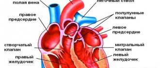
Echocardiography (EchoCG) is a test that uses high-frequency sound waves (ultrasound) to view the structure and study the function of the heart.
The common non-medical name for this test is cardiac ultrasound. The research is absolutely harmless to humans. Echocardiography uses reflected ultrasound waves to create images of the heart, its chambers, valves, walls and vessels (aorta, pulmonary arteries and veins). The sensor of the ultrasound machine is located on the chest and sends ultrasound waves that are reflected from the heart and again captured by the sensor, after which the signal is transmitted to the device, where it is converted into an image understandable to a specialist. If it is necessary to assess coronary reserve, stress echocardiography methods (stress echocardiography) are used.
Types and features of EchoCG
At the Family Doctor clinic you can undergo two types of echocardiography:
- a simple ultrasound examination of the heart to determine the health of the heart tissue, measure the volume of its cavities, wall thickness, correct operation of the valves, contractile activity of the heart muscle;
- stress echocardiography – a study with an artificial increase in the frequency of contractions of the heart muscle (physical activity). It is indicative when examining patients with coronary artery disease, since with increasing load, myocardial foci with impaired contractility are better visible.
By the nature of access, all ultrasound examinations are non-invasive - the integrity of the skin and tissues is not compromised. Acoustic gel is used during the procedure. Duration up to 20 minutes, no painful sensations.
Indications and contraindications for diagnosis
Echocardiography is prescribed to detect heart disease and evaluate its function. Most often, an echocardiogram is prescribed by a cardiologist; in preparation for major vascular operations, an ultrasound of the heart can be prescribed by the attending physician or an anesthesiologist-resuscitator.
Echocardiography can reveal:
- The size and shape of your heart, the thickness and movement of the heart walls.
- Assess the pumping function of the heart - ejection fraction
- Check the condition of the heart valves, whether the valves are closed, whether there is a narrowing in the valve area.
- The presence of cardiac aneurysms, blood clots in the cavities of the heart.
- Abnormal openings between the atria or ventricles.
- Identify the presence of an infectious process on the valves.
- During echocardiography, you can evaluate the pressure in the pulmonary artery and its size
- Identify aneurysms of the ascending aorta.
- Fluid accumulation or disease in the outer lining of the heart (pericardium).
- Heart tumors.
Echocardiography is a safe study and has no contraindications.
How to prepare properly?
With transthoracic ultrasound of the heart, preparation for the study is not required. In this case, it is better to quit smoking two hours before the ultrasound, alcohol, caffeine-containing drinks, and medications on the day of the examination.
However, it is worth knowing how to prepare for a cardiac ultrasound using the transesophageal technique: you must refuse to eat 2-3 hours before the procedure. Many young mothers are concerned about whether babies can eat before a heart ultrasound. Doctors recommend skipping one feeding before the ultrasound.
If there are diagnosed diseases, the attending physician gives recommendations to the sonologist on the procedure. Otherwise, preparation for cardiac ultrasound in women, men and children over 1 year of age is no different.
How is the diagnosis carried out?
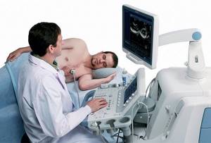
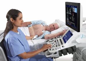
When performing an ultrasound of the heart, the patient is placed on his back or left side. During echocardiography, the sensor can be positioned in different planes for better visualization of the heart chambers. The sensor and the patient's skin are moistened with a special water-soluble gel, which ensures tight contact between the sensor plane and the body.
Our cardiac ultrasound machines allow us to perform various types of echocardiography. One-dimensional echocardiography in M-mode allows you to reproduce the movement of the heart walls and valves in the form of a graph, which allows you to evaluate the function of the ventricles.
Two-dimensional echocardiography shows a section of the heart in a certain projection and allows you to determine the size of the cavities of the ventricles and atria, the thickness of their walls, evaluate the movements of the valves and walls of the ventricles, and identify thrombosis of the cavities of the heart.
Using Doppler mapping, it is possible to identify the speed and direction of blood flow in the cavities of the heart, which makes it possible to determine valve insufficiency or stenosis, defects of the interatrial and interventricular septa.
The usual procedure for echocardiography involves first identifying the heart valves; heart septum. Next, the pattern of movement of the valve leaflets is revealed, the thickness of the walls and the size of the cavities of the heart are measured. Finally, Doppler echocardiography is performed to detect stenosis or insufficiency of the heart valves and pathological holes in the heart septa.
Indications
Symptoms of diseases of the cardiovascular system are varied and can be easily confused with problems of other organs. The patient must pay attention to:
- the appearance of interruptions in the work of the heart, increased heartbeat;
- shortness of breath, cough, pale skin, swelling in the lower extremities at the end of the day;
- weakness, dizziness, frequent headaches, loss of consciousness.
All these signs should not be ignored; they should serve as a reason to contact a specialist who will refer you for an ultrasound of the heart.
Direct indications for the study are an increase or decrease in blood pressure, the presence of pathological noises during auscultation of the heart, changes detected on the electrocardiogram, as well as pain in the heart area or behind the sternum (angina pectoris). Patients who have had a heart attack or heart surgery are required to undergo echocardiography periodically.
But not only people with health problems undergo echocardiography. It is recommended for athletes, men over 55, women over 50, and pregnant women. Heart ultrasound is also performed:
- children in the neonatal period to exclude heart defects;
- adolescents upon reaching 14 years of age, since during intensive growth of the body, pathology of the heart muscle may occur.
April 28, 2018
Modern diagnostic techniques make it possible to assess the functioning of the heart, identify disorders and causes of deviations in the functioning of this human organ. Ultrasound examination is one of them.
Ultrasound of the heart, or echocardiography, is painless and harmless. With the help of special equipment, the doctor is able to monitor the main functional processes of the organ. The progress of the examination is displayed on the computer monitor. Thanks to a sensor that transmits high-frequency sound waves, a clear picture appears on the screen. At the end of the diagnosis, the doctor gives the patient a conclusion, on the basis of which a diagnosis is made and a treatment program is developed. You can find out where to get a heart ultrasound in Moscow on our website.
Medical clinics are open from morning until late evening. Sign up for the procedure at any time convenient for you.
What does ultrasound of blood vessels and heart show:
- The work of the main muscle.
- The thickness of the walls of the organ.
- The speed of blood movement.
- Condition of valves and chambers.
- Parameters of cavities and pressure in them.
Why do you need an ultrasound of the heart?
To evaluate the characteristics of organ contractions.
Deterioration and disruption in the functioning of the cardiovascular system are visible even at the initial stage of the disease. During an ultrasound examination, congenital and acquired defects, blood clots, areas of asynergy, and valvular changes become apparent.
An ultrasound assessment of an organ is carried out not only if patients have symptoms of pathologies, but also if they do not. The method allows you to measure the pressure of the pulmonary artery. Repeated echo diagnostics demonstrates the dynamics of the pathology and the success of therapy.
Advantages of the technique
Patients are referred for echocardiography even in serious condition. Explanation for this:
- safety - no negative impact on the body and no side effects;
- information content - an accurate reflection of the functioning of the cardiovascular system;
- painlessness - physical and moral comfort.
Echocardia is good because it does not take much time, the patient immediately receives a conclusion. The duration of the procedure varies between 5–45 minutes. The duration depends on the purpose of the procedure, the patient’s condition and the symptoms of the pathologies.
List of indications
Symptoms of diseases of the cardiovascular system are the main reasons to visit an uzologist. Both adult and young patients receive referrals. Indications for echocardiography are as follows:
- heart rhythm disturbance;
- pain in the chest and ribs;
- heart murmurs;
- swelling, enlargement of the liver and other manifestations of failure;
- acute and chronic ischemia;
- shortness of breath, lack of air;
- increased fatigue;
- pallor;
- blue tint to the skin around the ears and eyes.
Patients are referred for ultrasound after heart surgery and chest injuries, and people suffering from frequent headaches are also recommended to undergo echocardiography in the therapist’s office. Why?
The causes of unpleasant painful sensations are sometimes microemboli. This disorder is treated by a cardiologist, drawing up a therapeutic program based on the ultrasound findings.
EchoCG is indicated for patients:
- hypertension;
- atherosclerosis;
- cancer;
- dystrophy;
- anorexia.
Ultrasound of the heart during pregnancy is performed mainly at the end of the first trimester. The study demonstrates the number of chambers, the rhythm of the fetal organ.
It is recommended that professional athletes undergo an ultrasound scan at least once a year. The explanation for this is the increased load on the heart. Based on the diagnostic findings, serious illnesses can be prevented and a preventive program can be drawn up.
Are there any contraindications
Diagnosis of the heart using ultrasound is safe and prompt, so there are no restrictions on its implementation. Over the many years of existence of ultrasound, no contraindications have been identified. However, in some cases the procedure is technically difficult. We are talking about examination of newborns. The cabinets are equipped with traditionally sized sensors. Using such equipment, it is difficult to diagnose the state of the heart of a small patient.
Carrying out an ultrasound examination
Let's take a closer look at how heart ultrasound is performed.
Once you receive a referral for an echocardiogram, there is no need to worry. Diagnostics will not cause any discomfort. There is no need to carry out any preparatory activities. However, you should not abuse fatty foods, smoke, or drink alcohol.
In the office, the ultrasound specialist will ask you to remove clothing from your upper body and lie down on the couch slightly on your left side. This position allows you to get a better picture. Before the examination begins, gel is applied to the chest and sensors are attached. After placing these pieces of equipment, all parts of the heart are viewed. Information about dimensions and functional features becomes available. Connecting the sensors is a painless procedure. There is also no discomfort during startup of the equipment.
The picture on the monitor screen is obtained due to the movement of acoustic waves. They are converted into electrical signals, which, after processing by an echocardiograph, become an image. At the same time, the doctor moves a sensor in the area of the heart.
This type of diagnosis should not be confused with electrocardiography. In the latter case, information about heart activity becomes known, presented in the form of a graphical recording. The ultrasound described earlier is called transthoracic.
There are the following types of echocardiography:
- Transfood type. This is not the most pleasant procedure, as the uzologist inserts a thin tube into the esophagus through the oral cavity.
- Stress analysis. Analysis of the organ’s functioning is carried out under conditions of increased stress on the body.
- Dopplerography. Allows you to monitor blood flow processes.
But most often patients are examined transthoracically.
Classification of areas of methodology according to the method of implementation
Echocardiography, as a diagnostic technique, can be one- or two-chamber. In the first case, the study is carried out in the “M” mode.
What is special about single-chamber technology? The ultrasound sensor emits waves along a single axis, which is determined by the doctor. A picture with a top view is displayed on the screen. If you change the direction of the sensor movements, the following become visible:
- ventricles of the heart;
- aorta;
- atrium.
Ultrasound in M-mode is performed on patients of any age.
Dual-chamber echocardiography differs from the previous technique in the type of image. The picture is obtained in two planes. The scanned plane is perpendicular to the 4-chamber position. With this type of ultrasound, a sensor with a wave frequency of 30 units. in 1 second they are directed in an arc of 90 degrees. When the trajectory of movement changes, the current processes of the cardiac structures are viewed.
During the previously mentioned Doppler assessment, turbulence and blood flow velocity become clear. What do deviations from the norm of such an ultrasound indicate? About the fullness of the left stomach and hidden heart defects.
The attending physician should decipher the conclusion of a particular ultrasound examination. During the analysis of a medical document, he compares the indicators with the established generally accepted norm. Discrepancies in values indicate the presence of diseases.
If the ultrasound specialist sees a clear deviation on the computer monitor, he displays this in the conclusion.
You can obtain more detailed information about the examination from the Delomedic administrators. Sign up for a cardiac ultrasound in advance or come during business hours. If necessary, our experienced doctors will decipher the conclusion.
Contraindications
There are no absolute contraindications to the study. However, testing may be difficult in some patients. Thus, it is more difficult to undergo echocardiography of the heart:
- women with large breasts;
- chronic smokers;
- people with bronchitis, asthma;
- patients with traumatic injuries of the anterior chest wall;
- people with abnormalities in the structure of the sternum (costal hump).