SPECIALISTS Gynecologist Gynecologist-endocrinologist Pediatric gynecologist Mammologist-oncologist Dermatologist Hirudotherapist Intimate plastic surgery Doctor Contour plastic doctor Ultrasound doctor SERVICES AND PRICES Gynecology Mammology Ultrasound diagnostics Paid tests Intimate surgery Contour plastic Treatment for women PROMOTIONS AND DISCOUNTS Students Teams Friends and subscribers am For residents of the region For pensioners Promotions in clinic
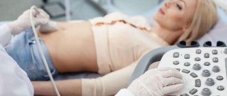
One of the most accurate and safe diagnostic methods used in medical practice is ultrasound of the pelvic organs. For women, obstetrician-gynecologists, general practitioners and family medicine specialists usually prescribe a complete ultrasound scan of the uterus, ovaries with fallopian tubes, and ligaments. For certain indications - ultrasound to determine the speed and nature of blood flow in the pelvic arteries and veins.
If you are in the center of Moscow, not far from Kutuzovsky Prospekt and the Kutuzovskaya, Studencheskaya, Victory Park metro stations, the Moscow City tower complex and are looking for where pelvic ultrasound is performed near me, you can sign up for our clinic ! Experienced doctors will help solve your problems. Prices for services and contact information are at the bottom of the page.
Announcement ✅
• Who gets a pelvic ultrasound: women, virgin girls, children; • Indications for carrying out: presence of complaints and for preventive purposes; • Procedure time: 15-30 minutes; • Contraindications: no; • Technique: transvaginal sensor - for women, transabdominal and/or transrectal - pelvic organs are examined on ultrasound for children, virgins, pregnant women; • Result of the study: photographs and a written opinion of a specialist, optionally recording a video clip on a flash card; • Price for pelvic ultrasound: from 2,500 rubles; transabdominal is lower, transvaginal is higher, screenings during pregnancy and 3D/4D are even higher.
Reference Information. ✅
Gynecologists perform pelvic ultrasound to assess the condition of the cervix, the structure of the uterus, ovaries, the condition of the periuterine space and the ligamentous apparatus. In clinical practice, ultrasound is a good and safe method of medical imaging, which allows timely identification of any pathology in the female reproductive system. This diagnosis is carried out for adults and virgin girls using acoustic waves that are completely harmless to the body, does not cause any pain, and can be done repeatedly without any consequences for the body. Ultrasound performed by a good specialist is rightfully considered the most effective and reliable method for detecting pelvic pathology.
For everyone who wants to have a pelvic ultrasound in Moscow, our clinic is open daily from 10-00, including on weekends (Saturday and Sunday). A list of services in this area and prices are presented at the bottom of the page.
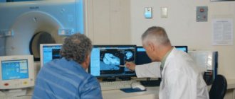
Ultrasound in gynecology and endocrinology
1. Ultrasound of the thyroid gland.
Ultrasound examination of the thyroid gland is the basis of endocrinology. It allows you to detect the presence of nodes or cysts, determine changes in the size of the thyroid gland or its structure. It is with the help of ultrasound that malignant neoplasms can be detected in time, which will allow the doctor to prescribe the necessary treatment and avoid serious consequences.
2. Ultrasound for pregnant women.
Ultrasound examination of pregnant women is carried out not only to determine gender, but also to monitor the development of the fetus. The harmonious development of the limbs, the absence of any pathologies - all this would be impossible to determine without ultrasound.
3. Ultrasound of the pelvic organs.
There are two types:
- Abdominal - i.e. without introducing an examination device into the body;
- Transvaginal - with the insertion of a sensor inside and a more detailed study of the internal organs of the small pelvis.
The decision to prescribe one type of ultrasound or another is made by the doctor. Examinations without insertion of a sensor are usually prescribed for children, whose anxiety may affect the results. At the same time, ultrasound with the introduction of a sensor is not prescribed to all adults, being limited only to special cases: for example, in the presence of pain in the lower abdomen or bleeding not associated with menstruation.
4. Ultrasound of the mammary glands.
Breast cancer is the most common cancer among women. And the cancer is getting younger. Today, women under 40 are beginning to encounter it more and more often.
The most reliable way to avoid serious consequences is to visit a gynecologist and mammologist annually and undergo an ultrasound examination. This is especially true for those women who have been diagnosed with mastopathy, a dishormonal disease that occurs in more than half of the female population.
5. Ultrasound of the pancreas.
Diabetes is a serious disease associated with improper or insufficient production of insulin in the pancreas. Ultrasound of the pancreas helps to diagnose malfunctions in its functioning in the early stages and prevent the development of dangerous diseases, including cancer.
6. Ultrasound of the adrenal glands.
The most common reason for prescribing an ultrasound examination of the adrenal glands is a suspicion of hormonal changes, which can manifest themselves in the form of the following symptoms:
- Sudden weight gain and problems losing it
- Sexual dysfunction, potency
- Sudden changes in the menstrual cycle
- Hair problems: excessive or insufficient
- Muscle weakness
Using the ultrasound results, the doctor will be able to determine the presence of pathologies: cysts, tumors, inflammation or enlargement of the adrenal glands, which will ultimately help to create an objective picture of the state of health and prescribe hormonal therapy or another type of treatment.
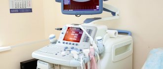
Ultrasound scanning (sonography) is one of the most informative diagnostic methods in modern medicine.
This type of research is based on the property of ultrasonic waves being reflected from tissues with different densities. The reflected signal is captured by the sensor and converted into a digital image, which is transmitted to the monitor. The received signal varies depending on the density and other characteristics of the tissue. Changes in tissue density and content indicate benign or malignant formations developing in the organ.
Ultrasound of the mammary glands is prescribed for diagnostic purposes if a mass formation is suspected, detected by palpation. Women over 35 years of age should undergo an annual ultrasound examination to prevent breast cancer. Ultrasound of breast tissue can be prescribed either as an independent examination or as an addition to mammography. In some cases, breast ultrasound is more informative than mammography. Ultrasound examination has advantages in identifying cystic formations and when examining young patients. Under ultrasound control, a targeted puncture biopsy of the tumor tissue is performed, and minimally invasive operations are performed to remove altered tissues.
Other advantages of ultrasound scanning include painlessness, non-invasiveness of the procedure (which eliminates infection of the body during the examination), low cost and lack of special training on the part of the person being examined. It has long been proven that ultrasound does not have a harmful effect on the human body, therefore ultrasound is used for the purpose of dynamic monitoring of previously discovered benign breast formations. This is necessary for timely correction of the course of treatment and suppression of further development of the pathology.
Why do you need an ultrasound of the mammary glands?
Ultrasound of the mammary glands is prescribed for the following purposes:
- diagnostics of cysts and neoplasms in the gland detected by palpation and mammography
- examination of the condition of the mammary glands of pregnant women and during lactation
- breast examinations in women over 30 years of age
- examination of the mammary glands in children and adolescents, regardless of gender
- assessing the condition of silicone breast implants
- male breast examinations
- additional examination to X-ray mammography in premenopausal patients
Ultrasound of the mammary glands can reveal the following changes in breast tissue: cysts, papillomas, fibroadenomas, infectious process, fibrocystic mastopathy, galactocele, oncological diseases of the mammary glands.
Ultrasound is most informative when examining breast tissue in the first phase of the menstrual cycle, that is, 5-14 days from the start of menstruation. An ultrasound of the mammary glands is performed with the woman lying on her back with her hands behind her head. If the mammary glands are large, the scan can be performed while lying on your side, standing, or sitting with your arms raised up. In order to better visualize tissue, a high-frequency sensor with Doppler ultrasound and complex scanning functions is used. When examining the gland, it is conventionally divided into the nipple area and 4 segments, similar to a watch dial. When a formation is detected, the doctor determines its position and distance from the nipple. The study of education is carried out in 3 standard planes. After diagnosing the breast tissue, the adjacent lymph nodes in the supraclavicular, subclavian, prothoracic and axillary zones are examined.
General rules for preparing for research
The optimal time for performing an ultrasound of the mammary glands is 5-7 days of the menstrual cycle (from the beginning of menstruation). No additional preparation is required for ultrasound examination of the mammary glands.
Attention! Ultrasound examination of the mammary glands does not replace consultation with a mammologist, but is one of the leading methods for diagnosing diseases.
Ultrasound in gastroenterology and urology
1. Ultrasound of the gallbladder.
It is prescribed if there are complaints of heaviness and pain in the area where the organ is located. It is important to properly prepare for the examination: exclude fatty and fried foods, yeast and fermented milk products, coffee and strong tea for 3 days. The procedure must be performed on an empty stomach. Even chewing gum is prohibited, because it provokes the secretion of bile and gastric juice, which significantly complicates the ultrasound process.
Sometimes during the procedure it becomes necessary to take a sorbitol solution orally to determine the contractility of the gallbladder.
An experienced doctor, based on ultrasound results, will be able to diagnose the presence or absence of most gallbladder diseases, including stones or sand.
2. Ultrasound of the abdominal organs.
The abdominal cavity is the location of most of the human organs. The doctor may prescribe a general ultrasound of the abdominal cavity, which usually includes examination of the liver, kidneys, pancreas, gallbladder, spleen, or conduct a separate examination of each organ.
As for the special preparation for an ultrasound, it is the same as for an ultrasound of the gallbladder: following a diet and performing the procedure on an empty stomach.
An ultrasound of the kidneys is prescribed for cases of lower back pain, problems with urination, and changes in the color of urine. The procedure allows early detection of kidney failure, cysts and tumors, and kidney stones.
3. Ultrasound of the spleen
prescribed in two types of situations:
- Emergency ultrasound of the spleen in case of bruises or injuries to the abdominal cavity. Splenic rupture is a dangerous condition that leads to internal bleeding and enormous blood loss.
- Routine ultrasound of the spleen. Normally, the spleen is located under the left hypochondrium. If there are pathologies, it increases in size and begins to extend beyond the edge of the rib. Ultrasound examinations are prescribed for patients with cirrhosis, suffering from pathologies of the circulatory system, infectious diseases, and if lymphoma or sarcoma is suspected.
4. Ultrasound of the bladder
The bladder is often examined together with the kidneys as a single system. As a rule, the following symptoms are the reasons for prescribing an ultrasound of the bladder:
- Nagging pain in the lower abdomen;
- Too frequent or infrequent urination;
- The presence of impurities in the urine: sand, blood clots;
- Change in urine color.
The examination is carried out in two ways: with or without immersing the sensor into the body through the genitourinary openings or rectum.
A timely examination helps to identify diseases such as urolithiasis, prostatitis, inflammation and abnormal development of the ureters in the early stages.
5. Ultrasound of the scrotum
Indications for use are usually quite serious: infertility, oncology, suspicion of the presence of a cyst or neoplasm. In diagnosing male diseases, ultrasound examination of the scrotal organs is sometimes the only reliable method.
No special training is required; standard hygiene procedures are sufficient. You must take with you all the available doctor’s reports and test results so that the doctor conducting the study has an idea of the nature of the preliminary diagnosis and can objectively evaluate the data obtained.
How is a vein ultrasound performed?
Ultrasound examination of the vascular system of the legs means a procedure for examining the anatomy of veins and arteries, assessing their functioning, the condition of the valves and the characteristics of blood flow. The technique makes it possible not only to find out why blood circulation is affected in a particular area of the vessel, but also to identify inflammation or blood clots. Ultrasound of the vessels of the lower extremities is an important procedure for diagnosing existing problems and helps in planning the correct treatment.
Types of sonography
To study the vessels of the lower extremities, a procedure based on the Doppler effect is used. In medicine, it means the reflection of ultrasound radiation from red blood cells flowing through the vessels.
As a result of a Doppler examination, the doctor gets an idea of the characteristics of the passage of the vascular cord, the speed of blood flow and other nuances of the functioning of the veins and arteries of the legs.
So what types of research are there, and what do they show?
USDG
The term “USDG” means “ultrasound Dopplerography”. This is a technique usually used to: – establish the patency of deep vascular collectors; – assessing the condition of superficial veins; – diagnostics of the condition of valves, including valves of typical main components of the venous system, that is, perforating veins.
UZDS
Duplex scanning combines the principles of Doppler and traditional examination methods and performs the following functions: – studies the operation of vein valves in real time; – makes it possible to assess the condition of the vascular walls; – allows you to analyze the patency of deep and superficial veins; – calculates the presence and location of blood clots.
Technologically, the method remains the most popular and accurate way to assess the performance of the venous system in all respects.
Online scanning
Online scanning is a complex that combines ultrasound and ultrasound scanning. It is intended to determine: – the condition of the walls of arteries and veins; – assessment of valve health; – patency of vascular collectors; – the condition of the veins connecting superficial vessels with deep ones; – the presence of blood clots and their characteristics, including size and location; – degree of blockage of the vessel.
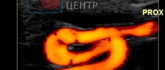
Ultrasound with color mapping
The most modern method of studying the veins and arteries of the legs is distinguished by color identification of the speed of blood flow in different areas. For example, shades of red characterize the flow of blood to the sensor, and blue tones characterize the flow of blood directed away from the sensor.
Important! The brighter the color, the higher the speed of blood movement.
In modern diagnostics of arterial-venous pathologies, the popularity of this method is growing due to its high information content and simplicity.
Preparation
When referring veins and arteries of the legs for ultrasound (as well as other soft tissues, as a rule), there is no need to carry out preliminary preparation for the procedure - it is not required.
How do they do it?
How is the deep veins and arteries of the legs diagnosed?
Ultrasound examination of blood vessels looks like this:
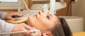
The patient enters the ultrasound diagnostic room, provides the doctor with a referral and frees the veins of the lower extremities. That is, he takes off tights or trousers, remaining in his underwear. The diagnostician applies a little conductive gel to the legs one by one, which will ensure better adhesion of the sensor to the surface of the skin.
During the manipulation, the doctor can change the frequency of the sensor for better visualization of deep vessels, but the patient will not feel this in any way.
The difference between an ultrasound scan is that during the examination the doctor will measure the pressure in both the upper and lower extremities. During the procedure, the patient will change his position from sitting to lying down and back.
During the examination of the leg veins, they are first examined with the patient lying down, then he will be asked to stand up. In addition, as part of the examination, special tests are carried out to determine the blood flow between the superficial and deep vessels. The test consists of taking a deep breath, without interrupting it, you need to make a strained effort.
The deep veins of the legs are examined in traditional and color modes. For a better examination, the patient is asked not only to perform stress tests, but also the areas where the veins are located are palpated with different intensities. Such tests (like the ultrasound itself) are performed in different positions: lying on your back or stomach, as well as standing.
Contraindications
In addition to the indications for ultrasound of the blood vessels of the legs, there are also contraindications.
Contraindications to duplex vascular examination are associated with the relatively long duration of the procedure - it takes about 40 minutes.
It may turn out that this examination turns out to be secondary and the study of adjacent tissues or internal organs comes to the fore.
Important! You should not perform an ultrasound in the area of the veins of the lower extremities if the skin is broken or damaged.
The main contraindications to studying vessels using the duplex method are:
– acute infectious processes; – any diseases accompanied by open wounds on the skin at the location of the sensor; – burns; – emergency conditions; – mental illnesses that make it impossible to carry out the procedure.
There are also conditions in which it is not recommended to scan blood vessels due to possible discomfort for the patient during prolonged immobility. These conditions include:
- excessive fullness;
- bloating;
- cystitis and some diseases of the genitourinary system;
- lymphostasis, causing severe swelling of the limbs.
Advantages and disadvantages
The advantages of the ultrasound method of studying limbs are:
- no pain;
- non-invasiveness, that is, the absence of punctures and other damage to the skin;
- economic accessibility of the procedure;
- ease of implementation;
- absence of radiation or ionizing load;
- linking the research picture to real time;
- the ability to perform a biopsy under ultrasound guidance;
- good visibility of all soft tissue features;
- the possibility of repeated repetition (for example, during treatment therapy to monitor its effectiveness).
Minuses:
- there may be insufficient data for a complete diagnosis;
- Adequate assessment of small vascular formations is not always possible;
- with atherosclerotic changes, the patency of the ultrasound wave may be impaired;
- is not a replacement for angiography;
- If carried out on old equipment or with insufficient qualifications of the doctor, the procedure may have low diagnostic value.
How much does it cost, where is the best place to do it?
It is best to contact your attending vascular surgeon, who will recommend a good specialist or tell you where and at what time he himself conducts ultrasound examinations.
In the Department of Vascular Pathologies, ultrasound can be performed free of charge, as prescribed by the surgeon. For a fee, in the absence of a referral, ultrasound examination of the extremities is carried out in phlebological clinics or multidisciplinary medical centers. You can find out the cost of the examination from the administrator by phone or by making a personal appointment. But it is better to refuse to perform vascular ultrasound in the offices of small diagnostic centers where any organs are examined.
Reference! The price of an ultrasound scan will depend on the specifics of the procedure and which vessels the patient needs to examine.
For example, for an ultrasound scan of the legs, the price will be from 1300 to 3500 rubles, and duplex angioscanning will cost 800–5000 rubles, color scanning using the duplex method will cost from 900 to 6500 rubles. On average, the cost of examining the vascular sections of the legs using ultrasound will cost about 2 thousand rubles.
There are also portable ultrasound machines, so ultrasound can be performed at home: this route naturally requires specific knowledge. Still, it is better to entrust this matter to a specialist.
Conclusion
Any patient, if indicated, can safely undergo angioscanning of blood vessels, because this manipulation is safe and painless. It does not require preparation and allows you to know exactly how healthy the vessels are an hour after the start of the examination.
Ultrasonography. The essence of the method
Ultrasound diagnostics is a method for studying the size, structure, shape, position, and movement of organs and tissues using ultrasound in real time.
The method is based on the ability of ultrasonic waves to pass through various tissues of the body, reflecting differently from structures of different densities. These “reflections” are recorded by a sensitive sensor and, using special software, are converted into an image on the monitor of an ultrasound scanner.
There are different types of ultrasound diagnostics. The most commonly used are scanning (what is traditionally understood as ultrasound) and Dopplerography (a type of ultrasound diagnostic that allows one to study the speed, direction and other characteristics of blood flow).
In advanced clinics today, ultrasound with elastography is also performed - a study of the stiffness and elasticity of tissues, which allows not only to detect neoplasms, but also to differentiate malignant from benign, thereby reducing the number of painful and unsafe diagnostic punctures (biopsies).
A9 Ultrasound examination helps to make a decision on performing TAB
RECOMMENDATION 7
- Fine needle aspiration biopsy is the procedure of choice for the qualitative evaluation of thyroid nodules when clinically necessary.
- FNA is the most accurate and cost-effective method for assessing thyroid nodules. Retrospective studies have reported a reduction in the proportion of both nondiagnostic and false-negative cytology following ultrasound-guided FNA compared with palpation.
- Therefore, for nodes with a high probability of non-diagnostic cytology (>25% - 50% cystic component) or difficulties in choosing a biopsy site (difficult to palpate or posteriorly located nodes), ultrasound-guided FNA is preferable.
Ultrasound diagnostics at MedikCity
Ultrasound diagnostics is one of the most informative and accessible methods for studying the health of patients. In some areas (eg obstetrics), ultrasound is the primary diagnostic imaging tool.
In addition to examining pregnant patients, this technique is unique for diagnosing certain organs and systems, for example, blood vessels (triplex vascular study), heart (echocardiography), etc.
Ultrasound is painless, has no harmful effects on the body and does not require significant material costs. It is also important that the procedure does not take long and is not tiring for patients, while it provides high-quality and complete information about the condition of the examined part of the body.
Advantages of ultrasound diagnostics:
- the ability to study most organs and systems;
- accessibility and speed of research;
- no contraindications;
- high information content;
- safety, non-invasive;
- the possibility of repeated procedures to clarify the diagnosis, monitor the course of the disease, and track the dynamics of treatment;
- the possibility of conducting screening examinations for prevention and early detection of pathology, etc.