Diagnosing coronary heart disease is not always an easy task. In some cases, the diagnosis is beyond doubt. But there are also situations when the patient’s complaints are ambiguous, and studies performed at rest do not completely exclude this serious disease. Then the attending physician prescribes stress echocardiography.
Stress ECHO-KG
Posted at 18:15h in Services by doctor
Stress echocardiography has become a generally accepted technique in recent years, as it is widely used in the diagnosis of heart diseases that lead to myocardial damage due to pathological changes in the circulatory system. The study is carried out to identify pathologies of the heart valves in patients who need urgent surgery, as well as to determine the dysfunction of diastole, recognized as one of the causes of heart failure. The essence of the method is the use of stress tests, with the help of which the contractility of the heart is accelerated, making it possible to determine areas of the myocardium experiencing oxygen deficiency. What are the indications for the test and what are the diagnostic features?
Characteristics of the method
Most patients, after being prescribed stress echocardiography, are interested in what it is and how the procedure is performed.
Determination of local disturbances in individual areas of cardiac tissue indicates the development of a pathological process. As a rule, an electrocardiogram is performed at rest, so it is difficult to identify minimal disturbances in the functioning of the organ. Stress echocardiography, on the contrary, is performed while the heart is stressed, causing an increase in its contractions, which helps to accurately detect coronary artery disease at an early stage of development.
- pharmacological medicines;
- bicycle ergometry;
- cold stimulation;
- transesophageal electrical stimulation;
- hyperventilation of the bronchopulmonary system.
The method also allows you to determine cardiac endurance, which is why it is used annually among professional athletes.
Indications for the study are as follows:
- Surgery on the heart;
- The need to evaluate its performance during the recovery period after a heart attack;
- ECG indicators, swelling and shortness of breath;
- Unsatisfactory results of load application;
- Ischemic disease in the chronic stage;
- Analysis of the effectiveness of therapy;
- Forecast for the development of pathology;
- Suspected angina;
- Work ability analysis;
- Routine examination of professional athletes.
Advantages and disadvantages of the test
The use of each type of stress test has both its advantages and disadvantages.
- Allows you to evaluate the activity of the heart in a state of increased physical activity.
- Possibility of performing diagnostics outside the hospital using portable equipment.
- Qualitative level of assessment of the state of the heart muscle.
- High sensitivity of the method.
- No biological effect on the patient and medical staff.
- The accuracy of the study depends on the skill level of the specialist.
- During exercise, blurred visualization of the left ventricle is possible.
- The development of side effects in the form of dizziness, rapid pulse, discomfort in the chest area.
Despite all the shortcomings, the technique is considered an effective and inexpensive procedure that can diagnose pathological changes in the functioning of the myocardium.
general information
Echocardiography is performed using a machine that produces ultrasound waves. Echocardiography is based on the use of the physical Doppler effect. Stress - EchoCG allows you to accurately assess functional changes in the heart muscle and valves during physical activity.
Stress - Echo CG is an alternative to ergometry. The doctor determines which procedure - ECG with exercise or ergometry - is preferable for the patient, taking into account his state of health and the clinical picture of the disease.
The duration of the procedure is 45 minutes. It is not accompanied by exposure to radioactive radiation on the body.
In what cases is it prescribed?
Experts recommend undergoing stress diagnostics for patients who have received normal ECG and Echo kg readings, but at the same time they have symptoms characteristic of heart disease.
- Diagnosis of myocardial ischemia.
- Assessment of the degree of damage to the coronary vessels.
- Assessment of the functioning of the heart muscle in patients with organ dysfunction.
- Identification of myocardial areas with a high risk of developing ischemic damage.
- Analysis of the status of chronic ischemic heart disease.
- Preparing the patient for minimally invasive procedures on the chest area.
- Analysis of the effectiveness of angioplasty, stenting and bypass surgery.
- Clarification of the possibility of developing complications after cardiac operations.
- Establishing the timing of surgery in the presence of valve defects.
- Determination of the patient's ability to work.
Features of the method
An early sign of weakened blood circulation in the myocardium is a decrease in the number of heart contractions in response to additional load, whereas in a normal physiological state contractions remain unchanged or increase.
- Deterioration in the contractility of the damaged area (visualized by ultrasound).
- Pathological changes during ECG registration (determined by stress test).
- The appearance of pain behind the sternum.
Myocardial movements are preliminarily assessed before testing. After this, the patient is given a medicine that increases the heart rate or is asked to do an exercise test.
Pharmacological testing is associated with more cardiovascular complications than exercise testing. In the case of a load test, it is recommended to rotate the pedals on a bicycle ergometer in a horizontal position. This makes it possible to quickly move the patient to the couch.
How to prepare for research?
At the preparatory stage of the study, the patient is prescribed medications containing nitrates, which can reduce the number of myocardial contractions and also lower blood pressure. The medication is prescribed to protect the heart muscle from the effects of adrenaline produced during stress, which can cause adverse reactions from various organs and systems.
To fully prepare the body for the procedure, you must follow these recommendations:
- The day before the procedure, drinks containing caffeine and alcohol should be avoided.
- It is necessary to avoid physical activity for several hours.
- The last meal is no less than 3-4 hours before the procedure.
- Stop smoking immediately before screening.
On the day of diagnosis, patients are allowed to take nitroglycerin to stop a possible attack of angina. However, the use of funds must be agreed with a specialist.
Load test
When carrying out the method, various stress tests can be used. The type of test used depends on the objectives being pursued and the intended diagnosis. Thus, to detect myocardial ischemia, as well as assess the degree of damage to individual areas after a heart attack, they resort to dynamic loading.
When using a load in the form of a treadmill, the initial readings are taken at rest, and then when regaining strength after stopping the load.
It is preferable to carry out the test with a bicycle ergometer, since the recording of indicators occurs directly during the period of load or at its peak. The best visualization of the organ is achieved when using bicycle ergometry in a horizontal position.
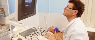
How is cardiac ultrasound performed?
During echocardioscopy, the doctor uses various modes of the ultrasound machine:
- one-dimensional (M-mode),
- two-dimensional (B-mode),
- Doppler mode (assessment of the speed of blood flow in chambers and vessels),
- color Doppler - CDK (to determine the direction of blood flows and identify pathological ones),
- power doppler (records the very fact of the presence of blood flow in the vessels),
- tissue Doppler (a deeper assessment of myocardial contractility, based on a study of the nature of the movement of the walls from the sensor and to it),
- 3D echocardiography (it brings maximum benefit before operations on the valves - they are almost completely visualized before the intervention, which is important for the surgeon to determine tactics).
Transthoracic ultrasound
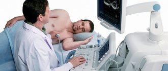
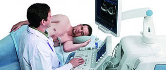
Echocardiography through the chest is performed in an ultrasound or functional diagnostics room. Sometimes in a hospital, due to the severity of the patient and the impossibility of transportation, a portable ultrasound machine is used to examine him. The nurse or doctor asks the patient to undress from top to waist, including women who need to remove their underwear. Next, the patient needs to lie on the couch on his left side and place his left hand under his head. In this case, the head end of the couch is slightly raised - this way, the maximum distance between the intercostal spaces is achieved for better visualization of the heart.
The position of the patient in relation to the doctor can be different, it all depends on the preferences of the latter and the arrangement of the office. The patient can be turned with his face or back towards the doctor, with his head towards or away from the device.
The specialist lubricates the sensor with ultrasound conductive gel for better contact with the skin, applies it to the left side of the chest, visualizes the heart, displaying its standard positions for measurements.
Transesophageal echocardiography
Transesophageal ECHO is performed strictly on an empty stomach in an ultrasound or functional diagnostics room. They take the patient’s consent to conduct the study, having first explained its entire essence and course of events. The throat is irrigated with lidocaine spray, the patient is asked to remove dentures and lie on the couch on his left side, bending his knees and placing his hands under his cheek or on his stomach. A mouthpiece is inserted into the mouth to prevent the patient from biting the probe. The doctor then inserts the endoscope. At the beginning, he asks the patient to make swallowing movements to move the device easier. Having reached a certain position, the doctor begins the examination itself. It lasts 10-20 minutes.
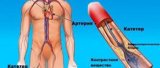
Echocardiography with contrast
When using a contrast agent, it is injected into one of the veins or arteries on the thigh - depending on the type of drug and the purpose of the study. In this case, the ultrasound sensor remains on the patient in order to register all data and take measurements in a timely manner.
Medication tests
In case of intolerance to physical activity, the use of a drug test is recommended, which is quite safe for the body and causes minimal side effects.
Dobutamine test
It is the most widely used test, during which the number of myocardial contractions increases, blood pressure increases, which causes an increase in the organ's need for oxygen. The difference between the heart's need for oxygen and the ability of the coronary vessels to supply it indicates the presence of local pathological processes in the myocardium.
Test with dipyridamole
The test is done with a gradual increase in the dose of the drug. At each stage, the degree of impairment of myocardial contractility is assessed. In the absence of pathological changes, in order to achieve the required heart rate, an additional 1 mg of atropine is administered. A couple of minutes after the administration of dipyridamole, an intravenous injection with aminophylline, which is an antidote, should be given.
What is stress echocardiography
Stress echocardiography is an ultrasound examination of the heart performed during physical exercise, medication, or exposure to electrical impulses. The purpose of the analysis is to monitor the work of the heart during the period of maximum contractions and when consuming more oxygen than at rest.
Stress echocardiography is a safe method for early diagnosis of ischemia and other heart diseases. The advantages of the test include the reliability of the results, lightness and portability of the equipment.
Evaluation of results
The results of the study are displayed in the form of a two-dimensional graph, which makes it possible to fully assess the quality of left ventricular functioning. Interpretation of the results includes an assessment of the degree of thickening and mobility of the heart muscle tissue in individual areas.
A preliminary analysis of the graphs is carried out by a specialist immediately after their registration. After the screening is completed, the cardiologist can view a video recording of diagnostic indicators in slow motion. The obtained data is stored on disks, creating an information base for the patient with further assessment of the dynamics of heart performance indicators.
Thus, stress echocardiography is a modern method for diagnosing coronary artery disease. The study allows us to determine the initial stage of the disease, when other methods reveal low effectiveness. However, before the procedure, possible cardiac complications associated with excessive load on the organ should be taken into account.
Are there any contraindications to this procedure?
Despite the fact that this examination is carried out under close medical supervision, it in any case includes stressful effects on the heart muscle. Therefore, such tests must be ordered with great caution.
All contraindications to stress echocardiography can be divided into two large groups:
Absolute contraindications
:
- acute myocardial infarction within a period of up to 3 weeks;
- unstable angina;
- heart rhythm disturbances that cannot be corrected with medication;
- severe aortic stenosis;
- severe forms of heart failure;
- pulmonary infarction or pulmonary embolism;
- suspicion of the presence of a dissecting aortic aneurysm;
- suspicion of inflammatory pathologies of the heart, such as pericarditis, myocarditis or endocarditis;
- serious infectious diseases in the acute stage;
- renal failure and other serious diseases that can affect test results or worsen during stress echocardiography.
Relative contraindications
which the doctor identifies. Potentially avoidable causes can usually be corrected before stress echocardiography.