Electromyography (EMG) is a mandatory study if damage to the neuromuscular system is suspected. Such disorders accompany various diseases diagnosed at any age. EMG is a painless procedure, does not require preliminary preparation, and its indicators are quite informative for making a diagnosis. This makes muscle electromyography an indispensable study in neurological practice.
At the Yusupov Hospital, EMG is performed using modern electromyographs, and highly qualified personnel interpret the study indicators in the shortest possible time.
Electromyography: what is it?
Electromyography is a method for diagnosing disorders of the neuromuscular system, based on indicators of bioelectrical muscle activity. The study is based on the ability of muscle tissue to create electrical activity with each contraction. Electromyography records these values, resulting in an assessment of the results obtained. Depending on the indicators, taking into account the accompanying clinical picture, the lesion and its location are determined.
EMG is performed using an electromyograph. The device records bioelectrical activity, transmitting it to monitor screens or recording it on paper. Any deviation from normal values indicates a violation of muscle conduction.
Several research methods are used to take electromyography. The choice of the type of EMG is made by the attending physician based on the individual characteristics of the development of the disease. This study of muscle nerve conduction is used in various fields of medicine: neurology, traumatology and orthopedics, cosmetology, dentistry, sports medicine. Electromyography makes it possible to identify the pathological focus in the early stages of the disease. In addition, EMG is used to monitor treatment.
Electromyography allows you to determine:
- Localization of the pathological focus.
- Nature of the pathology. Damage to muscle or nerve fibers is determined.
- The extent of the process.
- Stage of the disease.
- Damage level. Local or systemic disease may be present. Depending on this, the type of research is selected.
- Dynamics of the pathological process. The doctor monitors the prescribed treatment using dynamic electromyography. This allows you to timely adjust or continue previously prescribed therapy.
Types of EMG
There are several ways to perform electromyography. The choice of method is made by the doctor depending on the existing pathology. The following types of EMG are distinguished:
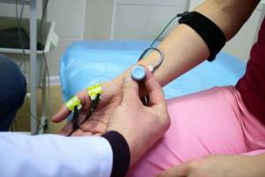
- Stimulation (surface) electromyography. It is a non-invasive and painless examination. This EMG method allows you to evaluate bioelectrical activity over a large area of muscle. Stimulation myography is performed on the lower and upper extremities to study weakness, fatigue, numbness, and decreased muscle sensitivity. In addition, surface EMG is performed to diagnose nerve damage. This type of study evaluates the condition of the masticatory and facial muscles, which is informative for cosmetologists and dentists.
- Needle (local) electromyography. Used for more accurate research. For this purpose, a needle electrode is inserted into the muscle. In this case, minor pain occurs, which soon disappears. Local electromyography is an invasive research method. In this regard, hematomas or infiltrates may occur after the procedure.
Any of the EMG methods is performed for diagnosis and treatment evaluation.
Electroneuromyography as a research method
Using the electroneuromyography (ENMG) method, the electrical activity of the muscles of the lower and upper extremities is checked to diagnose diseases in the spinal cord, spine, nervous disorders, and identify injuries to the muscular system.
During the ENMG procedure, it is possible to determine in which part of the neuromuscular plexus of the extremities there are lesions, this makes it possible to prescribe further therapy to patients.
Although the peripheral nerve bundles are resistant to damage, diseases or injuries of various types in the spinal region lead to disruption of the functioning of the nerve bundles; for this reason, impulses cannot pass through the affected nerves to stimulate muscle movement.
ENMG technology allows:
- Check the ability of the limb muscles to contract.
- Determine the speed of electrical impulses and their oscillation.
- Identify areas with pathology in the muscles and peripheral nervous system.
The results of such diagnostics are 95% reliable.
Indications for electromyography
The method of recording the bioelectrical activity of muscle tissue has become widespread in various fields. EMG is used in neurology, cosmetology, traumatology, dentistry, and sports medicine. The high accuracy and painlessness of the procedure make it mandatory in the presence of pathology of the neuromuscular system.
General indications for EMG include:
- The appearance of muscle weakness, increased fatigue.
- Presence of convulsive syndrome.
- Impaired sensitivity.
- Reduced muscle volume.
- Muscle pain of varying severity.
The presence of any of the above signs is an indication for electromyography.
Most often, changes in the conductivity of muscle fibers occur in neurological practice. Using EMG, the condition of the nerves innervating the muscles is assessed. Diseases requiring electromyography include:
- Polyneuropathy. Nerve damage causes changes in the EMG waveform. The oscillation amplitude values depend on the degree of damage to the nerve fiber.
- Muscle pathology. This group of diseases includes inflammation, dystrophy, and increased fatigue.
- Degenerative-dystrophic changes in the spine. The presence of a spinal hernia often causes compression of the nerve roots. Such conditions are reflected in electromyography.
- Hyperkinesis. Involuntary muscle movements cause EMG changes characteristic of this syndrome. Using diagnostics, it is possible to establish the localization of the pathological focus.
- Parkinsonism. The tremor that appears in Parkinson's disease significantly changes muscle conductivity. Such a condition must be examined using electromyography.
- Radiculopathy. Nerve root lesions are diagnosed using EMG.
In dental practice, electromyography is prescribed to study the masticatory muscle. Diagnosis is carried out both for diagnostic purposes and to monitor previous treatment. Indications for EMG are:
- Bruxism (teeth grinding).
- Jaw injury.
- Inflammatory dental diseases.
- Damage to the facial nerve.
- Prosthetics.
Sports medicine is another area where electromyography is actively used. Assessing the degree of muscle damage plays an important role in the recovery of athletes after injury. EMG makes it possible to identify a lesion at the initial stage, preventing the development of serious complications.
When selecting a prosthesis, traumatologists and orthopedists always prescribe electromyography to assess the lost functions of the limb. Diagnostics of muscle conductivity is actively used in cosmetology for the administration of Botox.
Types of ENMG
ENMG research is carried out using three types of techniques, with the help of which a complete picture is created to accurately determine the pathology in the lower and upper extremities.
The functional combination of the three types makes it possible to identify diseases at the initial stage of development and promptly prescribe appropriate treatment. Patients must have x-rays, general analysis results from MRI and CT procedures, and provide a medical record.
If a patient is suspected to have only one nerve ending with pathology, he undergoes a procedure according to an abbreviated program with the preparation of a protocol corresponding to the disease.
Superficial
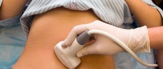
This technique is considered non-invasive, that is, during the procedure the integrity of the body’s structure is not violated. During a superficial examination, sensor electrodes are attached to the patient’s lower extremities.
When the device is connected, impulses begin to emanate from nerve endings that are examined without stimulation, which are reflected on the device monitor. The study is carried out to record voluntary nerve contractions and evaluate the signals emanating from them.
Superficial electroneuromyography
Needle
This is done very carefully using sensors with electrodes with thin needles installed at the ends:
- The needles of the device are inserted into the area of the plexus of the muscles of the lower or upper extremities.
- Patients may experience slight itching or a burning sensation.
- The needle method is effective for pathologies of the spinal cord and skeletal muscles, and gives a more accurate result in the diagnosis of myelopathy and inflammatory muscle diseases.
Contraindications
Electromyography is a procedure performed on patients of any age. There are no specific contraindications for its implementation, but there are common ones for all diagnostic procedures. Conditions in which EMG is not prescribed include:
- Acute pathology. Infectious or non-infectious diseases in the acute stage are an absolute contraindication for electromyography.
- Presence of a pacemaker.
- Skin lesions. Inflammatory processes on the skin, accompanied by pustular rashes, exclude the possibility of performing an EMG.
- Epilepsy.
- Cardiovascular pathology. This group of restrictions includes hypertensive crisis, myocardial infarction, and unstable angina.
- Mental disorders. Contraindications are conditions in which the patient cannot control his actions.
Needle electromyography is contraindicated in cases of blood clotting disorders, as well as increased pain sensitivity.
Contraindications for ENMG studies
There is a category of patients for whom the procedure of electroneuromyography of the extremities is contraindicated.
These are patients diagnosed with:
- cardiovascular diseases, hypertension;
- ARVI;
- schizophrenia and epilepsy;
- hemophilia (blood clotting disorder).
ENMG is not prescribed for patients with hepatitis or HIV infection, who are undergoing physiotherapy, pregnant or breastfeeding women. To this list are added those who have trophic ulcers on the lower extremities or severe damage to the skin.
Attention! Those using an endoprosthesis and a pacemaker should know that ENMG is also contraindicated for them.
Electromyography: preparation for the study
The advantage of electromyography over other diagnostic methods is the lack of special preparation before the study. However, there are several recommendations, adherence to which will ensure the most accurate recording of bioelectrical muscle activity. These include:
- Refusal to take certain medications. Such medications include tranquilizers, muscle relaxants and other drugs that affect the nervous system. It is recommended to cancel them the day before the study.
- Observe dietary restrictions. For a more accurate diagnosis, it is necessary to stop eating foods that increase nervous excitability several hours before electromyography. These include tea, coffee, caffeinated drinks, chocolate, and carbonated drinks.
In cases where discontinuation of medications is not possible, the attending physician must be notified in advance.
How is electromyography performed?
Electromyography takes from 30 to 60 minutes. The time depends on the number of areas examined, as well as the severity of the lesion. Electromyography is performed using an electromyograph. With its help, the bioelectrical activity of muscle fibers is registered and recorded.
The EMG procedure can be performed in an inpatient or outpatient setting. For this, the patient is asked to take a comfortable position (sitting, lying, half-sitting). The area to be examined is treated with an antiseptic. After this, I do not apply the electromyograph electrodes. In cases where needle EMG is indicated, a needle electrode is inserted into the muscle being studied. This is the only type of electromyography in which minor pain is felt. All other methods are painless.
At the very beginning of the procedure, muscle conductivity at rest is assessed. After this, she is asked to strain, after which the bioelectrical activity is recorded again. The results obtained are an electromyogram, which reflects all the changes occurring in the neuromuscular system. Based on the data obtained, a diagnosis is made or treatment is assessed.
Indications for the procedure
A direct indication for EMG is pain. Sudden or frequent muscle pain is an alarming sign that should be responded to immediately. Intense muscle pain and muscle twitching require additional examination of muscle tissue. Using the EMG procedure, diagnoses are confirmed: myasthenia gravis, myoclonus or amyotrophic sclerosis. Electromyography is prescribed if the development of polymyositis is suspected.
It is advisable to diagnose muscles in case of loss of muscle tone (dystonia) or after injury to peripheral nerves. Damage to the central nervous system, brain or spine is a reason for a complete examination of muscle tissue using EMG.
Diagnostics with the introduction of diodes is prescribed for suspected multiple sclerosis, botulism, or after polio. For facial neuropathy or carpal tunnel syndrome, invasive electromyography is used. The direct indication for the procedure is diseases: micro-stroke or tremor. To safely administer Botox, EMG is used first.
The patient is prescribed the required number of procedures that do not harm surrounding tissues. The first examination occurs at the initial stage of diagnosis before treatment is prescribed. During therapy, EMG is performed repeatedly. For preventive purposes, electromyography is also used for adults and children.
EMG interpretation
The electromyography technique is based on recording muscle activity. The results obtained form an interference curve, reflecting any changes in conductivity. There are several types of EMG curve:
- First. In this case, the curve registers rapid fluctuations in potential. The frequency is about 50-100 Hz. Such an EMG is considered a normal variant. Indicators may vary depending on age, weight and physical development.
- Second. Fluctuations on electromyography occur with a frequency of less than 50 Hz. This indicates decreased muscle conductivity. Such changes are characteristic of neuropathy.
- Third. The EMG curve records a significant decrease in oscillation frequency. On average it reaches 4-10 Hz. These results indicate pathology of the extrapyramidal system.
- Fourth. Electromyography does not cause any hesitation. Bioelectrical activity is completely absent. A similar condition occurs with muscle paralysis. Another name for type 4 EMG is “bioelectric silence.”
The following main diseases are identified in which changes in the amplitude of oscillations are recorded on electromyography:
- Polyneuropathy. The results of the curve depend on the degree of damage to the nerve fiber. With a mild degree, the oscillation frequency is significantly reduced. In cases where the pathology of the nerve has caused its complete death, “bioelectric silence” is recorded.
- Myositis. Inflammation of muscle fibers causes a decrease in their conductivity. Indicators of bioelectrical activity depend on the degree and stage of myositis.
- Amyotrophy. The pathology is characterized by loss of muscle mass. In this case, an increase in the amplitude of oscillations is recorded on the EMG. The curve has the shape of a “picket fence”.
- Myasthenia. Muscle fatigue syndrome significantly affects the electrical conductivity of fibers. On the EMG there is a decrease in the amplitude of oscillations. The values depend on the stage of the disease.
- Tremor. The symptom is characteristic of various neurological pathologies. In this case, a sharp increase in the amplitude of oscillations is recorded. Their frequency depends on the location of the lesion.
- Myotonia. Syndromes associated with delayed muscle relaxation are an indication for electromyography. It identifies curves with low amplitude and high frequency.
Highly qualified doctors interpret EMG results. Based on the data obtained, the specialist is able to establish the localization of the pathological focus, its degree and stage.
Electromyography – price in Moscow
Electromyography is a mandatory study in the presence of damage to the neuromuscular system. The Moscow Yusupov Hospital has the latest equipment that allows diagnostic procedures to be carried out with high precision. The many years of experience of the institution’s specialists allows us to decipher the results of the study and make a correct diagnosis in the shortest possible time. Identifying pathology of muscle fiber conduction at an early stage allows you to begin timely treatment and avoid serious complications. You can sign up for electromyography and learn more about the cost of the procedure by phone.