When the phrase “soft tissue bruise” is heard, the average citizen is not particularly frightened - this is not a fracture or, God forbid, a concussion. It will hurt and stop. However, not all so simple.
Half of a person's weight comes from soft tissue. This category includes skin, subcutaneous tissue, fat, glands, muscles, tendons and fascia, as well as blood vessels, nerves, and lymph nodes.
This is an extremely complex interconnected system, which, despite its apparent strength, is quite vulnerable. Any ill health caused by injury, infection or other pathology can cause serious damage to health. Thus, muscles injured, for example, due to necrosis or extensive traumatic damage, do not recover. As a result, a person may lose the ability to perform some movements and remain disabled.
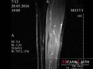
Timely and accurate diagnosis of the condition of soft tissues allows you to avoid many dangers. At the same time, magnetic resonance imaging is one of the most preferred methods. The non-invasiveness, harmlessness and high information content of the study made it the gold standard for diagnosis. At MedicCity you can undergo MRI of muscles and superficial structures and other types of tomography around the clock by appointment.
1 MRI of muscles
2 MRI muscles
3 Muscle MRI
What does an MRI of the leg reveal?
MRI of the leg allows you to determine:
- various soft tissue injuries of the lower leg;
- localization of foreign bodies;
- injuries and damage to the ankle and knee joints;
- inflammatory changes in the tissues of the lower leg;
- post-traumatic processes;
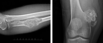
- benign and malignant tumors;
- metastases in the soft tissues of the leg;
- condition of blood vessels and nerves;
- degree of severity of the inflammatory process;
- damage to tendons and ligaments.
Advantages of the method
The 3D model obtained during MRI shows not only the condition of the suspected lesion, but also the surrounding area. This will allow you to see how much the pathology has developed. Even small lesions as small as 1 mm are visible on MRI.
The nature of the pathology (for example, a benign tumor or cancer), the degree of development, and size are assessed Diseases that you may not even be aware of are recognized, thereby eliminating complications and getting rid of the disease faster.
MRI of soft tissues is a safe study . In an MRI scanner, you will be exposed to electromagnetic waves in a range that does not harm the body.
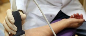
A conventional X-ray will not cope with the task of checking soft tissues, since it is focused on the joints. Ultrasound examines the superficial layers, and CT, with all its capabilities, is undesirable due to x-ray radiation. MRI can be performed many times: for example, to make a correct diagnosis and monitor the progress of treatment.
Indications for MRI of the leg
The study is prescribed in the following situations:
- unknown cause of pain in the foot or leg;
- numbness in the lower leg/foot;
- weakness, decreased strength and endurance of the lower leg muscles;
- loss of sensation in the lower leg area;
- changes in skin color of the lower leg (redness, bluishness or pallor);
- complex dislocations and fractures of the lower leg bones;
- inability to move the joints;
- clarification of the characteristics of a malignant tumor (shape and size, exact localization, stage and extent of spread);
- performing a biopsy of tumors under MRI control.
MRI is also indicated for:
- osteomyelitis (in the initial stages MRI is more informative than CT);
- atherosclerosis;
- arthrosis and arthritis of the lower leg joints;
- inflammatory processes (phlegmon, abscesses);
- pinched nerves;
- ruptures of tendons, ligaments and muscles.
MRI with contrast is indicated to identify vascular pathology:
- benign and malignant vascular formations;
- aneurysm of the arteries and veins of the leg;
- thrombosis in the lower leg area;
- vascular abnormalities;
- establishing the cause of obstruction of the arteries of the leg and venous insufficiency.
Who is prescribed MRI of soft tissues?
MRI of superficial structures and muscles may be recommended for diseases and conditions such as:
- severe pain;
- edema, swelling of tissues;
- vascular disorders;
- traumatic damage to muscles, tendons, subcutaneous fat, etc.;
- pinched nerve fibers;
- suspicion of the presence of neoplasms;
- suspicion of inflammatory or infectious processes;
- preoperative or postoperative examination;
- atrophic, dystrophic and other degenerative changes, etc.
1 MRI of muscles
2 MRI muscles
3 Muscle MRI
How the study is performed
No special preparation is required for the research procedure. In the case of an MRI with contrast, the patient is warned not to eat 4–5 hours before the examination (to avoid nausea and vomiting). The patient is placed on the tomograph table in a supine position, the legs and head are fixed (complete immobility is required to obtain high-quality images) and the table is moved into the MRI machine.
The duration of the procedure is 30 minutes, 45 if performed with contrast. Its implementation does not cause discomfort to the patient and does not cause pain. After completing the study, the radiology doctor describes the images and issues a conclusion. If a pathology is identified, with recommendations on which specialist should be contacted.
If the patient wishes, the images are copied onto electronic media or printed on film.
Contraindications Preparation
You can see prices for services
Contrast tomography
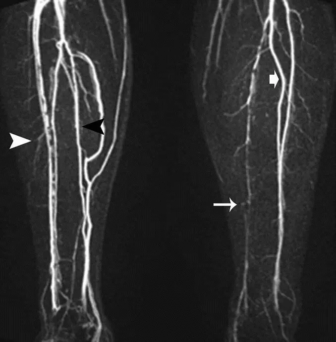
For better visualization of small vessels, it may be necessary to administer contrast. The contrast agent allows you to obtain an image with a clearer visibility of the blood network, which is justified when diagnosing tumors that usually have a good blood supply.
Correct interpretation of MRI results of extremity vessels requires high qualifications. This diagnostic method is carried out by top-level specialists, with the issuance of an opinion, based on the results of which prescriptions are made by the attending physician.
Contraindications
There are a number of restrictions on performing MRI. These include:
- the presence of metal in the body, because it may affect the image or attract the magnet;
- the presence of electronic devices (artificial heart valve; clamps for the treatment of brain aneurysms; cochlear (ear) implant; drug or insulin pump; pacemaker);
- first trimester of pregnancy.
Contraindications for MRI with contrast include:
- chronic renal failure;
- allergy to the drug;
- any stage of pregnancy.
It is recommended to leave metal objects outside the MRI machine room. Tell your doctor if you have tattoos or permanent makeup. Some inks contain iron, which may become hot during the test.
Prices for CT angiography of vessels of the lower extremities
Prices for computed tomography of the lower extremities can vary significantly among different medical institutions. When choosing a clinic for examination, you need to focus on the quality of diagnostic equipment and the experience of specialists.
The medical center on Yauza offers services for angiography of the arteries of the lower extremities at an average price. At the same time, we perform the procedure using the latest equipment used by leading clinics in the world. As alternative diagnostic methods, the clinic uses ultrasound examination of blood vessels and MRI.
CT scan with contrast in the clinic on Yauza is a safe procedure. Modern equipment allows you to take high-quality images and create a 3D model of the vessels of the study area.
When is it necessary to perform angiography of the vessels of the lower extremities?
The study is used in preparation for surgical treatment and to monitor the effectiveness of therapy. For diagnostic purposes, MSCT angiography of the lower extremities is performed on the recommendation of a doctor. A procedure is prescribed if the doctor, based on the symptoms, suspects the development of diseases that impair blood flow:
- embolism;
- Takayasu;
- varicose veins;
- angiopathy;
- thrombosis;
- thrombophlebitis;
- venous insufficiency;
- physiological changes in blood vessels.
Tomography of the lower extremities is recommended for all patients suffering from diseases that cause vascular damage. For example, patients with diabetes.
Types of MRI examination of joints
MRI of the knee joint
The price of an MRI of the knee joint in Moscow is noticeably higher than the cost of an X-ray examination, but the diagnostic capabilities of this method are disproportionately greater. It is difficult to compare the level of safety of procedures. And given that the knee joint is one of the most problematic in terms of the frequency of injuries and various disorders, the demand for MRI can hardly be overestimated.
To increase the accuracy of diagnosis, it is possible to conduct an examination with the introduction of a contrast agent, which will highlight small bone tissues, individual ligaments and tendons in the images.
MRI of the ankle
The ankle joint is one of the leaders in the number of sports injuries that require extremely accurate diagnosis. This is due to the anatomy of this joint. The most complete and detailed results of the study are provided by MRI of the ankle joint in Moscow.
Shoulder MRI
Tumors, dislocations, fractures, sports injuries, arthritis and arthrosis are just some of the reasons to do an MRI of the shoulder joint in Moscow, prices for which usually range from 5,600 rubles and above. It is important to note that when diagnosing this area, a contrast agent is used much more often, because it allows one to achieve unprecedented detail in the image, showing the bones and soft tissues surrounding the joint.
MRI of the hip joint
Timely MRI of the hip joints is an indispensable tool in diagnosing fractures, arthritis, displacements and other pathologies. It is especially important in this case that scanning of the pelvic area occurs without ionizing radiation - it is completely safe for the pelvic organs and abdominal cavity.
MRI of the elbow and wrist joint
For many reasons, the upper extremities are especially vulnerable to various types of injuries and pathologies. Timely examination of the connective, bone and muscle tissues of this area allows us to determine the true cause of pain or motor limitations, and therefore prescribe the most effective treatment and/or rehabilitation. Like the diagnosis of the shoulder girdle, MRI of the wrist joint and MRI of the elbow joint in Moscow are often performed with contrast.
Side effects of contrast
Usually the only phenomenon that accompanies this process is the appearance of a metallic taste in the mouth. However, gadolinium contrast agents sometimes have side effects:
- lacrimation;
- vomiting;
- allergic reactions (itching, urticaria);
- nausea;
- rush of blood, pain in the injection area.
If the patient has an allergic reaction, the administration of the contrast agent and the study itself are stopped, and the subject is administered antihistamines.
Dislocation and rupture of ligaments
The ankle joint is a complex structure made up of four bones, with two shin bones enclosing the foot bones.
Due to this, the joint is able to move flexion, extension, abduction and adduction. All these bones are connected and secured by ligaments. When the ankle is injured, it is the ankle that is most often injured. The ligaments can either tear, tear, or tear off. Doctors also call this pathology subluxation or dislocation of the ankle joint. You can treat a torn ankle ligament at home by bandaging the foot, applying ice, and using anti-inflammatory and pain-relieving ointments. But, if you cannot step on your foot without pain or see that your leg is very swollen from inflammation, you should definitely contact an orthopedic traumatologist, since there is a high probability of an ankle fracture. Also, severe pain will indicate that the ligaments may have ruptured or been torn off, and doctors will need to perform an operation to stitch them together. Initial appointment with an ORTHOPEDIST
ONLY 1800 rubles!
(more about prices below)
Decoding
At the end of the procedure, you receive a series of digital images of the ankle joint with a description of the detected abnormalities, as well as an official report with a detailed explanation and recommendations from the diagnostician. Medicine is an area where we want to be 100% sure. Therefore, at your request, we will be happy to offer the service of a second independent opinion.
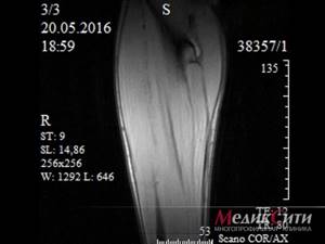
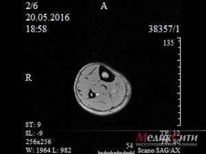
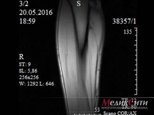
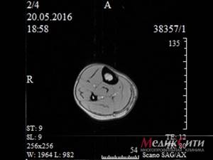
An example of an MRI transcript of the ankle joint
On a series of MRIs of the left ankle joint, the relationships in the joint were not disturbed.
The bones that form the ankle joint have a normal configuration and are positioned correctly. The notch formed by the ankles is well developed.
The cartilage covering of the ankle, subtalar and talonavicular joints is thinned, the joint spaces are not narrowed.
The synovial membrane is hypertrophied.
In the anterior outer parts of the tibia and the inner parts of the lateral malleolus, areas of local low-intensity bone marrow edema are noted.
An increase in the intensity of the MR signal from the anterior talofibular ligament, signs of partial damage, and swelling of the fatty tissue in the adjacent sections are determined. Other ligaments of the joint without reliable signs of rupture. The interosseous ligament between the talus and calcaneus is intact.
The Achilles tendon has normal thickness and course; in the distal parts there is a slight increase in signal, with the general tension of the tendon remaining intact. The adipose tissue surrounding the tendon is unchanged.
The tendons and plantar aponeurosis are unremarkable.
Paraarticular soft tissues are not changed.
An accumulation of fluid is detected in the cavity of the ankle and subtalar joints.
Conclusion:
MRI signs of grade 2 deforming arthrosis of the ankle and subtalar joints; damage to the anterior talofibular ligament; Achilles tendinopathy. Intra-articular effusion.
It is difficult for an ordinary person to independently understand and interpret the results that, after an MRI, will be given to him on a digital medium. Therefore, with the conclusion of the radiologist and the photographs, he should go for a consultation with the attending physician, who will make a final diagnosis based on the summary data of the examination, medical history and tomography data. In our clinic , after an MRI, you can have a free consultation with a neurologist or orthopedist . Doctor
- will answer all questions based on the results of the research and the conclusion received
- Helps explain tomography results without using complex radiological terminology
- will conduct an examination and, if necessary, offer treatment.
“Second independent opinion” service Medicine is an area where we want to be 100% sure . Therefore, at your request, we will be happy to offer you the service of a second independent opinion from the leading consultant of our clinic, Candidate of Medical Sciences , a doctor of the highest category with 18 years of experience in the field of tomography and radiology N.V. Marchenko.
Preparation
In order for the examination to be effective, you should take the list of contraindications and preliminary preparation seriously. Before starting the MRI procedure, it is imperative to get rid of all, even the smallest, metal objects, glasses, watches and hairpins, as well as electronic devices. Remember, MRI and metal are two things that don't go together. The powerful magnetic field of the tomograph is capable of moving metal objects, even implanted into the body. Therefore, it is imperative to inform medical personnel about the presence of any implants and provide documents describing what material they are made of. Most modern medical alloys do not have magnetic properties and are approved for examination using MRI.
Doctors performing MRI of joints

Lisenkova Irina Vladimirovna
Doctor - radiologist, doctor of the highest category
Make an appointment

Uchevatkin Andrey Alekseevich
doctor - radiologist, doctor of the highest category
Make an appointment