Ultrasound examination of the hip joint is a mandatory component of newborn screening. Screening (dispensary examination) is a set of measures aimed at identifying/preventing diseases. It must be carried out at the age of one or two months. The main purpose of ultrasound is to confirm or refute the diagnosis of hip dysplasia. What do you need to know about the disease, how exactly is the study carried out and is ultrasound safe for the baby’s body?
Peculiarities of pediatric diagnostics
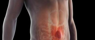
The hip joint is a ball-and-socket joint in the human body. It is formed by the lunate surface of the pelvic bone and the articular surface of the head of the femur. The main properties are circular rotation, flexion, extension, abduction, adduction of the hip. The functionality of the musculoskeletal system largely depends on intrauterine development. Initially, the baby is born with “soft” bones in order to safely pass through the birth canal and be born. But already in the first months of life, the bone skeleton becomes stronger, which makes it possible to track norms and pathologies.
Content:
- Peculiarities of pediatric diagnostics
- How does an ultrasound machine work?
- Indications/contraindications for
- What are the advantages of the method
- Preparation and conduct of the study
- Norms and pathologies
Ultrasound of the hip joint is a mandatory procedure as part of newborn screening. It is taken every 1 or 1.5 months to confirm/refute dysplasia. Diagnostics helps to identify changes in the structure, position of the glenoid cavity, the degree and features of the formation of the femoral head, flexibility of the ligaments, etc.
In the treatment of dysplasia, the age of the child and timely diagnosis are fundamentally important. In the period from the first to the sixth month, therapy will consist of massage, soft swaddling or no swaddling, wearing the baby in a sling and other comfortable manipulations. After six months, to get rid of dysplasia, the child will have to wear hard plaster spacers. They limit mobility, and with prolonged wear they trigger the process of muscle atrophy. This disrupts the natural development of the baby and can affect psychological health.
Do not ignore preventive examinations and provide your child with qualified medical care. Timely diagnosis will speed up the treatment process without affecting the baby’s quality of life.
Hip dysplasia

Dysplasia is a congenital defect of the hip joint. The pathology is caused by improper development of the mobile bone joint and leads to dislocation/subluxation of the femoral head.
According to statistics, the disease is diagnosed in 2-3% of newborns worldwide. In 80% of cases, dysplasia occurs in girls. Most often, problems arise with the left hip joint (60%), less often with the right (20%) or with both at the same time (20%).
Dysplasia is diagnosed as a change in the shape/size/structure of the joint. The load is distributed unevenly, which determines the slowdown/acceleration of bone growth. Manifestations, final shape and general condition of the bones are determined individually. In newborn babies, the hip joint is an immature biomechanical structure. His ligaments are too elastic, and the glenoid cavity is thick and vertical. The overall functionality of the body depends on the nature of the development of the mobile bone connection. This is why early diagnosis is so important for overall musculoskeletal health.
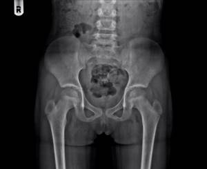
There are several stages of dysplasia - preluxation, subluxation, dislocation. With preluxation, the joint cannot be held within the boundaries of the glenoid cavity. Subluxation is characterized by partial displacement of the femoral head. When a dislocation occurs, the head of the femur is completely displaced. Ultrasound examination helps to identify all stages of dysplasia. If diagnosis and treatment are ignored, the child begins to limp, feel severe pain, and may have impaired growth/development of the body, which leads to serious problems in the future.
What does ultrasound of the hip joints reveal in newborns?
This diagnostic method allows you to identify a wide range of abnormalities. With its help, the shape and structure of joints are studied in detail, congenital or acquired pathologies are determined. The doctor can assess the condition of the articular cartilage, the quality of the joint fluid, and detect narrowing of the joint spaces. A high-quality ultrasound of the hip joints in newborns will reveal hip dysplasia, the presence of inflammatory processes or rheumatic diseases. The method is absolutely harmless and allows you to monitor the baby’s condition over time. It is recommended to undergo the first examination within one month. If necessary, it can be prescribed several more times until the child reaches 6 months of age.
The earliest possible ultrasound of the joints (1 month) will help identify complex forms of dysplasia that require immediate treatment. In the following months, when the bone head begins to form, tireless monitoring of the formation of the hip joints is necessary, since early and most importantly timely detection of pathologies means a positive result, that is, a complete cure through conservative methods, that is, physiotherapy, abduction splints, specially designed massage, the use of vitamins (B) and calcium preparations.
Late diagnosis (6 months or more) of pathology in the development of a child’s hip joints is unacceptable, since it entails severe complications, to avoid which it is necessary to visit a specialist within a strictly established time frame. Late treatment of a child who has already begun to walk (1.5 years old) may even involve surgical intervention. Almost 98% of dislocations acquired at birth are eliminated without a trace if, thanks to ultrasound, early diagnosis and timely therapy are carried out.
How does an ultrasound machine work?
Ultrasound is powerful sound waves. The human hearing organs can perceive frequencies from 16 to 20 kHz, so ultrasonic vibrations (from 20 kHz) are beyond our acoustic perception. However, some groups of animals (whales, dolphins, bats and others) communicate using ultrasound. Each wave is characterized by an oscillation period, frequency and length. They all depend on the elasticity/density of the medium through which this wave propagates. Any environment, including the tissues of the human body, prevents the propagation of sound vibrations. This is called acoustic impedance. The speed and density of sound waves depend on the magnitude of acoustic resistance.
As soon as a sound wave reaches the boundary of two media with different acoustic resistance (for example, soft and hard tissue), one part of it propagates in the new medium and is absorbed by it, and the other is reflected. The intensity of reflection depends on the magnitude of the acoustic impedance. The higher the indicator, the brighter and lighter the signal that the ultrasonic equipment receives. Additionally, the technique records the distance to the separation boundary, the travel time of the wave, the speed of movement, the difference in densities, and so on.
Human skin reflects 99.99% of sound vibrations, making ultrasound examination impossible. That is why the scanned area is lubricated with a special aqueous jelly, which acts as a transition medium.
The reflected sound wave enters an amplifier and special reconstruction systems. They process the information received and transform it into sections of the body part being studied. The picture is painted in black and white, which uses no less than 64 different gradients of the black and white scale. The maximum intensity of sound vibrations is recorded in white, and the minimum - in black.
There are three operating modes of ultrasound - A, B, M. A-mode provides a one-dimensional image, B-mode provides a two-dimensional image of anatomical structures in real time. M-mode is a one-dimensional image with a time coordinate. It is used to diagnose the functionality of the heart.
To make the image as informative and accurate as possible, contrast agents (echo contrast) are used. The contrast agent contains free gas microbubbles (less than 5 microns in diameter). They improve visualization of blood flow or individual organs, increase contrast between tissues and increase diagnostic accuracy.
Classification of normal angles and interpretation of ultrasound results of TSB in newborns
The doctor compares the average normal values and the results obtained for a number of parameters, determining the absence or presence of pathology and its stage. One of the evaluation criteria is the echogenicity of the structures. Thus, if there is hyperechogenicity of the femur and the dome of the acetabulum, the formation of the TSB occurs correctly. In this case, the femoral head and cartilage plate will be hypoechoic.
Another criterion by which the assessment is made is the angle of the femoral head in relation to the acetabulum. In this case, the following classification has been adopted, the indicators of which are the norm for children aged two to three months:
- Angle α (“Alpha”) – is more than 60° and determines the level of elevation of the acetabulum;
- Angle “β” (“Beta”) is less than 55° and determines the development of the cartilaginous space of the acetabulum.
Experts distinguish 4 types of hip joints and 3 degrees of dysplasia:
| Types of hip joints and degrees of dysplasia | Alpha | Beta |
| Norm | No violations were found. | The cartilaginous plate is wide and short. |
| Formation delay | Delay up to three months. | Delay of more than three months. |
| Subluxation | Characterized by changes in the structure of the cartilaginous protrusion. | There are violations in the structure. |
| Dislocation | The joint is not formed correctly. | The head of the femur is not covered with a cartilaginous protrusion. |
Indications/contraindications for
Indications for examination of the hip joint in newborns (in addition to screening):
- birth of a baby at less than 37 completed weeks;
- increased tone of the lower extremities;
- heterogeneity of symmetry/depth of skin folds on the baby’s body (particular attention should be paid to the area of the hips and buttocks);
- unequal length of limbs;
- disembryogenesis (disorders of embryonic development that arise due to genetic or teratogenic factors);
- excessive clicking or crunching in joints;
- limited functionality of the hip (for example, it is impossible to fully spread the hips during a massage);
- neurological abnormalities;
- the birth of twins/triplets (one fetus gets unlimited access to the resources of the mother’s body, and several children have to share nutrients among themselves).

There are no contraindications for ultrasound examination. Ultrasound is sound waves that the human ear simply cannot perceive. They do not affect the performance of our body in any way, which means they are absolutely harmless.
What are the advantages of the method
Since 1989, ultrasound has been used in pediatrics. It is noteworthy that the first studies were aimed at studying the hip joint. The problem of dysplasia was encountered everywhere, which means it required an effective solution and constant monitoring. Ultrasound examination still does not lose popularity and remains the safest, most informative and accurate diagnostic method. The production and use of ultrasound equipment is strictly regulated by the World Health Organization, so parents can be confident in their quality and functionality.
The main advantage of ultrasound is safety. Unlike X-rays or computed tomography, after which radiation doses accumulate in the body, ultrasound does not affect a person in any way. It is this type of diagnosis that is suitable for the most vulnerable categories of patients - newborns, pregnant/lactating women and the elderly.
The second advantage is maximum patient comfort. During the examination, the baby does not need to be fastened with soft belts or forcibly held in your arms for the device to record the state of the body. On the contrary, activity within normal limits will help the doctor better study the hip joint and will not darken the mood of the little patient. Additionally, a specialist can track the dynamics of changes, record inflammatory processes or defects in muscles, ligaments, tendons, joint capsule and cartilage.
Preparation and conduct of the study
No specific preparation is required. The main thing is that the baby is well-fed and calm, since excessive activity, crying or fear simply will not allow the doctor to diagnose. Parents should think through the child’s outfit in advance - the lower part of the costume should be easily and quickly removed so as not to waste time.
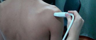
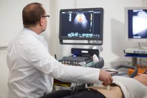
The baby is placed on a couch, previously covered with a diaper, lubricated with a special gel and the ultrasound machine sensor is placed against it. The doctor carefully moves the sensor from side to side, examining the joint. The device simultaneously emits sound waves, records their characteristics and displays an image of the internal cavity on the computer. The doctor and parents can watch what is happening in real time through the computer screen. The ultrasound specialist scans both hip joints with adjacent soft tissue and bone areas. Periodically, the doctor turns the baby onto the left or right side/back/stomach, raises or lowers the limbs, and slightly turns them in order to study the condition and functionality of the bone skeleton in more detail.
Best materials of the month
- Coronaviruses: SARS-CoV-2 (COVID-19)
- Antibiotics for the prevention and treatment of COVID-19: how effective are they?
- The most common "office" diseases
- Does vodka kill coronavirus?
- How to stay alive on our roads?
To diagnose the hip joint, a convex sensor is used. Its frequency varies from 1.8 to 7.5 MHz. The size of the device is minimal, which ensures complete adherence to the patient’s skin. A special feature of the sensor is the size of the final image. Its width is several centimeters larger than the sensor itself. Doctors must take this discrepancy into account in order to navigate the anatomical structures and understand their real size. Typically, convex sensors are used to diagnose deep-lying organs (hip joint, organs of the gastrointestinal tract, reproductive or urinary systems).
The examination lasts only a few minutes. Once all the information has been collected, the doctor completes the examination, wipes off the liquid gel, prints the necessary photographs and talks with the parents about the norms and pathologies of the hip joint.
Carrying out a diagnostic procedure
During an ultrasound, the child is placed on his side so that the examined joint is on top and bent at an angle of 20°. A gel is applied to the hip joint to facilitate the sliding of the sensor and improve the conductivity of acoustic waves. During manipulations, all articular and periarticular structures are displayed on the screen. The doctor takes pictures in the positions necessary to assess the condition of the tissues. Usually there are five of them: in the initial position, when bending and extending the leg, when abducting and bringing it to the body.
To prevent the procedure from being interrupted, pediatricians who prescribe a referral for sonography recommend not feeding the baby immediately before the examination. The child must be well-fed so as not to be capricious, but the process of regurgitation can interfere with diagnosis.