Ultrasound examination or ultrasound has long been firmly established in medical practice and is widely used to diagnose a wide variety of pathological changes, including in blood vessels. It is highly informative and absolutely safe. Therefore, the procedure can be carried out as many times as necessary without fear of harming the body, including children and pregnant women. One of the areas of ultrasound diagnostics of blood vessels is the study of blood vessels of the neck and head - brachiocephalic vessels.
Brachiocephalic vessels
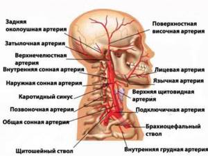
Brachiocephalic vessels (BCVs) are arteries that are responsible for supplying blood to the brain, tissues of the head, neck and shoulder girdle. This:
- 2 carotid arteries;
- 2 vertebral arteries, which at the base of the brain stem merge into one artery - the basilar;
- brachiocephalic trunk;
- 2 subclavian arteries.
They all form the so-called Circle of Willis at the base of the brain, thanks to which blood is evenly distributed throughout all parts of the brain. Therefore, the occurrence of pathological changes in any of the brachiocephalic vessels leads to disruption of the blood supply to the brain.
The Circle of Willis got its name in honor of the English physician and anatomist Thomas Willis, who became the first to describe it back in the 17th century.
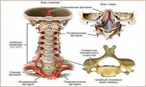
Quite often, one or both vertebral arteries, which run along the lateral surfaces of the cervical vertebrae in a special bone canal, are affected. The main reason for impaired blood flow in them is pathobiomechanical changes in the cervical spine. They can lead to mechanical compression of the vertebral arteries and thereby create difficulties for the flow of blood through them into the circle of Willis and, accordingly, the brain. Also, with pathologies of the cervical spine, the cervical sympathetic nerve plexus can be irritated. This causes a spasm of the walls of blood vessels and also causes a decrease in their lumen, and therefore a decrease in the amount of blood entering the brain. In addition, when the nerves of the cervical plexus are irritated, limited neck mobility and acute pain are observed. All this together is called vertebral artery syndrome.
Thus, the cause of a decrease in the lumen of one or both vertebral arteries may be:
- functional blocks, i.e. the occurrence of a slight limitation in the mobility of the spine, including with osteochondrosis;
- instability in spinal motion segments of different locations of the cervical spine;
- tension in individual neck muscles, especially the anterior scalene and oblique muscles of the head;
- protrusion and herniation of the intervertebral disc;
- thickening of the atlanto-occipital membrane;
- bone growths, including those caused by uncovertebral arthrosis;
- spondyloarthrosis (a chronic disease accompanied by degenerative processes in the facet intervertebral joints).
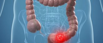
Also, the cause of circulatory disorders in the brachiocephalic vessels can be atherosclerosis, i.e. the formation of plaques of different sizes in the walls of blood vessels, which also leads to a decrease in their lumen. In such cases, patients are found to have high levels of cholesterol in their blood. But often this is combined with disorders in the cervical spine.
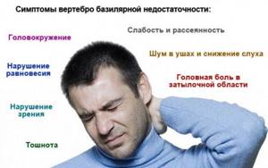
In any case, if blood flow in the BCS is disrupted and vertebrobasilar insufficiency develops, the following may occur:
- headaches in the occipital, parietal and temporal regions;
- dizziness when turning the head;
- unsteady gait;
- visual disturbances;
- noise in ears;
- numbness of one half of the face;
- weakness, numbness of one or both hands;
- sleep disorders;
- fluctuations in blood pressure;
- heaviness in the head after sleep;
- deterioration of memory, ability to concentrate.
Often the vertebral arteries are pinched during movement, in particular turning or throwing back the head. Therefore, symptoms of cerebrovascular accident often occur after performing a trigger movement.
Dangers of cerebrovascular accident
For normal functioning of the brain, a large amount of energy is needed, which it receives from nutrients and oxygen delivered to its cells by blood. It enters the brain through 4 main or main arteries: 2 carotid and 2 vertebral. Therefore, if the blood flow in one of them deteriorates, it is possible to compensate for this due to the work of the remaining three blood vessels. But the body’s compensatory capabilities are not limitless.
Initially, when blood supply to the brain decreases, headaches, dizziness, memory loss and increased fatigue occur, which leads to deterioration in performance. After some time, patients experience more pronounced neurological symptoms, which already indicate multiple brain damage. And the reason for such phenomena is chronic cerebral circulatory failure, which is also called discirculatory encephalopathy.
Circulatory disorders provoke the gradual death of neurons (nerve cells) in different parts of the brain, which causes the development of neurological symptoms with a predominance of asthenic manifestations: weakness, irritability, sleep disturbances. This is often accompanied by depressive states, which may subsequently give way to apathy.
Once it begins and is not diagnosed in time, cerebrovascular accident progresses. During its course, there may be periods of sharp deterioration and gradual worsening of symptoms. The main danger of such phenomena is a sharp increase in the risk of developing severe diseases, one of which is a stroke or acute disruption of the blood supply to the brain.
Every year, strokes are diagnosed in more than 400,000 residents of Russia, and 35% of patients die within the first 3 weeks, and only 50% live longer than a year after suffering a stroke.
Prevention measures
Intervertebral hernia is the result of improper loads on the spine. The conditions for the normal functioning of the spine are the preservation of its mobility and the strength of the surrounding muscles (muscle corset).
To keep your back healthy, patients are recommended to:
- Take daily walks in comfortable shoes.
- Do regular exercises to strengthen your back muscles.
- Eat properly and balanced to maintain optimal weight.
- Sleep on the right orthopedic mattress (medium, high degree of hardness).
- Maintain an optimal work and rest schedule.
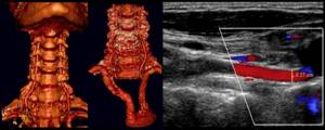
Duplex scanning of BCS
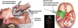
To assess the condition of the brachiocephalic vessels and the quality of their functioning, ultrasound diagnostics, namely Doppler ultrasound (USDG), is used. The method is based on an effect discovered by physicist Christian Doppler. The essence of the diagnostic procedure is that the ultrasound wave is reflected from different tissues with different intensities. This allows you to determine the density and homogeneity of the studied area of the body or an individual organ from the image displayed on the monitor of the ultrasound machine. Based on this, the correct structure of a particular tissue or organ and the quality of its functioning are assessed.
Doppler was able to develop special programs that were able to recognize ultrasonic waves reflected from moving objects, namely from erythrocytes, i.e. red blood cells. This discovery significantly expanded the capabilities of ultrasound diagnostics and made it possible to analyze the speed and direction of blood flow, as well as provide data on the quality of blood supply to the tissues and organs being studied.
The first and most important advance was the development of Doppler ultrasound, or Doppler ultrasound. It is used to assess and determine the characteristics of blood circulation and allows you to obtain a two-dimensional image of blood flow in the examined area, assess its speed, the presence of blood flow deficiency, and the nature of vascular resistance. The disadvantage of this method is the inability to assess the condition of the vessel walls, so it is usually prescribed to determine the effectiveness of treatment for an already established diagnosis.
Duplex scanning of neck vessels (USDS) has wider capabilities, so this type of diagnosis is used most often. With its help, it is already possible to study the structure of the walls of the brachiocephalic vessels, their course, as well as the characteristics of blood flow dynamics in real time. Therefore, duplex scanning makes it possible to accurately detect various circulatory disorders in the BCS at the early stages of development and select their most effective treatment. Although recently the concepts of ultrasound dopplerography and ultrasonic scanning doppler scanning have already merged into a single concept, and doctors themselves, when prescribing ultrasound of the vessels of the neck, mistakenly indicate ultrasonography and ultrasound doppler scanning, implying ultrasonography.
Ultrasound scanning is often used to study the arteries and veins of the neck in combination with triplex scanning of blood vessels. The latter method allows you to study the direction of blood flow, coloring it red and blue depending on the direction of movement: towards or away from the ultrasound sensor.
Ultrasound examination of the vessels of the neck and head, in particular the brachiocephalic vessels, can detect:
- wall-occlusive pathologies of blood vessels, i.e. leading to a decrease in their lumen or complete closure;
- level and degree of vascular lumen obstruction;
- deformations, tortuosity, abnormalities in the development of blood vessels;
- aneurysms (protrusion of walls) and vascular spasm;
- venous blood flow disorders;
- changes in the condition, elasticity of blood vessels and their hemodynamic significance.
An x-ray examination of the cervical spine is also prescribed.
The results of the study make it possible to accurately determine the cause of impaired blood supply to the brain and the appearance of other symptoms that bother the patient, assess the severity of existing changes and select the optimal treatment tactics. In the future, ultrasound examination is prescribed to monitor the dynamics of changes and evaluate the effectiveness of the therapy.
Complications and consequences of intervertebral hernia
If there is no timely treatment for a hernia, this is fraught with the development of consequences and complications that negatively affect a person’s life. The most striking complications of intervertebral hernia are:
- paresis of the back or limbs;
- paralysis of limbs;
- chronic pain syndrome;
- dysfunction of the bladder and intestines. This happens when the nerve bundles that are responsible for nerve patency are compressed.
Intervertebral hernia is a serious disease that requires a careful approach to treatment. It is important to consult a doctor on time, undergo appropriate examinations, and follow all medical prescriptions. This is the only way a doctor can prescribe competent therapy. With the right approach to treatment, it is possible to stop the growth and development of the disease and prevent serious consequences and complications.
Indications for ultrasound scanning
Ultrasound duplex scanning of brachiocephalic vessels is indicated for:
- frequent headaches;
- the occurrence of signs of acute or chronic vertebrobasilar insufficiency, i.e. transient ischemic attack or discirculatory encephalopathy (dizziness, nystagmus, imbalance, numbness of the face, limbs, pain in the cervico-occipital region, tinnitus, cognitive impairment);
- systemic diseases, in particular vasculitis or inflammation of the walls of blood vessels;
- the presence of factors contributing to the development of cerebrovascular diseases (smoking, sedentary lifestyle, obesity, atherosclerosis, diabetes mellitus and arterial hypertension);
- receiving a traumatic brain injury, damage to the cervical spine.
Every person should undergo the procedure after 40-45 years of age, especially if there are cases of stroke in blood relatives, and then repeat it once a year for preventive purposes.
Also, ultrasound scanning of the brachiocephalic vessels is mandatory to assess the effectiveness of the prescribed treatment for previously discovered diseases.
Device structure
The electrophoresis apparatus is an alternating current rectifier, semiconductor (now), electron tube (formerly). It operates from a standard network with a frequency of 50 Hz and a voltage of 220 V and is equipped with a milliammeter.
Electrophoresis devices operate primarily using galvanic current. It is continuous, low voltage, constant intensity. Always passing in one direction, the current is able to maintain polarity, strength, and voltage. The action is comparable to a blow of wind that does not change its strength. Thanks to this, the electrophoresis device is able to “deliver” drugs deep into the body, affecting the tissues and mucous membranes of the body. For direct current, even dense tissues can be considered permeable.
Contraindications
Ultrasound diagnostics is absolutely safe and cannot harm the body. Therefore, there are no absolute contraindications for its implementation.
Experts may recommend delaying the examination if the following is present on the skin in the examination area:
- rashes;
- burns;
- damage of various types.
Also, a relative contraindication for performing ultrasound scanning of brachiocephalic vessels is the patient’s severe condition. But if necessary, the procedure can also be carried out.
Establishing diagnosis
Diagnosis of intervertebral hernia is of key importance for choosing a treatment regimen and is carried out using the following methods:
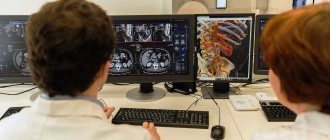
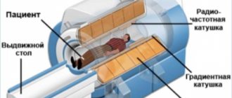
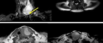
- MRI of the spine. “Gold standard” for identifying pathological changes in muscles, ligaments and nerve structures.
- CT scan of the spine. Allows you to assess the condition of bones and soft tissues.
Ultrasound for intervertebral hernias is not the main way to diagnose spinal diseases. X-rays are prescribed only to determine the traumatic or non-traumatic nature of existing disorders.
Features of ultrasound examination of neck vessels
The study does not require special preparation. Although to increase its accuracy and information content it is worth:
- Avoid drinking tea, coffee, alcohol and energy drinks on the day of the ultrasound examination;
- 2-3 hours before diagnosis, you must stop smoking and thermal procedures;
- Coordinate your medication intake with your doctor.
Some experts recommend doing an ultrasound scan of the neck vessels on an empty stomach, since after eating there is an active flow of blood to the gastrointestinal tract and its outflow from the brain. Therefore, this may affect the accuracy of diagnosis.
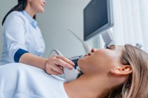
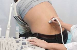
Direct ultrasound diagnostics are carried out in a special room. The patient needs to lie on the couch on his back, raising his chin and throwing his head back a little. The doctor applied a special conductive gel to the neck, which will create an optimal environment for transmitting and reading ultrasound waves. He will then apply a sensor to the area being examined. Performing smooth sliding movements with the sensor along the flow of blood, the doctor examines all the large blood vessels of the neck, accurately measures the size of their walls, diameter, degree of narrowing, evaluates the speed of blood flow and other parameters. During this time, the patient is prohibited from turning his head or talking.
In some cases, the ultrasound doctor may ask the patient to stand up, hold his breath, or vice versa, breathe deeply. Sometimes the doctor can press the test vessel with a finger or sensor and evaluate the nature of blood circulation in this condition. The changes detected in this case will help to more accurately understand the characteristics of blood flow and assess the degree of its disturbance, which will increase the information content of the study.
One of the most important stages of ultrasound examination is the study of the extracranial part of the cervical spine, since it is often compressed due to excessive tension in the neck muscles. This may be due to a spasm that becomes a reflex response to pain impulses from the intervertebral discs, in which degenerative changes occur, i.e., osteochondrosis develops. Therefore, a detailed study of the extracranial section of the brachiocephalic vessels allows not only to detect signs of a number of chronic diseases, but also to assess the likelihood of an ischemic stroke caused by a malnutrition of the brain.
In general, the duration of the procedure is 30-50 minutes, which depends on the purpose of the study (primary or to evaluate the results of treatment), what pathological changes are detected, their severity, etc.
How is it carried out?
Ultrasound of the spine does not require preliminary preparation. When studying the lumbosacral region, it is recommended not to consume foods that cause increased gas formation for one to two days before the procedure.
The examination is carried out from the abdomen (anterior approach) and from the back through the spaces between the vertebral arches (posterior approach).
Decoding the results
After an ultrasound scan of the neck vessels, the patient receives a study protocol. It indicates all the parameters obtained during the examination and the expert’s conclusion. Based on these data, the attending physician, taking into account the characteristics of the patient’s complaints, his gender, age, as well as the results of other studies, can already establish a diagnosis and select treatment.
The diagnosis cannot be established solely based on the results of ultrasound scanning.
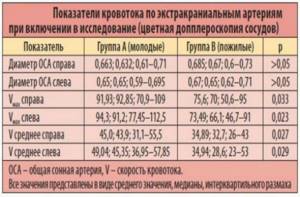
Normal indicators of BCS are:
- absence of pathological changes and anomalies in the structure of the great vessels of the neck, their thickening, compression by surrounding anatomical structures;
- full cross-country ability;
- absence of blood flow turbulence in areas with missing capillaries;
- artery wall thickness – at least 10 mm;
- systolic blood velocity - at least 50 cm/s, diastolic - 9-36 cm/s;
- the same diameter of the vertebral arteries, which is at least 3 mm;
- localization of the carotid artery to the left of the aortic arch;
- the diameter of the common carotid artery is in the range of 4.2–6.9 mm, the external carotid artery is 3–6 mm, and the internal carotid artery is 3–6.3 mm.
Even in the complete absence of pathological changes, some indicators may differ from the norm. In such situations, this is regarded as an individual characteristic. Therefore, you cannot independently diagnose yourself, much less begin treatment, only on the basis of ultrasound results.
The truth about treating lumbar disc herniation
Let's consider whether a lumbar hernia can be cured with medications, exercises and physical therapy.
Do medications help?
After the doctor makes an accurate diagnosis, the patient is prescribed conservative therapy. An effective treatment method is drug therapy. With the help of medications, it is possible to relieve pain in the acute stage of the disease. It is also recommended to take medications that make you feel better.
The most effective medications in the treatment of intervertebral hernia are:
- Non-steroidal anti-inflammatory drugs. They have a good therapeutic effect; after taking them, significant relief occurs within a few days. First, the patient is given drugs by injection, then tablets or capsules, ointment, gel or cream are prescribed. Such medications include Diclofenac, Melixicam, Nimesil, Ketoprofen.
- Corticosteroids. Prescribed if non-steroidal anti-inflammatory drugs do not give the desired result. To relieve pain, injectable forms of medications are administered. This is a short-term therapy, since such medications have a large number of side effects. Corticosteroids include Diprospan, Dexamethasone, and Prednisolone.
- Muscle relaxants. Used to relieve pain caused by muscle spasms. When muscle fibers are compressed, the course of the disease worsens. After using muscle relaxants, it is possible to relieve spasms, relax tense muscles, and thereby eliminate pain. Such medications are used in a course. The most effective are Tolperil and Movalis.
- Chondroprotectors. The effectiveness of these drugs has not been proven. According to manufacturers and doctors, such drugs are designed to restore damaged cartilage tissue and help relieve pain. Among them are Teraflex, Chondroxin, Artru.
- B vitamins – thiamine, pyridoxine, cyanocobalamin. They help improve nerve conduction and activate the functions of the central nervous system. Due to this, the severity of painful sensations is reduced. Such drugs include Neurobex, Milgamma.
The effect of gymnastic exercises
An important stage in the rehabilitation period is performing therapeutic exercises. With the help of gymnastics you can get rid of muscle atrophy and weakness. If there is weakness in the muscle fibers, the load on the back is distributed unevenly. So the spine suffers from increased stress. As a result, the risk of developing osteochondrosis and intervertebral hernia increases. If there is a hernia, physical therapy is mandatory.
With the help of simple exercises, it is possible to restore blood circulation, evenly distribute the load on the spine, increase the distance between the discs, strengthen the muscle corset and make the body more resilient.
In each specific case, specialists select individual exercises. They must be done carefully and slowly. There should be no sudden movements. Be sure to start a set of exercises with a warm-up and finish with a cool-down. Therapeutic exercises should be performed regularly.
Lumbar physiotherapy
Physiotherapeutic procedures are effective in the treatment of intervertebral hernia. With their help, you can prolong the period of remission, speed up tissue restoration, and get rid of chronic pain. The most effective types of physiotherapy are:
- Electrophoresis. This procedure makes it possible to overcome chronic pain syndrome, improve blood circulation, and get rid of the inflammatory process. To enhance the effect, doctors use additional medications. Under the influence of current, medications penetrate deeper into the inflamed tissues and begin to act actively.
- Magnetotherapy. Assumes exposure to a magnetic field. After completing a course of treatment for the hernia, the patient's condition improves.
- Phonophoresis. Involves exposure to ultrasound. As a result of the course of treatment, blood microcirculation improves, the inflammatory process is stopped, and the patient’s condition improves.
- Laser therapy. Under the influence of a laser beam, metabolic processes are activated, microcirculation of blood and lymph is improved, the body's natural defense mechanisms are activated, and tissues are restored faster.
- Massage. Helps get rid of muscle spasms, relax tense muscles, and improve blood circulation in a specific area. By taking regular massage courses, you can get rid of chronic inflammation and chronic pain in the spine. It is important to remember that this procedure should only be performed by a qualified specialist with a medical education. During the period of exacerbation, the procedure is prohibited.
- Manual therapy. When using this method, special techniques are used, thanks to which it is possible to relax muscle tissue and eliminate pain. This therapy involves influencing certain points located on the surface of the back. This is not a massage; this procedure is used periodically.
- Acupuncture. This procedure uses special needles. Under their influence, the muscles relax and it is possible to get rid of pain.
- Hirudotherapy. Medicinal leeches are used to conduct the session. With their help, swelling is eliminated, the severity of the inflammatory process and pain are reduced.
- Paraffin therapy. Liquid paraffin is used. After completing the course of treatment, the patient’s condition improves, inflammation and swelling decrease, lymph outflow and blood circulation accelerate.
Only a doctor can prescribe any procedures. You cannot use this treatment on your own. You must first undergo diagnostics, establish an accurate diagnosis and determine whether there are any contraindications. It is important to undergo comprehensive treatment for a lumbar hernia in Moscow in a specialized clinic.
PRICES FOR OUR PROCEDURES
(select the section you are interested in)
Shock wave therapy
Prices
UVT prices
Before starting the procedures, consultation with a specialist is required!!!
SHOCK WAVE THERAPY PRICE
| Name of procedure | Price, rub. |
| Name of procedure | Price, rub. |
| Shock wave therapy (SWT) procedure using the enPuls Zimmer device for the treatment of heel spurs | 800 |
| Shock wave therapy (SWT) procedure with enPuls Zimmer device for one anatomical zone | 1200 |
Areas of procedures for medical UVT treatment: Hand - 1 zone Forearm - 1 zone Shoulder - 1 zone Back - 2.5 zones Buttocks - 1 zone Thigh - 1 zone Lower leg - 1 zone Foot - 1 zone
Zones for cosmetic UVT procedures: Hand -1.5 zones Forearm -1.5 zones Shoulder -1.5 zones Back -3.5 zones Buttocks -1.5 zones Thigh -1.5 zones Lower leg -1.5 zones Foot -1.5 zones
Laser therapy, ILBI
Prices Laser Vlock
Prices Laser therapy ILBI
Before starting the procedures, consultation with a specialist is required!!!
| Name of procedure | Price, rub. |
| Laser therapy device "Mustang -2000+" one field | 500 |
| Laser therapy device "Mustang -2000+" two fields | 700 |
| Intravenous laser blood irradiation (ILBI) 1 procedure | 1000 |
| Intravenous laser blood irradiation (ILBI) 6 procedures | 5000 |
| Intravenous laser blood irradiation (ILBI) 10 procedures | 8000 |
Magnetotherapy
Prices Magnetotherapy
Prices Magnetotherapy
Before starting the procedures, consultation with a specialist is required!!!
| Name of procedure | Price, rub. |
| Vortex magnetotherapy apparatus “Magnetoturbotron” EOL | 1000 |
| Running magnetic therapy device "Almag-02" | 500 |
Ultrasound, Electrotherapy
Ultrasound Electrotherapy
Prices Ultrasound Electrotherapy
Before starting the procedures, consultation with a specialist is required!!!
| Name of procedure | Price, rub. |
| Ultrasound therapy device “SoleoLine” (1 procedure) | 500 |
| Ultrasound therapy device “SoleoLine” (6 procedures) | 2500 |
| Ultrasound therapy device “SoleoLine” (10 procedures) | 4000 |
| Medicinal electrophoresis apparatus “SoleoLine” (1 procedure) | 700 |
| Medicinal electrophoresis apparatus “SoleoLine” (6 procedures) | 2500 |
| Medicinal electrophoresis apparatus “SoleoLine” (10 procedures) | 4000 |
| Pulse currents apparatus “SoleoLine” (1 procedure) | 500 |
| Pulse currents apparatus “SoleoLine” (6 procedures) | 2500 |
| Pulse currents apparatus “SoleoLine” (10 procedures) | 4000 |
Lymphatic drainage, Khivamat
Prices Lymphatic drainage hivamat
Prices Lymphatic drainage, Khivamat
LYMPH DRAINAGE
| Service | Prices / rub. |
| LYMPHODRAINAGE Morning Life Premium device: | |
| Lymphatic drainage of legs 30 min certified device (South Korea). | 800 |
| Hand lymphatic drainage 30 min certified device (South Korea). | 800 |
| Lymphatic drainage of the abdomen 30 min certified device (South Korea). | 800 |
| Lymphatic drainage of the thighs 30 min certified device (South Korea). | 800 |
| Procedures using the Hiwamat 200 device (Germany): | |
| Procedure apparatus Khivamat 200 one zone. | 1000 |
| Procedure apparatus Khivamat 200 two zones. | 1500 |
Others
Other prices
Prices Other
Before starting the procedures, consultation with a specialist is required!!!
| Name of procedure | Price, rub. |
| Vegetative resonance test (VRT) Parasites (40 min.) | 2000 |
| Vegetative resonance test (VRT) General (2 hours) | 4800 |
| Vegetative resonance test (VRT) Allergies (50 min.) | 2500 |
| Vegetative resonance test (VRT) Nutrition (40 min.) | 2500 |
| Vegetative resonance test (VRT) Drugs (50 min.) | 2800 |
| Initial appointment with a homeopath + selection + provision of a drug + bioresonance therapy session | 5000 |
| Repeated appointment with a homeopath + selection of a drug + provision of a drug + bioresonance therapy session | 3500 |
| Consultation with a homeopathic doctor regarding the possibility of using homeopathic treatment (without selecting medications) | 1500 |
| Bioresonance therapy, electropuncture (1 procedure) | 450 |
| Checking the compatibility of a drug using the ART method (1 drug) | 200 |
| Detensor therapy (1 procedure without instructor 45 min.) | 400 |
| Detensor therapy (1 procedure with instructor 75 min.) | 2200 |
| Khivamat medical massage for the back Khivamat medical massage for the lower back Khivamat medical massage for the collar area Khivamat medical massage for the extremities Khivamat anti-cellulite massage for the buttocks Khivamat anti-cellulite massage for the hips Khivamat anti-cellulite massage for the abdomen Anti-cellulite massage for the shoulders (arms) | 1500 1000 1000 1000 1000 1500 1000 1500 |
| Consultation with a physiotherapist, neurologist of the highest category (repeated consultation is free) | 900 |
FREE TREATMENTS
|
FAQ
Can electrophoresis be used for children?
Galvanization is often used in pediatrics. There is no need to be afraid of this procedure. It does no harm, is non-invasive, absolutely painless. But only a doctor should select the necessary medications, frequency and amplitude of the current.
Is it possible to combine electrophoresis with other procedures - acupuncture, massage, paraffin baths?
Electrophoresis is compatible with many other procedures, such as acupuncture, various therapeutic and cosmetic massages, and paraffin baths. But a complex treatment regimen is developed by a doctor, since between some manipulations it is necessary to take a break from several hours to several days.
Literature:
- ICD-10 (International Classification of Diseases).
- L. O. Neuropathology. - M.: Education, 1982. - P.307-308. Bogolyubov.
- Medical rehabilitation (manual, 3 volumes). // Moscow - Perm. - 1998. Popov S. N.
- Physical rehabilitation. 2005. - P.608.
- State Register of Medicines (GRLS) of the Russian Federation.
Themes
Intervertebral hernia, Spine, Pain, Treatment without surgery Date of publication: 12/23/2020 Date of update: 01/11/2021
Reader rating
Rating: 5 / 5 (1)