Almost anyone can experience pain, limited mobility, swelling, crunching and other joint complaints. There are many reasons - from injuries (they occur more often at a young age or in professional athletes) to diseases (they can develop at a young age, but are more common in people over 40-50 years old). Joint diseases (there are about two hundred of them) cause significant discomfort to patients, reduce performance, limit mobility and cause disability.
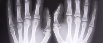
One of the main methods for diagnosing diseases, pathologies and traumatic deformations of bone and cartilage tissue is x-ray of joints. It is indispensable for diagnosing many pathologies. X-rays clearly show bone structures, salt deposits - osteophytes, and an experienced doctor is able to assess the condition of the joint using X-rays and determine further treatment tactics and the necessary additional studies. Especially if radiography of the joint was carried out using modern high-precision digital equipment.
Indications for X-ray
Traumatologists, orthopedists, and rheumatologists can prescribe x-rays after joint surgeries, injuries, or for chronic diseases, when it is necessary to monitor the dynamics of changes and the effectiveness of treatment.
Most often, problems arise in large joints, which are subject to heavy daily stress and are often injured.
The main indications for x-ray examination of the joint are diagnosis and assessment of the effectiveness of treatment for:
- Injuries of various types - dislocations, ligament ruptures, fractures, cracks in bones and other injuries.
- Congenital or acquired pathologies - arthritis (inflammation in the joint), arthrosis (destruction of cartilage tissue), necrosis (tissue death), bursitis, dysplasia and many other joint diseases.
An X-ray of the hip joint is required for dysplasia and coxarthrosis, an X-ray of the knee joint for a torn meniscus, gonarthrosis, an X-ray of the ankle joint for flat feet, heel spurs, gout, an X-ray of the shoulder joint for bursitis, glenohumeral periarthritis and tendinitis. The list of situations and pathologies is far from complete.
The main symptoms of the disease and reasons to take a photo:
- Pain or discomfort in any joint at rest or with movement.
- Limitation of the range of motion - when the joint does not bend or extend completely (even if this happens in the morning after rest, and later the movement returns to normal).
- Swelling, deformation or resulting asymmetry in paired joints.
- Redness of the skin in the joint area.
- A crunching sound in the joints (especially with dry clicks) during flexion/extension.
- Lack of stability and stability in the joint after injury, which may be a sign of ligament rupture.
- Severe bruises, blows, falls, dislocations, when there is suspicion of cracks or broken bones.
- Suspicion of joint damage in a patient with tuberculosis.
- Diagnosis of cysts and tumors.
- Assessing the effectiveness of treatment or surgery, monitoring the dynamics of changes in chronic diseases.
Indications and contraindications for radiography of the hip joints
Indications
Diagnosis of hip joints is carried out for many diseases of the musculoskeletal system. It allows you to identify pathological conditions of the joint that occur as a result of:
- Injuries suffered: dislocations, cracks and injuries;
- Degenerative-dystrophic disease - osteoarthritis;
- Death of the bone tissue of the femoral head - necrosis;
- Neoplasms of hip bone tissue;
- Pathologies of inflammatory etiology: arthritis, osteomyelitis;
- Congenital anomalies in the form of hypoplasia or dysplasia;
- Metabolic disorders, which are represented by diseases such as osteoporosis and gout.
In addition, radiography of the pelvis and hip joints is carried out as part of preoperative preparation in order to determine the scope of the intervention and its tactics.
Contraindications
Despite the fact that the radiation dose during diagnostics is minimal and does not cause harm to the human body, the procedure is used only in extreme cases - if:
- the patient is pregnant or breastfeeding;
- the patient is in serious condition;
- if the patient is under 15 years of age;
- if the patient has received a high dose of radiation in the last 12 months;
- for diseases of the thyroid gland.
As for contraindications when using radiopaque agents, in addition to the above, they are as follows:
- individual intolerance to contrast components;
- severe renal and liver failure;
- intestinal obstruction;
- blood clotting disorders;
- diabetes mellitus in severe form.
How to prepare for a joint x-ray
A procedure that requires preliminary preparation is an x-ray of the hip joint. In this case, the patient must take measures to minimize gas formation in the intestines. The doctor will recommend 2-3 days before the procedure to follow a light diet without cabbage, legumes and other foods that promote fermentation and gas formation in the intestines. On the day of the examination, you need to cleanse your intestines and come to the clinic on an empty stomach. If such preparation is neglected, the resulting images may be unclear and insufficiently informative due to accumulated gases, which look like darkened areas.
In all other cases, no preparation is required, and x-rays can be taken immediately after consulting a doctor and ordering an examination. Before taking the picture, you will need to remove all metal jewelry and accessories, and in some cases, clothing from the area of the joint being examined. To protect the internal organs of the abdominal cavity and chest, special protective aprons are used, which will be offered to be worn in the radiologist’s office.
Preparing patients for radiography:
Special preparation of patients for x-ray examination is generally not required, however, the following preparation methods are available for examination of the digestive organs:
- Previously, special diets were carried out, foods that contributed to flatulence were excluded from the diet, and a cleansing enema was performed, but now it is generally accepted that RI of the stomach and duodenum of patients with normal intestinal function does not require any preparation. However, in case of severe flatulence and persistent constipation, a cleansing enema is performed 2 hours before the test. If there is a large amount of liquid, mucus, or food debris in the patient’s stomach, gastric lavage is performed 3 hours before the test.
- Before cholecystography, the possibility of flatulence is also excluded and a radiopaque iodine-containing drug is used (cholevid, iopagnost 1 g per 20 kg of live weight). The drug enters the liver and accumulates in the gallbladder. To determine the contractility of the gallbladder, the patient is also given a choleretic agent - 2 raw egg yolks or 20 g of sorbitol.
- Before cholegraphy, the patient is injected intravenously with a contrast agent (bilignost, bilitrast, etc.), which contrasts the bile ducts.
- Before irrigography, it is carried out using a contrast enema (BaSO4 at the rate of 400 g per 1600 ml of water). On the eve of the study, the patient is given 30 g of castor oil, and in the evening a cleansing enema is given. The patient does not eat dinner, the next day a light breakfast, two cleansing enemas, a contrast enema.
How to do joint x-rays
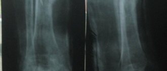
The procedure will take a few minutes and is completely painless, except in cases where it is necessary to take photographs with a load - when bending the joint, which can cause pain (for example, in case of injury). For a more complete diagnostic picture, two paired joints are examined simultaneously. This will allow you to assess the degree of impairment in the diseased joint, comparing it with a healthy one.
The patient can stand, sit or lie down - the position depends on which joint is being examined and in what projection the images are needed. For clear and comprehensive visualization of the joint, images are usually taken in several projections:
- X-ray of the ankle joint - an X-ray of the joint is taken lying down (in oblique and lateral projections).
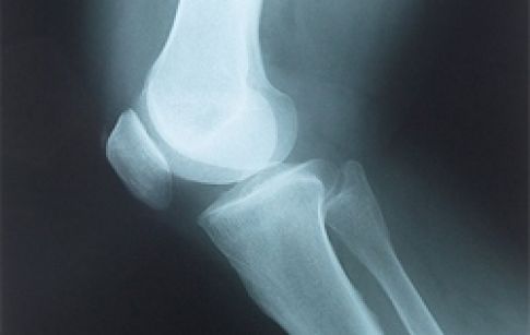
- X-ray of the knee joint involves two projections: direct and lateral. The patient lies on his back or side with his leg straight. Sometimes there may be a need for special styling - with the leg bent at an angle of 30 or 45 degrees.
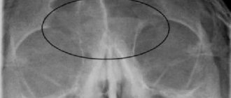
- X-rays of the hip joint are taken while lying down in two positions. In the direct projection, the subject lies on his back with his feet turned inward and his legs spread apart. In the side position - the legs are extended forward, and the person lies on his side.
- X-ray of the shoulder joint and X-ray of the elbow joint - the patient is sitting or standing, and the image should include neighboring anatomical formations - scapula, collarbone and others.
The entire procedure will take no more than 10 minutes, and based on the images obtained, the radiologist prepares a report describing the condition of the joint, bones, and soft tissues around them.
Based on the conclusion, the doctor can make or clarify the diagnosis. In some cases, one x-ray will not be enough, especially if the examination takes place during the so-called x-ray negative period. At this time, pathological processes exist only in soft tissues, which cannot be visualized by x-rays. Then diagnostics will be carried out using other methods: ultrasound, MRI and others.
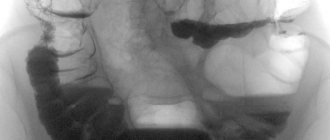
Pathologies of the sacroiliac joint
In the presence of pathology of the sacroiliac joint, the patient always feels pain in the back, which can radiate to the legs or spread higher up the spine. To eliminate pain, and most importantly, the cause of its occurrence, it is necessary to correctly determine the group of pathology.
Inflammatory – infectious and non-infectious
Among the main inflammatory processes that can negatively affect the functioning of the SIJ are:
- tuberculosis;
- brucellosis;
- pyogenic flora.
The proportion of disturbances in the functioning of the SIJ of infectious origin is quite small. Treatment consists primarily of eliminating the primary infection.
Degenerative (osteoarthritis of the SIJ)
The most common cause of pain in the SIJ area is osteoporosis or joint dysfunction. Arthritis of the SIJ can be caused by diseases such as psoriasis, Reiter's syndrome or ankylosing spondylitis.
Sacroiliac joint dysfunction (SIJD)
SIJ dysfunction most often develops in older people due to abrasion of tendons and ligaments. DKPS can also develop in pregnant women, in this case the pathology is explained by the hormonal effect on the connective tissue of the semi-joints themselves and their ligamentous apparatus. Women with this diagnosis may be contraindicated for natural delivery, that is, it is preferable to perform a cesarean section (as prescribed by the attending physician).
Injuries (pelvic fractures, rupture of SIJ ligaments)
Pain in the SIJ area can also be of traumatic origin. Pathological childbirth, road accidents, falls from height, sports injuries. All this can cause deformation of the sacroiliac joint, pinching of nerves in this area, displacement or destruction of bones, and so on.
Contraindications
X-rays are not recommended or should be done with caution:
- During pregnancy at any stage, so as not to harm the development of the child;
- Children under 14 years old;
- Patients with diseases that cause uncontrollable body tremors - in this case, the image will be “blurred”.
The issue of harm from X-rays is no longer as relevant as it was 10-15 years ago. Modern digital equipment operates with low radiation doses - about 0.03 mSv per shot, with an acceptable level of up to 1.4 mSv per year. Thus, even dozens of x-rays per year are allowed, even if they were prescribed in such quantities.
X-ray and fluoroscopy in the Volyn hospital
Digital X-ray equipment used in the Volyn hospital is highly sensitive and allows you to reduce the radiation dose by 5-10 times for radiography and 2 times for fluoroscopy, i.e. make the procedure as safe as possible for humans.
In X-ray rooms, radiographic and fluoroscopic examinations of any degree of complexity are carried out:
- Examination of the chest organs (fluorography);
- Examination of the gastrointestinal tract;
- Retrograde cholangiopancreatography;
- Examination of the genitourinary system;
- Mammography;
- Fistulography;
- Radiography and fluoroscopy of skeletal bones;
- X-ray of the paranasal sinuses;
- X-ray of the nasopharynx, etc.
X-ray of joints in Moscow
The examination can be done at a time and day convenient for you in the diagnostic and treatment center. Our main advantages:
- Fast diagnosis - thanks to the availability of all necessary equipment in the clinic and developed comprehensive examination programs;
- High accuracy of results - x-rays are taken on a modern digital device;
- Professionalism and experience of doctors of various specialties;
- No queues or need to wait when making an appointment in advance.
You can ask questions, find out the cost of x-rays or other examinations, or make an appointment by calling: +7 (495) 478-10-03.