What does an ultrasound of the lungs show in adults?
Most often, a study is prescribed to clarify the data obtained from radiography, but just as often, a doctor may prescribe a study for the following symptoms:
- with a rough, prolonged cough;
- if there is unusual sputum discharge (impurities of various shades, thick consistency, etc.);
- at high temperature accompanied by cough;
- inflammation or pneumonia;
- rales in the sternum when listening with a stethoscope;
- with the possible development of lung cancer;
- if there is a suspicion that a foreign object is stuck in the lungs and adjacent tissues;
- if the patient complains of pain in the chest area not related to the gastrointestinal tract;
- if there is shortness of breath or shortness of breath for no apparent reason;
- as an additional method for diagnosing patients with chest injuries.
You can often hear patients ask whether an ultrasound of the lungs shows pneumonia or whether tuberculosis is visible on the screen of an ultrasound machine. So, with the help of ultrasound diagnostics of the chest, the following pathologies can be recognized:
- pneumonia (with significant lesions and in the initial stages of development, an experienced diagnostician can also see pus in the plaques);
- with almost 100% probability you can see neoplasms;
- metastases from other organs or tumor nodes;
- fluid in the lungs, which can lead to an abscess;
- a specialist may suspect tuberculosis based on certain characteristics, for example, significantly enlarged lymph nodes with fluid inside and changes in tissues.
Ultrasound examination of the lungs for the new coronavirus infection COVID 19
Return to section:
Department of Radiation Diagnostics
Ultrasound examination of the lungs for the new coronavirus infection COVID 19. Literature review.
The new coronavirus infection, acute respiratory syndrome (SARS-Cov2), is known to cause severe lung damage in a number of patients, which spreads bilaterally, involving the basal and peripheral parts of the lungs1.

To date, chest CT is the recognized standard of imaging in this situation2. However, this method has a number of disadvantages, namely: the need for serious disinfection measures after use in patients with the highly contagious COVID-19 virus, significant cost, and radiation exposure.
Lung ultrasound has been used previously to diagnose and monitor the development of pneumonia and distress syndrome. Therefore, compared with CT, lung ultrasound has potential as a cheap and effective method in patients with SARS-Cov23.4.
Let us immediately note that there is not much literature on this issue. Let us dwell on a number of, in our opinion, interesting publications.
Huang et al5 showed in a small pilot study that 75% of patients with COVID-19 they observed had detectable bilateral lesions in the lower lobes of both lungs. This study included 20 non-critically ill patients. The following typical features were identified: multiple bilateral B-lines, subpleural consolidation foci, and depleted blood flow. Importantly, these findings were clearly consistent with the CT findings. In addition, it was found that the nature of subpleural lesions in COVID-19 was significantly different from those in bacterial pneumonia, abscesses, tuberculosis, atelectasis and cardiogenic pulmonary edema. An example of this is the fact that B lines in COVID-19 appear more fixed, wider, and confluent compared to cardiogenic pulmonary edema.
Peng et al6 also conducted a study in 20 patients with COVID-19 and described similar features that typically presented bilaterally and multisegmentally: local B-lines emerged as the main early feature, followed by alveolar interstitial syndrome in progressive stages, and then A-lines. lines as a sign of recovery. Pleural effusion was observed very rarely.
Another preliminary study was performed by Poggiali et al7 using ultrasound and CT scans in 12 patients with symptomatic COVID-19 disease. They noted a good correlation between B-lines on ultrasound and ground-glass appearance on CT. Both methods determined the consolidation of pneumonia in 4 patients. As noted in early clinical observations, lung ultrasound in patients with COVID-19 was able to identify lesions that were consistent with those on CT.
Cameron W. Pierce8 in his critical article says that, firstly, ultrasound signs of COVID-19 are extremely nonspecific and can be detected not only in other viral pneumonias, but also in non-viral pneumonias and a wide range of non-infectious processes, including interstitial lung lesions and acute respiratory distress syndrome.
Secondly, the nature of ultrasound findings must be clarified, in particular with regard to multiple B-lines. It is stated that if 3 or more B-lines are detected in the acoustic window, then this indicates an “intestinal” or “alveolar-intestinal” syndrome. However, if such a picture represents homogeneous changes, this may indicate the cardiogenic nature of the lesion. If we see a heterogeneous nature of the lesion, especially in combination with subpleural consolidation, thickening of the pleura and decreased lung mobility, this is more consistent with pneumonia and acute respiratory distress syndrome.
The author concludes that ultrasound findings in COVID-19 are nonspecific and similar to those in other respiratory diseases. Therefore, in general, pulmonary ultrasound in critically ill patients should be evaluated as complementary to radiographic techniques. A unique advantage of pulmonary ultrasound in this context is the ability to perform it directly at the patient's bedside, especially when chest radiography is not available or is not possible for infection control reasons.
Laith R. Sultan and Chandra M. Sehgal9 in their large review note that early experience with ultrasound diagnostics looks very promising. Almost all studies regarding the use of ultrasound diagnostics in patients with COVID-19 note that this study can be used for diagnosis and follow-up. Typical findings include bilateral posterobasal lung involvement and multiple B lines that may be focal or diffuse, reflecting thickening of the subpleural interlobular septa. Doppler flow studies show the relative avascularity of the lesions. Alveolar consolidation, which has a “tissue” appearance with dynamic and static air bronchograms, is often associated with severe and progressive disease. The restoration of aeration and the return of bilateral A-lines indicate that recovery has begun.
The authors note that despite the encouraging data, further research is needed to optimize the use of this method. This involves using a standard vocabulary to describe ultrasound features similar to those described for, for example, breast, liver and thyroid cancers. The use of such a vocabulary will add objectivity in differentiating COVID-19 from other diseases based on the presence or absence of typical ultrasound signs. It is also possible to add other ultrasound techniques, such as Dopplerography or elastometry.
Matthew J. Fiala10 in his work says that although CT is still considered the preferred diagnostic method, ultrasound can be used for early diagnosis of changes in the lungs in the emergency department in patients with suspected COVID-19, or for follow-up of already confirmed cases. Additionally, the author notes, if CT resources are limited, ultrasound may be used more broadly as a screening tool to identify those patients for whom CT is indicated first. This also reduces the number of infected patients that enter the scanner.
Of course, the author concludes, despite the fact that more research on this problem is needed to confirm the effectiveness of the presented technique, this should not stop clinicians from early application of this method in everyday clinical practice during a pandemic.
Thus, according to the literature, we can confidently say that ultrasound examination of the lungs has its place in the diagnosis and dynamic monitoring of patients with the new coronavirus infection COVID-19.
- Kanne P., Little B., Chung J., et. al. Essentials for radiologists on COVID-19: an update—Radiology Scientific Expert Panel.
- National Health Commission of the People's Republic of China: Diagnosis and treatment of novel coronavirus pneumonia (trial, the fifth version)
- Mayo PH, Copetti R., Feller-Kopman D., et. al. Thoracic ultrasonography: a narrative review. Intensive Care Med 2019; 45: pp. 1200-1211
- Pagano A., Numis F.G., Visone G., et. al. Lung ultrasound for diagnosis of pneumonia in emergency department. Intern Emerg Med 2015; 10: pp. 851-854
- Huang Y., Wang S., Liu Y., et. al. A preliminary study on the ultrasonic manifestations of peripulmonary lesions of non-critical novel coronavirus pneumonia (COVID-19) (February 26, 2020).
- Qian-Yi Peng, Xiao-Ting Wang, Li-Na Zhang. Findings of lung ultrasonography of novel corona virus pneumonia during the 2019–2020 epidemic. Chinese Critical Care Ultrasound Study Group (CCUSG). Intensive Care Medicine volume 46, pages 849–850(2020)
- Erika Poggiali, Alessandro Dacrema, Davide Bastoni, et.al. Can Lung US Help Critical Care Clinicians in the Early Diagnosis of Novel Coronavirus (COVID-19) Pneumonia? RadiologyVol. 295, No. 3
- Cameron W. Pierce. Canadian Medical Association Journal, 2020-04-20, Volume 192, Issue 16, Pages E436-E436
- Laith R. Sultan, Chandra M. Sehgal. A Review of Early Experience in Lung Ultrasound in the Diagnosis and Management of COVID-19. Ultrasound in Medicine & Biology, Copyright © 2021 (in print)
- Matthew J. Fiala. A Brief Review of Lung Ultrasonography in COVID-19: Is It Useful? Annals of Emergency Medicine, 2020-06-01, Vol 75 – 6, pp. 784-785.
Ultrasound diagnostics doctor, candidate of medical sciences Sivashinsky Mikhail Semenovich
← Back
How to do an ultrasound of the lungs
No preliminary preparation is required to conduct the study; it can be done at any time of the day. What an ultrasound of the lungs will show does not depend at all on the time you ate or whether you have smoked recently. Lung ultrasounds are performed equally for women and men. The big advantage of the study is that it is safe for both children and pregnant women.
On X-rays, there are often cases when smoke settled in the lung from recent smoking is mistaken for a pathology and additional studies are prescribed, including repeated radiation exposure, which is dangerous for a person in large quantities.
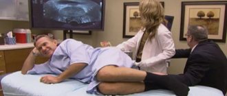
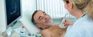
All devices have the same principle of operation and design, so a person needs to lie down on a couch, and the doctor will move a special sensor across the chest area from all angles, examining the readings. He may also ask the patient to take a deep breath, hold his breath, or turn to his side - all this is necessary to obtain the most complete information about the condition of the lungs and adjacent tissues.
How much does an ultrasound of the chest (heart, lungs and bronchi) cost in a medical clinic?
Prices for ultrasound of the heart and chest organs at Art Medic in Khabarovsk are shown in the table:
| Code | Name of procedure | price |
| 7.30 | Ultrasound of pleural cavities | 600 |
| 7.34 | Echocardiography with color Doppler mapping | 1600 |
| 7.36 | Ultrasound of the thymus | 600 |
Our clinic often holds promotions during which the cost of some tests is reduced. Follow the information on our website carefully and get a discount on the type of diagnostics you need!
Interpretation of ultrasound of the lungs
What an ultrasound will show strongly depends on the qualifications and experience of the doctor who conducts the study, therefore, based on the results of this study alone, no diagnosis is made; additional studies are prescribed.
The patient should ask the doctor who conducts the diagnosis to issue not only a report sheet with his assessment of what he saw, but also a photo from the device for confirmation.
For a high-quality and accurate assessment of the condition of the lungs and pleural area, contact the Amadeus Clinic medical center in Gomel. We offer high-quality lung ultrasound [/anchor] using modern equipment, our doctors are experienced and have all the necessary approvals to carry out this diagnosis, in addition, we pursue a moderate economic policy and here you will find affordable prices for everything. To explore the service or appointment we offer, go to this page.
Where in Khabarovsk can you get an ultrasound of the chest (heart, bronchi, lungs)?
If you are interested in where an adult can undergo an ultrasound of the heart and other chest organs for a fee, then call 8 (4212) 50-30-03; (landline and mobile). You can also make an appointment for an examination with a transcript of the result by filling out the feedback form on the website.
Our medical facility is located at Khabarovsk, st. Panfilovtsev, 38.
Many patients choose to undergo an ultrasound of the heart and chest in the hospital, however, this can take a long time. Typically, registration for a free examination is carried out several weeks in advance and, however, does not eliminate the need to spend 2-3 hours in line outside the office. In our medical center, all procedures are carried out according to modern world protocols, and pre-registration one day in advance allows us to save time for our patients. In urgent cases, it is possible to undergo diagnostics without an appointment. To do this, you need to contact the clinic administrator.
Symptoms of bronchitis
The signs of bronchitis are similar to those of other respiratory diseases. Only a qualified doctor can make an accurate diagnosis.
Symptoms of bronchitis in adults usually appear at the very beginning of the disease.
A cough appears, the patient feels general weakness, fatigue, fever, and the temperature rises. All these are manifestations of general intoxication of the body caused by inflammation of the bronchi.
Symptoms of acute bronchitis
In addition to bouts of dry, painful cough, acute bronchitis is accompanied by symptoms such as:
- runny nose and nasal congestion
- pain and sore throat
- hoarseness of voice
- chills
- high body temperature
- weakness
- headache
- sweating
- muscle soreness in the limbs and back.
Symptoms of chronic bronchitis
Symptoms of chronic bronchitis in adults usually worsen during cold and damp seasons.
Chronic bronchitis has two main symptoms:
- dry or more often wet cough
- fatigue.
Definition and risk factors
Bronchitis (obstructive bronchitis) is an inflammatory disease of the respiratory system that affects the bronchi.
Swelling of the bronchi and excessive mucus in the respiratory tract due to the inflammatory process prevent the flow of air to the lungs, which leads to difficulty breathing.
Depending on the etiology there are:
- viral bronchitis
- bacterial bronchitis.
Also, this disease is classified depending on the duration and characteristics of the course into acute bronchitis and chronic bronchitis . The symptoms and treatment of these two forms of bronchitis have some differences.
Factors influencing the development of bronchitis
The main reasons why bronchitis may develop:
- smoking
- unfavorable climatic living conditions
- harmful working conditions
- frequent hypothermia
- inhalation of toxic substances.
Smoker's bronchitis is especially common . Symptoms of bronchitis caused by exposure to tobacco smoke are particularly difficult to treat.
Indications for the procedure
The examination is necessary to identify pathologies of the lungs and pleura. Effective when studying:
- adhesive process;
- pneumonia;
- tumors;
- hydrothorax;
- pleurisy;
- metastasis;
- pleural empyema.
Ultrasound is often performed urgently. For example, if a patient is brought to a medical facility with a penetrating chest injury.
The examination method is non-invasive and effective. It is considered an excellent alternative to radiography. Ultrasound is preferable if it is necessary to conduct multiple examinations of the pleural cavity and lungs. Often this approach is carried out while monitoring the stages of treatment.
Materials and methods
The process of formulating recommendations consisted of the following sequential stages:
- review and selection of literature
- database creation
- formulation of statements
- analysis of the reliability of literature data
- discussions according to the Delphi method
- secret voting of experts in three rounds.
Two independent supervisors observed the progress of each stage. The process of preparing recommendations was accompanied by a methodologist and a librarian.
A systematic review of the literature was conducted independently by 13 individuals. Relevant publications were searched in the following databases: PubMed, OVID, Embase, and Medline.
The literature review included prospective, retrospective and observational studies, as well as meta-analyses with their full texts or abstracts published in English up to November 2021.
Exclusion criteria for selecting studies included case reports, reviews (except meta-analyses), letters to the editor, and publications in pediatrics, neonatology, anesthesiology, and surgery.
The database prepared for the previous version of the recommendations was replenished with 253 new publications. The initial selection of publications was based on screening of titles and abstracts, followed by analysis of full texts.
When analyzing the reliability of the literature data, the following parameters were taken into account: age, gender, number of patients examined, homogeneity of groups of patients participating in the study, inclusion and exclusion criteria, type of publication (prospective, retrospective, meta-analysis), sensitivity and specificity of the method used, true positives, false positives , true negatives, false negatives, and imaging modality recognized as the diagnostic gold standard.
| Grade | Interpretation |
| A | High – data from several meta-analyses, it is unlikely that further studies will affect the reliability of the effectiveness or accuracy of the method. |
| IN | Moderate – data from isolated large non-randomized studies, further testing may have a significant impact on the reliability of the effectiveness or accuracy of the method. |
| WITH | Low or very low—consensus of experts and/or data from small studies, retrospective studies, registries, case series, or case reports, it is very likely that further testing will have an important impact on the validity of the effectiveness or precision of the method. |
Methodology
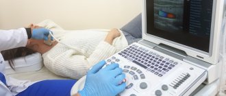
Ultrasound diagnosis of the pleural cavity does not require preparation. The patient is asked to release his torso and take a supine position on the couch.
Taking into account the clinical indications and complaints of the patient, the doctor alternately examines the pleural cavity and pulmonary lobes from the front, side and back. As a rule, to complete the picture, a total of 14 zones are examined and high-frequency linear and low-frequency sector or convex ultrasonic sensors are used. The former provides excellent visualization of the pleural line and is used to determine its thickness and condition, as well as to evaluate pneumothorax. The second transducers are preferable for visualizing structures located in the chest.