Ultrasound examination of the thyroid gland is an informative diagnostic method. It helps to determine the structure, identify pathological changes and formations in the organ, without having a negative effect on the body. The number of such examinations is not limited, unlike other hardware diagnostic methods.
Ultrasound is used to dynamically monitor the effectiveness of therapy. During a biopsy, the needle is used to accurately hit the node, thereby reducing the risk of injury to nearby tissues.
Why do you need to do the research?
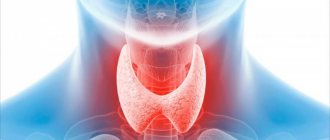
The thyroid gland (TG) is one of the largest endocrine glands in the body. The primary tasks of the organ are storing iodine reserves and producing thyroid hormones.
In turn, thyroid hormones perform the following functions:
- affect metabolism, heat exchange, and the level of other hormones in the body;
- participate in the formation and maintenance of immunity;
- regulate the functioning of the cardiovascular, reproductive system, gastrointestinal tract, brain;
- activate protein synthesis, cell and tissue regeneration processes.
In addition, parafollicular cells of the thyroid gland produce calcitonin, a hormone that regulates the level of calcium and phosphorus in the blood and promotes the deposition of calcium in bone tissue.
Based on the multifunctionality of the thyroid gland, we can conclude that there is not a single internal organ that is not affected by it. Therefore, any failure in its operation can lead to serious consequences. It is possible to diagnose organ diseases in a timely manner using ultrasound.
Clinical endocrinology uses thyroid ultrasound as a primary screening method. The undoubted advantages of the technique include:
- non-invasive;
- availability;
- absolute safety for health;
- high information content.
There are several ultrasound examination methods:
- Classic ultrasound. Provides information about the structure, location, and size of the organ.
- Dopplerography. It is used to study blood flow and assess the condition of blood vessels.
In addition, ultrasound is used for differential diagnosis, assessing the effectiveness of treatment of endocrine diseases, and as an additional technique for fine-needle biopsy of the thyroid gland.
How to prepare for an ultrasound
The procedure for performing an ultrasound of the thyroid gland does not require special preparation. It is necessary to free your neck from jewelry and remove your scarf or neckerchief. It is recommended to wear clothing with an open collar or neckline. You may want to bring a towel with you if the scan is being done at a public health facility. It is recommended to place it under the head, and then use it to remove the remaining gel from the neck.
There are no restrictions on meal time, foods and spices. The recommendations apply only to older people and children. They do not need to eat shortly before the test to avoid nausea that can be caused by pressing on the thyroid gland with the sensor.
All preparation of the child for the procedure takes place in conversation, his parents prepare him psychologically: they tell him that it doesn’t hurt at all, what the doctors do, why it is necessary.
If a scan is performed to determine the effectiveness of treatment, it is necessary to bring the results of a previous ultrasound so that the doctor can assess the dynamics of changes in the gland.
Who is the study for?
Many thyroid diseases are asymptomatic. However, there are some signs that may indirectly indicate organ pathology. An ultrasound scan of the thyroid gland is recommended for people who exhibit the following symptoms:
- sore throat when swallowing;
- causeless cough;
- depression, apathetic state;
- decreased performance;
- sudden weight loss or, conversely, rapid weight gain;
- increased body temperature of unknown origin;
- dry skin;
- brittle nails and hair.
An endocrinologist may prescribe an ultrasound examination if there are lumps in the throat area, visual enlargement of the organ, or changes in the normal level of thyroid hormones in the blood.
As a rule, endocrinologist patients are more often women than men. This is due to the higher need of the female body for iodine, which is actively consumed during pregnancy and lactation. Often the cause of thyroid dysfunction in women is menopause.
For preventive purposes, regular ultrasound examinations are recommended:
- people living in areas with unfavorable ecology;
- workers in hazardous industries;
- all persons over 40 years of age;
- women planning pregnancy;
- patients who are registered with an endocrinologist.
Contraindications
Ultrasound examination has no strict contraindications. Diagnosis can be carried out for all people.
A limitation for performing the procedure may be damage to the skin at the site of application of the gel:
- pustular formations;
- painful rash;
- burn;
- eczema.
Once the problem has been treated and resolved, an examination can be performed. The procedure is not recommended to be performed in the first days of the menstrual cycle, since during this period the gland is enlarged, which can distort the results.
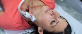
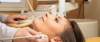
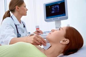
How is the research going?
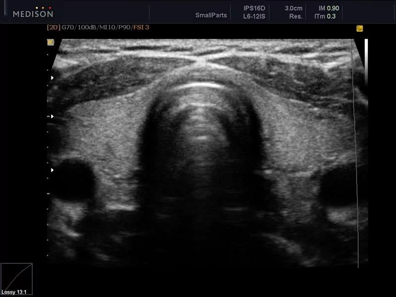
During the examination, the patient is placed on a couch. A special gel is applied to the skin of the neck in the area of the thyroid gland. The organ is scanned using an ultrasound probe, and the image is displayed on the monitor screen.
During the examination, the ultrasound doctor sees black and white images on the monitor screen. All tissues of internal organs reflect ultrasound in their own way: the higher the density of the tissue, the brighter the image. This property of tissues is called “echogenicity”.
The condition of the thyroid tissue can be assessed by echogenicity:
- tissues without pathologies (isoechoic) are displayed on the monitor in gray;
- structures with reduced density (hypoechoic) look like dark spots;
- tissues with high density (hyperechoic) are displayed in light shades.
There are also anechoic structures that absorb ultrasound completely. On the monitor they appear as black spots.
Who draws conclusions about the effectiveness of the examination and prescribes additional tests?
Only an endocrinologist should make a conclusion based on the results of ultrasound and all the studies listed above. Even detected deviations in hormone levels and anomalies on ultrasound do not mean serious health problems. Age over 65 years, pregnancy, hidden infections, inflammation change test results to one degree or another. Neck injuries and laryngeal surgeries also affect the functioning of the thyroid gland. Therefore, the study is always repeated if questions or suspicions arise.
Interpretation of ultrasound results: normal options
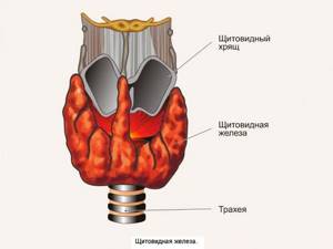
Normally the thyroid gland has:
- horseshoe shape;
- two lobes separated by an isthmus. An additional pyramidal lobe, an embryological remnant of the thyroglossal duct, may also be present;
- typical location. The upper border of the organ should be at the level of the middle of the thyroid cartilage, the lower - at the level of 5-6 cartilaginous rings of the tracheal wall;
- homogeneous structure. Organ tissues should be displayed on the monitor in a uniform color;
- smooth and clear contours.
Measurements of the parameters of the thyroid lobes will be performed in the transverse and sagittal planes. The volume of each lobe of the organ and the total volume of the thyroid gland are calculated. The thickness of the isthmus in the anteroposterior direction is also determined.
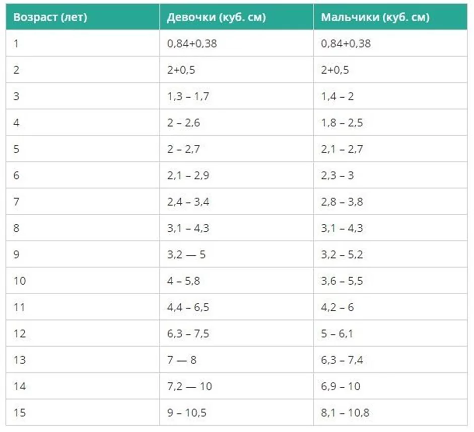
There are separate normative indicators for each age group of patients. The volume of the thyroid gland in women should not exceed 18 cm³, in men - 25 cm³. Standard indicators for children are presented in table ⇓⇓⇓
What diseases and disorders can ultrasound detect?
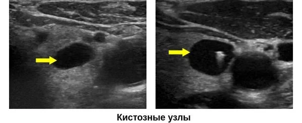
Thyroid cysts
A thyroid cyst is a hollow formation containing fluid inside. In most cases, cysts do not pose any danger to humans. Thus, the inactive form of thyroid hormones is stored in reserve. If the formations are small, there are no signs of their malignancy, or an increase in the volume of the organ, then the cysts do not require treatment. They are simply monitored with regular ultrasound examinations.
However, large formations can cause pathological symptoms and lead to compression of neighboring internal organs. With large cysts, a person experiences difficulty breathing, hoarseness, neck deformation, bursting pain in the cervical spine, difficulty swallowing, and an unreasonable cough. In such situations, conservative and surgical treatment methods are used.
Ultrasound signs of thyroid cysts include:
- correct form;
- smooth, thin walls;
- anechoic contents;
- the presence of an acoustic shadow - an anechoic strip.
During an ultrasound examination, the risk of malignancy of cysts is assessed in points according to the ACR TI-RADS classification:
- 0-3 points - the risk of malignancy is low, additional diagnostic methods are not required;
- more than 3 points - the risk of malignancy is high, a fine-needle biopsy of the node is required.
Diffuse toxic goiter
A disease of an autoimmune nature, which is based on hyperfunction of the thyroid gland. Clinically manifested by an increase in neck circumference, bulging eyes, rapid heartbeat, and difficulty swallowing.
Ultrasound signs of pathology:
- uniform increase in the volume of both lobes of the thyroid gland;
- smoothed and rounded edges of the organ;
- hypoechogenicity. The decrease in organ density is due to the high water content in the tissues;
- homogeneous structure;
- dilated vessels;
- in Doppler mode, increased blood circulation is visible;
- Cysts and calcifications may be observed.
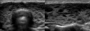
Thyroiditis
Thyroiditis is an inflammation of the thyroid tissue of an acute, chronic or autoimmune nature. The inflammatory process can affect a specific area or the entire organ. Depending on the type of thyroiditis, the pathology can lead to increased or decreased production of iodine-containing hormones, occur with clear signs or be asymptomatic.
Ultrasound signs of the most common types of pathology are presented in the table.
| Type of thyroiditis | Ultrasound signs |
| Acute thyroiditis is an inflammation of the thyroid tissue, which occurs with high fever, chills, and severe pain in the throat. In some cases, inflammation is accompanied by the formation of purulent exudate. | ● normal or enlarged size of the thyroid gland; ● heterogeneous echogenicity of the organ; ● abscesses (cavities with purulent contents) may be observed - hypoechoic areas; ● increased blood supply; |
| Autoimmune thyroiditis (AIT) is a chronic inflammatory process of autoimmune origin. | ● the size of the thyroid gland can be normal, reduced in atrophic AIT, increased in hypertrophic AIT; ● heterogeneous structure; ● reduced echogenicity of organ tissues; ● abnormal shape of blood vessels; ● increased blood supply. |
AIT on an ultrasound image looks like this ⇓⇓⇓
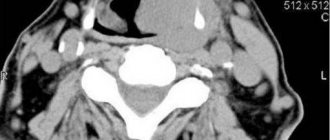
Thyroid oncology
Thyroid cancer is rarely diagnosed: it accounts for about 1% of all diagnosed cancer cases. The risk group includes people over 50 years of age, residents of regions with high radiation, and people with iodine deficiency.
The outcome of the disease depends on the timeliness of diagnosis. If the disease is detected at an early stage, the prognosis is favorable. The last stage of oncology reduces the patient’s chance of survival to zero.
Ultrasound signs of malignant thyroid formations include:
- uneven and unclear contour;
- hypoechogenicity;
- increased blood flow if the tumor is actively growing;
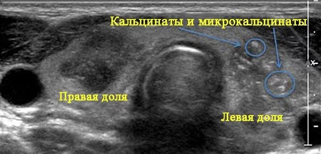
- in some cases, microcalcifications may be observed - white dots on a dark background of the node.
The photo below shows what papillary thyroid cancer looks like compared to a benign formation.
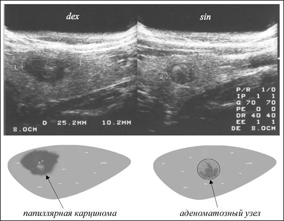
If an ultrasound reveals nodes with signs of malignancy, the patient is referred for a fine-needle biopsy.
History of ultrasound
Ultrasonic vibrations were discovered by Italian scientist Lazzaro Spallanzani back in 1794. While watching the bat, he wondered how it was possible to navigate in space at night while flying. It turned out that this is carried out using high-frequency sound vibrations, inaudible to humans. But animals, particularly bats, perceive them very well. The principles of echolocation are also used by marine animals - killer whales, dolphins, whales, etc. Over time, they became known as ultrasonic waves.
In 1942, for the first time, the German doctor Theodor Dussick and his brother, scientist and physicist Friedrich Dussick, tried to use ultrasound to diagnose a tumor in a patient’s brain. The first medical ultrasound device was created in 1949 by the American scientist Douglas Haury.
Doppler Christian Anders made a great contribution to the development of ultrasound diagnostics. While working on his scientific work “On the collometric characteristics of the study of double stars and some other stars in the sky,” he noticed that the frequency of the signal reflected from an object depends on its speed and direction of movement. This property is called the Doppler effect, which is widely used in Dopplerography to examine blood flow parameters.
Comprehensive examination of the thyroid gland
Despite its high information content, in some situations ultrasound alone is not enough to make a correct diagnosis. The most effective solution for a thorough diagnosis is an endocrinological check-up - a comprehensive examination of the thyroid gland.
Check-up allows you to quickly obtain the necessary information about the condition of the thyroid gland and correctly select a treatment regimen. As a rule, in addition to ultrasound, a comprehensive examination includes a consultation with an endocrinologist and laboratory tests.
When to do an ultrasound of the thyroid gland
Basically, ultrasound is prescribed by an endocrinologist as a result of the detection of tangible changes in the structure of the organ during examination. In addition, diagnosis of the thyroid gland is carried out in the following cases:
- abnormalities in the results of blood tests for hormones;
- painful or difficult swallowing;
- a sharp decrease or, conversely, increase in weight;
- shortness of breath, headaches, mood swings, weakness;
- an increase in temperature that persists for a long time;
- taking hormonal drugs.
Also, for the purpose of prevention, a thyroid gland examination is carried out when:
- pregnancy planning;
- living in areas characterized by iodine deficiency;
- congenital pathologies and chronic thyroid diseases;
- presence of a profession associated with environmental hazards.