Ultrasound examination of the breast is one of the graphic methods of imaging breast tissue, prescribed for the purpose of early detection of its pathologies, especially malignant changes. Ultrasound is a safe and painless method, unlike mammography, in which the woman receives little radiation.
Ultrasound examination has its advantages and disadvantages. It is used mainly for young women whose mammary glands are dominated by glandular tissue, which is clearly visible using ultrasound. The amount of such tissue decreases with age.
In women, breast cancer is the leading cause of cancer death in the world and is the second leading cause of death after lung cancer in all people. Early diagnosis is important for early initiation of therapy, which provides better prognosis for cure.
Calcium deposits in the breast - what does it mean?
Small calcium deposits (calcifications) found in tissue are usually benign, although in some cases they may be an early sign of breast cancer.
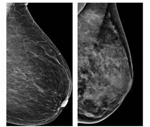
These deposits are better visible on mammography as white dots, but they can also be seen on ultrasound. The causes of calcifications in the breast are not entirely clear; they can occur after injury or infection in the tissue. There is an assumption that in those areas where calcium accumulates, there may be precancerous cells.
If in doubt, the doctor sends the patient for a breast biopsy - removing a small amount of breast tissue with a needle for histological analysis.
Ultrasound of the mammary glands during pregnancy
Pregnancy leads to hormonal changes in the female body. Changes also affect the mammary glands. During pregnancy, breast sensitivity increases (sometimes to the point of pain). The breasts increase in size, and vascular networks may appear on them. The nipples may darken slightly and there is usually nipple discharge.
All this is within normal limits, but ongoing hormonal changes can lead to the development of pathologies. To maintain breast health, you need to monitor its condition. If lumps appear, especially painful ones, you should consult a mammologist. In this case, as a rule, an ultrasound of the mammary glands is prescribed, which is a basic diagnostic procedure for examining the mammary glands during pregnancy.
Shadows detected during ultrasound examination
Shadows that are visible on a breast ultrasound may, but should not, be a sign of breast cancer. There are several criteria by which the nature of the found formations is determined, of which, in addition to the existence of a shadow, the shape of the edge, structure, and position in comparison with the skin are examined.
A shadow on an ultrasound scan can give:
- fibroadenoma;
- diabetic mastopathy;
- necrosis (cell breakdown) of breast tissue as a result of injury or surgery;
- scars;
- and etc.
When is a breast ultrasound prescribed?
Indications for ultrasound of the mammary glands are:
- pain;
- compactions in the gland itself or in the axillary lymph nodes;
- nipple discharge;
- injuries;
- preventive examination.
Ultrasound is also used to determine the condition after plastic reconstruction of the gland and the condition of silicone breast prostheses. For timely detection of possible pathologies, women under 35 years of age are recommended to undergo ultrasound of the mammary glands twice a year for preventive purposes. Under ultrasound control, puncture of the mammary gland can be performed.
Which is better - MRI or ultrasound?
Breast MRI, also known as magnetic resonance mammography, is one of the most accurate tests in diagnosing breast cancer. The examination method works on a completely different principle than ultrasound or mammography. Instead of ultrasonic waves and x-rays, a strong electromagnetic field is used. The resulting energy is converted into an image visible on the monitor. This examination allows you to accurately locate the lesion and determine its structure and degree of vascularization.
Magnetic resonance mammography
However, it should be remembered that not every woman will be able to perform such a test. First of all, this applies to patients who have some metal objects in their bodies. An absolute contraindication for magnetic resonance imaging would be, for example, a pacemaker, a neurostimulator, an implanted hearing aid, an insulin pump, the presence of a foreign body in the eye or a metal clamp in the skull. In addition, MRI should be performed with extreme caution in pregnant women and only when absolutely necessary. There are no such contraindications for ultrasound. As a rule, this is an absolutely safe method, as the only one among all available diagnostic methods.
Ultrasound of the breast during pregnancy and lactation
A certain difficulty in diagnosing the mammary glands is present in women during pregnancy and during breastfeeding. The difficulty is that already in the first trimester of pregnancy, the mammary glands begin to rebuild so that breast milk appears in them in the future. Therefore, doctors prescribe ultrasound diagnostics of the breast during this period only if a serious illness is suspected.
This procedure is completely safe for women during pregnancy and lactation.
Decoding the results
Decoding of the received information can be carried out either immediately after diagnosis or after a certain period of time. This depends on where the study was conducted, in a private medical clinic or a government institution, as well as on the employment and workload of the specialist.
The following are some pathological conditions that can be detected during ultrasound diagnosis of the mammary glands.
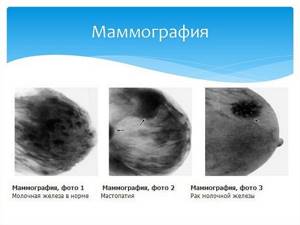
Mastopathy on ultrasound
Mastopathy is a pathological painful condition that develops in the tissues of the mammary gland. Characterized by excessive proliferation of glandular tissue. It is a hormone-dependent disease.
Cyst on ultrasound
It is a disease of the mammary gland in which a pathological formation is detected, which has a capsule and liquid contents in its structure. Clearly determined by ultrasound with the possibility of taking measurements.
Lactostasis and mastitis
Lactostasis is characterized by congestion in the ducts of the mammary gland and occurs due to a violation of the outflow of milk from these ducts. Often complicated by mastitis (inflammatory process).
Breast cancer on ultrasound
Breast cancer is a pathological process characterized by uncontrolled cell division and constant progression. There are several stages of development of this pathology, various symptomatic pictures. This disease requires immediate contact with an oncologist to choose an individual and optimal treatment method.
What to do if your cycles are irregular and you become pregnant
An irregular cycle is not an acceptable phenomenon, but is a common case that specialists have to work with. It is also necessary to remember about menopause, when menstruation stops completely.
In this case, the question regarding the time of the ultrasound loses its importance; it can be done on any day that happens to be free.
The exception is during pregnancy. As practice has shown, a significant hormonal surge will not make it possible to accurately determine the presence of changes or breast diseases, so there is little point in performing the procedure on pregnant women.
Order of conduct
The examination is carried out in a room where special equipment is located. Takes approximately 20 minutes. Painful sensations are usually absent, appearing only in the presence of inflammatory processes. During the study, both the chest and the axillary, prothoracic zone, areas above and below the collarbones are checked. If additionally necessary, Doppler sonography is performed (to study the blood flow to the organ).
If we talk about ultrasound and mammography, they cannot be compared, since they do not replace, but complement each other. For example, using a mammogram, they analyze the condition of the adjacent tissues and whether there are any formations located near the chest. Using ultrasound, the full dynamics will be determined; in addition, the condition of the lymph nodes can be checked.
The examination can be carried out both in public medical institutions and in private clinics by appointment.
When to do an ultrasound if menstruation is not regular
On what day should an ultrasound be done if menstruation occurs sporadically? You can undergo the procedure on any day. If we talk about pregnant women, then there are hormonal surges that are an obstacle to an accurate diagnosis. It is for this reason that manipulation during the process of bearing a child is inappropriate. As an exception, cases are considered when a pregnant girl is suspected of having cancer. But here, too, other studies may be prescribed.
Ultrasound scan for suspected malignant tumors
The reliability of ultrasound in the diagnosis of malignant neoplasms reaches 95% (without specifying the nosological form). When the tumor diameter is more than 5 cm, the accuracy reaches 100%, and less than 2 cm - 63%.
Main ultrasound signs of carcinoma:
- “blurred” uneven contours;
- heterogeneity;
- low echogenicity;
- blurred anterior wall;
- lack of visualization or low echogenicity of the posterior wall;
- presence of dorsal echo.
Scirrhous carcinoma, which occurs in 34% of patients, appears on scans as an irregularly shaped heterogeneous formation with uneven contours. A typical symptom is a dorsal acoustic shadow. Difficulties in differential diagnosis are caused by solid and medullary carcinomas, since they have ultrasound signs characteristic of benign neoplasms.
Due to the similarity of the ultrasound picture, differentiation of many pathologies can be difficult. In doubtful situations, the doctor should recommend mammography. There is a widespread opinion among experts that when diagnosing cystic diseases, the informativeness of ultrasound reaches 100%, and in the presence of small, up to 1 cm, tumors, preference should be given to X-ray examination. The latter is also indicated for studying abnormal areas during fatty degeneration of the gland. At the same time, mammography is not recommended for females under 35 years of age and pregnant women.
On what day of the cycle should I do a breast ultrasound?
In women of childbearing age, the condition of internal organs is very dependent on the menstrual cycle. Scientifically speaking, a woman’s breasts are nothing more than glandular tissue interspersed with fat, and these substances are largely dependent on hormones, which vary at different periods of the cycle. Depending on the hormonal state, the mammary glands can change their density, and therefore echogenicity.
It should be understood that in critical situations you should not delay and wait for the appropriate period of the cycle; in this case, it is necessary to undergo a study at any time.
So on what day is it best to perform a breast ultrasound? Gynecologists and mammologists agreed that the ideal period would be day 5-14 of the female cycle (day 1 of the cycle is the day the period begins). During this period, healthy breast tissue is the densest and has the least sensitivity. And entering the second phase of the cycle (starting from day 15), the breasts slowly swell, its tissues increase, and their density decreases; you can often observe swelling and cysts, which then go away, but can give an unreliable vision of the condition of the mammary glands.
If deviations from the norm were found during the examination, then a repeat ultrasound of the mammary glands is prescribed and the doctor looks at what it shows.
If you are interested in the question of how often you can do an ultrasound of the mammary glands, then the answer is simple: due to the fact that ultrasound technology is harmless to the body, then such a study can be carried out as often as the diagnosis of this organ is required.
Breast examination at Seline
You can undergo ultrasound and mammography in many clinics in Moscow. But the study at Seline Medical Center has several advantages:
- Diagnostics are carried out using modern high-precision equipment. A foreign-made ultrasound machine allows you to obtain a clear image, including 3D. The existing PINKVIEW-RT mammograph compares favorably with analogues in its minimal radiation dose, high image clarity, fast image display speed and the ability to record examination results on a digital medium.
- The examination is carried out by diagnostic radiologists and ultrasound specialists with extensive experience. They are able to recognize even small tumors, which makes it possible to begin treatment of pathology at an early stage.
- If necessary, you can undergo additional research and get advice from specialists.
It’s always a pleasure to be at the Seline clinic - impeccable cleanliness, premium interior, friendly staff. Sign up for a breast examination and come at the appointed time. Your health is in good hands here!
| Author of the article: | Sukhorukov Evgeniy Anatolievich |
| Speciality: | Chief physician of the aesthetic medicine clinic Seline Clinic (Belarusian), phlebologist surgeon, arthrologist, ultrasound doctor |
| Experience: | 18 years |
Make an appointment
What does an ultrasound of the mammary glands show?
In recent years, there has been an upward trend in breast diseases in all countries, including European ones. There are enough reasons for such dynamics. Among them: genetic factors, stress, unfavorable environmental conditions and others.
Today, with Anna Viktorovna Poskrebysheva, ultrasound diagnostics doctor at Clinic Expert Orenburg LLC, we are talking about a “female” topic - breast diseases and their ultrasound diagnostics.
— Anna Viktorovna, at what age should a woman become concerned about the condition of her mammary glands?
From the moment a girl’s mammary gland begins to form until her life continues.
The mother needs to be in close contact with her daughter so that the girl is not afraid to be frank with her parents about sensitive issues. It happens that the glands are formed asymmetrically or the process of development of the mammary glands causes severe discomfort to the child. In this case, you need to contact your pediatrician, family doctor or pediatric gynecologist.
There is a widespread myth that during menopause a woman does not have to worry about the condition of her mammary glands. This is wrong. The older a woman is, the higher the likelihood of developing breast cancer. Younger women are susceptible to the development of benign breast tumors, mastopathy and other diseases.
Therefore, protecting your symbol of femininity must be remembered at any age. This means undergoing preventive examinations on time.
— How can a woman understand that she needs to see a doctor and undergo ultrasound diagnostics of the mammary glands?
A visit to a gynecologist once a year is mandatory for every woman. If necessary, the doctor will refer the woman to a mammologist. But, of course, experiencing discomfort in the area of the mammary glands, a woman can decide to undergo an ultrasound on her own, before contacting a doctor.
Indications for an ultrasound of the mammary glands are symptoms such as breast tenderness, hardening of the nipples, any discharge from the nipples (both transparent, yellowish, greenish, and bloody); change in size, shape of the breast, skin of the mammary gland. Nodules should also alert the woman and bring her to the ultrasound room.
The test is often prescribed to breastfeeding women to rule out mastitis. Diagnostics are also carried out after injuries or inflammations, to monitor the condition after treatment by specialists. Women are also referred to us after plastic surgery for breast augmentation - ultrasound diagnostics allows us to assess the condition of the implants.
This type of diagnosis is also indicated when planning pregnancy.
For men, ultrasound of the mammary glands is also performed. Indications are hormonal changes leading to gynecomastia. This diagnosis, as a rule, results from the uncontrolled use by amateur athletes of various drugs and nutrition to increase muscle mass. But the condition can occur without exercise or sports nutrition in men of any age.
— Mammography or ultrasound of the mammary glands: which is better?
A large number of patients come to us after mammography, with a conclusion that says “ultrasound of the mammary glands is recommended.” To make a diagnosis, for example, when a tumor is detected, at least two studies are required.
But, despite the high efficiency of using mammography, when it comes to young patients, the ultrasound method has a number of advantages. This is due to the fact that breast tissue in young women is usually high in density. And this, with severe diffuse mastopathy, as well as the presence of multiple fibroadenomas, implants or post-inflammatory (after mastitis) changes, disrupts the structural background. In these cases, an ultrasound scan is necessary.
— On what day of the cycle is it better to do an ultrasound of the mammary glands?
If there is an urgent need, for example, an injury or some acute condition, then diagnostics are carried out at any time. Also, regardless of the day of the cycle, ultrasound is performed for women taking hormonal contraceptives and during menopause.
But ideally, ultrasound of the mammary glands is performed from the 5th to the 10th day of the cycle, counting from the first day of menstruation. It is on these days that the hormonal background is most stable, which reduces the risk of tissue swelling and expansion of the milk ducts and, as a result, the possibility of making an inaccurate diagnosis.
— How is breast ultrasound performed and what does it show?
The procedure is absolutely painless and lasts from 15 to 30 minutes, depending on the size of the mammary gland and the detected pathology. To carry out the diagnosis, a woman needs to completely undress to the waist and lie down on the couch. The examination is usually carried out lying on her back, but personally I can ask the woman to turn around so that it is convenient for me to examine the mammary gland on the ultrasound machine from all sides.
Diagnostics is carried out by a sensor with the application of a special gel. The study allows you to view the entire mammary gland, as well as the axillary, supraclavicular and subclavian lymph nodes. Ultrasound is an informative method that allows you to see almost any pathology of the mammary glands. Among them: cysts and tumors, both benign and malignant, various types of mastopathy and other diseases.
— How often can you undergo an ultrasound of the mammary glands?
The method is completely safe, does not carry any radiation exposure and can be carried out as long as necessary.
— Anna Viktorovna, as a doctor and as a woman, do you think that ultrasound of the mammary glands should be done for prevention?
Undoubtedly. Every woman needs to undergo a diagnosis once a year. I, my relatives and friends are no exception and strictly follow this rule.
— In order to do an ultrasound of the mammary glands at the Expert Orenburg Clinic, do you need a doctor’s referral?
No. A woman can make a decision about undergoing diagnostics on her own. The main thing is to arrive on the right days of the cycle and on time. If a woman feels some kind of formation on herself, this is not a reason to panic. Sometimes it hides a banal mastopathy. That’s why it’s so important not to be afraid, not to withdraw into yourself, but to come to the doctor and find the answer to the question “what’s wrong with me?”
Other interviews with Anna Poskrebysheva:
How to prepare for an abdominal ultrasound?
How to do an ultrasound of the mammary glands
Ultrasound of the mammary glands does not require special preparation. However, it is important in what period the research is carried out. It should be carried out in the first phase of the menstrual cycle (on the 5-12th day from the start of menstruation). Menopausal women can be screened any day.
You can get an ultrasound of the mammary glands in Moscow at the clinics of JSC “Family Doctor”. The duration of the study is approximately 10 minutes.
Sign up for diagnostics Do not self-medicate. Contact our specialists who will correctly diagnose and prescribe treatment.
What a breast ultrasound sees and doesn’t see. Which ultrasound sensors and machines are better?
Regular clinics and hospitals are equipped with ultrasound machines of standard quality, equipped with several universal sensors, including for breast examination. They have high resolution clarity, display echogenicity and contours, but only detect fairly large neoplasms measuring 5 mm or more.
The tumor reaches this size 8 years after its appearance. This means that an important moment when it was still possible to cure the pathology quickly and without significant interventions may be missed. But other pathologies in the chest - cysts, fibroids, blocked ducts, etc. Ultrasound sees 5 points.
For this reason, ultrasound diagnostics of the mammary glands should be complemented by other methods if oncology is suspected (sunken nipple, breast asymmetry, lump upon palpation). In these cases, mammography or, in special cases, MRI is additionally prescribed.
Standard ultrasound clearly displays structural changes in breast tissue: the presence of nodules, cysts, deterioration of blood circulation, and the occurrence of infiltration. The doctor’s experience helps to suspect oncology and refer the patient for a more detailed examination.
Today, there is already an ultrasound method that detects tumors at an early stage. These are ultrasound machines of the ABVS (Automated Breast Volume Scanner) system, developed by Siemens specifically for mammological studies.
A special sensor installed on a woman’s chest allows you to obtain a 3D image on the monitor screen. Screening is carried out automatically and does not depend on the experience and qualifications of the doctor. The information received is processed by a special program for 15 minutes, while the sensor lies on the woman’s chest and takes pictures in different sections from the chest wall to the tip of the nipple. The ABVS system allows you to examine women who have a hereditary predisposition to breast cancer and who have several blood relatives who suffered from or died from this disease.
FAQ
Lisa : Currently I am breastfeeding my baby. Hardenings appeared in the left breast. Is it possible to undergo examination, and which is better?
Administrator : You can perform an ultrasound of the breast. This method is absolutely safe and allowed during lactation.
Vladimir : The doctor sent me for an ultrasound of the breast after surgery. Is this procedure necessary for men?
Administrator : Ultrasound examination is prescribed regardless of gender. The cause may be hormonal imbalance or surgery to remove the testicles (for men).
When is ultrasound recommended for patients?
The procedure is prescribed by the attending physician, for example, a therapist or a highly qualified doctor, for example, a gynecologist or mammologist. Much depends on which doctor the woman decided to go to with her problem. The medical worker will interview the patient about complaints and symptoms, conduct an examination, and advise on how to conduct the examination and on what day to perform an ultrasound of the mammary glands in each specific case.
The doctor will also tell you what results can be determined using ultrasound, voice the features of the manipulation, and confirm that it is completely safe. As a rule, ultrasound is prescribed in the following cases:
- confirmation or refutation of the neoplasm;
- preventive research;
- if necessary, confirm the gynecological diagnosis;
- if a woman needs oral contraceptives;
- before in vitro fertilization;
- for pain in the mammary glands that occurs between menstruation;
- change in skin color;
- different sizes of mammary glands;
- discharge from the breast;
- diseases of the endocrine system;
- to check for unusual results obtained during mammography;
- to monitor the condition of the breast with implants installed inside;
- for benign or malignant neoplasms in the pelvic organs;
- during the rehabilitation period after surgical interventions associated with breast surgery;
- for mechanical injuries;
- for diagnosing mastopathy.
A little about the research
The ultrasound examination method itself appeared about half a century ago. Today it is widely used in diagnostics.
Ultrasound of the mammary glands is a fairly simple procedure that should be performed by an experienced specialist.
The patient who came for the examination should take off her clothes to the waist and lie on the couch with her hands behind her head so that the specialist can examine both the breast and the lymph nodes near it. The ultrasound doctor applies a little special gel to the chest so that the waves coming from the device pass better through the body tissues. Next, the doctor moves a special device over the area of the mammary glands and looks at a picture on the screen in which the internal tissues are visible. Based on these images, he evaluates the structure of the glands, their size and contour, ducts and lumens, the presence of unusual seals or protrusions, as well as neoplasms.
A gynecologist, mammologist or oncologist can correctly assess the condition of the mammary glands using ultrasound.
How to interpret an ultrasound result
Since 2010, many European countries have used a special BI-RADS classification to interpret sonomammography results. Standards based on this classification have standardized breast ultrasound descriptions and terminology for abnormal breast changes.
The BI-RADS classification takes into account the morphological elements of the breast structure and their abnormal focal lesions. Thanks to this, we can distinguish between benign and potentially malignant changes. It also contains information about the risk of malignant neoplasms of the lesion and suggestions for diagnostic and therapeutic procedures in case of detection of lesions of various nature.
According to the BI-RADS classification, breast condition can be classified into one of 7 categories:
- BIRADS-usg 0 category – means that there is not enough data. Additional imaging tests such as mammography, ultrasound, or biopsy are necessary to make a diagnosis.
- BIRADS-usg category 1 – means that the result is correct. No alarming changes were observed in the study.
- BIRADS-USG category 2 - means that the study revealed benign changes in the mammary glands, for example, cysts, fibroadenomas, lipomas, inflammatory and post-traumatic changes in the mammary gland. Changes classified as Category 2 do not require additional imaging tests, only regular checks.
- BIRADS-usg category 3 - means that the test identified lesions that are possibly benign with a risk of malignancy of less than 2%. Depending on the patient's age and individual preferences, as well as family issues, the doctor may recommend a repeat ultrasound scan after 6 months or a biopsy.
- BIRADS-USG category 4 - means that the examination revealed suspicious lesions that do not have the classic signs of a malignant tumor, but the risk of developing a malignant tumor ranges from 2 to 90%. In this case, a fine or core needle biopsy is required. Depending on the result, further treatment such as surgery is suggested.
- BIRADS-usg category 5 – means that the examination revealed lesions with a risk of malignancy >90%. In this case, histopathological examinations (puncture, mammotomy or open surgical biopsy), oncological assessment are carried out and the most appropriate treatment is planned.
- BIRADS-usg category 6 - indicates a malignant tumor confirmed by biopsy.
Remember, a systematic examination of the mammary glands allows you to detect tumor changes at an early stage of development. Breast cancer is completely curable if detected at an early stage! So, if you don't want to get cancer, get your breasts examined on time!
ONLINE REGISTRATION at the DIANA clinic
You can sign up by calling the toll-free phone number 8-800-707-15-60 or filling out the contact form. In this case, we will contact you ourselves.
If you find an error, please select a piece of text and press Ctrl+Enter