Medical editor: Strokina O.A. - therapist, functional diagnostics doctor. July, 2021.
Ultrasound examination of the stomach is a simple, but rarely prescribed diagnostic method by doctors. To obtain the most complete information about the state of the main digestive organ, there is another way - fibrogastroduodenoscopy (FGDS) or, more correctly, esophagogastroduodenoscopy.
Indications for ultrasound of the stomach
Ultrasound of the stomach is performed if there are contraindications to FGDS or the patient’s categorical refusal of it, as well as as a primary examination in a comprehensive diagnostic program.
A gastroenterologist may prescribe an ultrasound examination of the stomach if the patient has complaints of:
- pain and heaviness in the epigastric region;
- bloating;
- frequent belching and heartburn;
- nausea and vomiting;
- change in appetite;
- dry or bitter mouth.
The procedure is also carried out in the following situations:
- suspicion of polyps or stomach cancer;
- suspicion of stenosis (narrowing) of the antral and pyloric (final) parts of the stomach;
- analysis of the motor-evacuation function of the stomach;
- assessment of the functionality of the cardiac sphincter in reflux disease.
Examination of the gastrointestinal tract using a flexible endoscope
FGDS (gastroscopy)
is one of the options for endoscopic examination, which allows for an external examination of the mucous membranes of the intestines, esophagus and stomach. The procedure is performed using a flexible endoscope equipped with a fiber-optic system, a manipulator and a small video camera. The image transmitted to the monitor screen can be saved in the computer memory as a file.
In addition to photo and video recording, the endoscope allows you to perform the following operations:
- removal of tumors;
- stopping bleeding;
- administration of medications.
Fibrogastroduodenoscopy is performed as prescribed by the treating or consulting doctor. FGDS is used to diagnose gastritis, cancer, duodenal and gastric ulcers, inflammation of the esophageal mucosa and duodenitis. Gastroscopy may be prescribed as an additional procedure to clarify the diagnosis of allergic reactions, pain in the upper abdomen and cardiovascular pathologies.
Preparing for an ultrasound of the stomach
It is necessary to properly prepare for an ultrasound of the stomach, only in this case the examination will be informative.
2-3 days before the study, the patient must follow a special diet aimed at reducing gas formation in the intestines. To do this, you must exclude from your daily diet:
- black bread;
- sweet bakery and confectionery products;
- raw vegetables and fruits;
- black tea and coffee;
- alcohol and carbonated drinks;
- dairy products;
- legumes
You are allowed to eat:
- boiled or steamed fish;
- chicken or beef;
- soft-boiled eggs;
- low-fat cottage cheese;
- grain porridges cooked in water.
The night before, the last meal should be no later than 19.00-19.30.
People suffering from flatulence can take medications that improve digestion and reduce gas formation in the intestines (activated carbon, festal, mezim-forte). For those who suffer from chronic constipation, it is advisable to use laxatives.
On the morning of the procedure, you are not allowed to have breakfast, drink liquids or smoke.
In some cases, when the patient experiences severe “hunger” pain, you can drink a glass of tea.
Specifics of the gastrointestinal tract examination: why the stomach and intestines need to be examined in detail
Despite the close relationship between the stomach and intestines, the doctor examines both organs in detail, since they not only have similar diseases. For example, ulcers can be localized in any part of the gastrointestinal tract or form in all parts at once. The same applies to oncological tumors, inflammation and other processes.
Depending on the patient’s complaints, the specialist examines the intestines and stomach separately. Having received data indicating dangerous processes, the doctor refers the patient for additional diagnostics.
Along with an ultrasound, it is recommended to simultaneously take a breath test for Helicobacter pylori. This analysis is also not traumatic - the patient will only need to exhale air a few times. A complex ultrasound plus analysis for Helicobacter will allow literally in 15-20 minutes to identify the cause of heartburn, pain and cramps in the abdomen, diarrhea or constipation, bloating and other symptoms, establish the extent of the processes and prescribe treatment without resorting to unpleasant diagnostic methods.
Methodology
You can undergo an ultrasound of the stomach at any private or public medical institution. However, it is best to choose specialized medical centers that specialize in the diagnosis and treatment of diseases of the gastrointestinal tract, because there, immediately after the examination, you can get a consultation with a gastroenterologist.
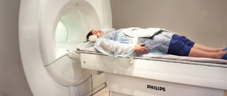
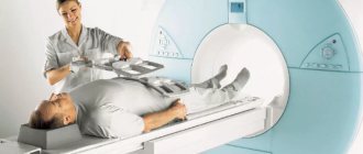
There are 2 ways to perform ultrasound of the stomach: standard transabdominal (through the anterior abdominal wall with an external sensor) and endo-ultrasound (using an external sensor and a microscopic camera that is placed in the stomach cavity). Both types of ultrasound are performed strictly on an empty stomach.
Carrying out endo-ultrasound
During the procedure, the patient is in a supine (on his back) or semi-sitting position.
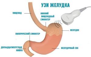
A sensor installed in the epigastric region allows you to obtain images in several projections at once. During the study, the specialist evaluates the location and shape of the organ, the size and thickness of its walls, and identifies deformations and pathological foci.
After the initial examination, the doctor asks the patient to drink about 500 ml of liquid and analyzes peristalsis and the rate of gastric emptying (motor function) in real time. Some specialists use special contrast agents, such as barium, to improve visualization.
The endo-ultrasound method combines gastroscopy and conventional ultrasound. The information obtained from the camera and from the external sensor gives the most complete picture of the condition of not only the stomach, but also nearby organs: the esophagus, duodenum, pancreas, liver, gallbladder and bile ducts.
In addition to assessing the condition of the organs themselves, endo-ultrasound makes it possible to monitor benign and malignant formations and identify other pathological processes occurring inside the stomach and inaccessible for visualization by transabdominal ultrasound.
Comparison of gastric ultrasound and FGDS: which is better? Pros and cons of both procedures

, only endoscopic and radiological methods were used in medicine to diagnose stomach diseases . Ultrasound diagnostics in gastroenterology was used for other purposes - examination of parenchymal organs (liver, kidneys, pancreas) and biliary tract. But, as experience accumulates and science develops, it has been proven that ultrasound can also be used to study the condition of the gastrointestinal tract, in particular the stomach. In this regard, the question arises: what is better: FGDS or ultrasound? How to choose the right method for examining the stomach? We will try to answer these questions in our article, and also consider:
- what is a stomach ultrasound?
- what is FGDS;
- how they are carried out and how to prepare for them;
- what are the indications and contraindications for these diagnostic procedures;
- what are the advantages and disadvantages of both methods;
- Let's briefly look at alternative research methods.
CONTENT
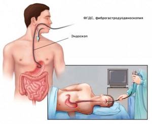
FGDS (fibrogastroduodenoscopy) is an endoscopic examination of the upper gastrointestinal tract. To better understand the meaning of the abbreviation “FGDS”, you should translate it into Russian: fibra – fiber, gastro – stomach, skopeo – watch.
The essence of gastroscopy is of a fibrogastroscope into the digestive tract through the oral cavity . The latter has the form of a tube with a diameter of about 1 cm and is a flexible endoscope equipped with a fiber-optic system. Using this device, you can sequentially examine the esophagus, stomach, and duodenum from the inside.
tissues using ultrasonic waves, which have the ability to reflect from tissue separation boundaries with different acoustic impedance . As a result, an image of an organ (in this case, the stomach) is formed on the monitor screen, the parameters of which are analyzed by a specialist.
Ultrasound of the stomach (ultrasound examination of the stomach) is an instrumental diagnostic method that allows, using special equipment and an ultrasound sensor, to examine the stomach and, if necessary, nearby structures. This method involves the study of biological
Ultrasound of the stomach and FGDS are completely different diagnostic techniques, however, they have common goals:
- assess the condition of the stomach ;
- its functioning;
- identify the pathological process (if any).
Both methods have good resolution. The main difference between them is the main approach to research.
- FGDS allows the doctor to examine the organ from the inside and makes it possible to perform various diagnostic and therapeutic procedures (for example, take tissue for analysis or stop bleeding).
- Ultrasound visualizes the stomach through the anterior abdominal wall without penetrating the body cavities . With its help, the specialist receives a spatial image of the organ, can examine it in different projections and study the entire thickness of its wall.
When prescribing a particular test, the doctor always takes into account: what information it can provide, whether this information is sufficient to make a diagnosis, and how safe it is for the patient. As a result, the most optimal diagnostic procedure is selected for each patient.
Table 1. Advantages and disadvantages of FGDS and ultrasound of the stomach.
| Advantages | Flaws | |
| FGDS |
|
|
| Ultrasound of the stomach |
|
|
Examination of the stomach using instrumental methods is indicated if there is a suspicion of the presence of a pathological process in this organ, which can manifest itself with the following symptoms:
- pain in the upper abdomen, especially associated with eating;
- heaviness and discomfort after eating;
- nausea;
- single or repeated vomiting;
- belching;
- heartburn;
- bloating.
For patients with the above complaints, both ultrasound and FGDS are indicated. As a result, the following pathological conditions can be identified:
- erosive and ulcerative lesions (peptic ulcer) ;
- gastropathy;
- polyps;
- stomach tumors;
- duodeno-gastric reflux;
- stenosis of the gastric outlet, etc.
How to choose which study is best? First of all , it is more advisable to perform an ultrasound , because it is a safe and non-invasive diagnostic method. And then, if necessary, prescribe an FGDS. also performed if the patient refuses FGDS or if contraindications are identified.
Please note Despite the information content of ultrasound, according to the recommendations of the Ministry of Health, this diagnostic procedure is not included in the list of mandatory ones for stomach pathology.
If it is necessary to collect tissue for morphological examination (biopsy of a tumor or long-term non-healing ulcer, confirmation of the diagnosis of gastritis), an FGDS must be performed . This study is also extremely necessary if gastric bleeding or a foreign body in the stomach is suspected.
Gastroscopy is not used in all patients, even if indicated. For some of them, it cannot be used, as it can be harmful to health. Absolute contraindications to FGDS are:
- impaired swallowing due to diseases of the esophagus (burns, cicatricial strictures);
- acute complicated forms of ischemia of the heart muscle and brain (myocardial infarction, stroke);
- severe cardiac and respiratory failure;
- acute infectious processes;
- violation of the integrity of the digestive tract (perforation of the esophagus, stomach);
- a significant increase in the size of the thyroid gland (grade 3 goiter).
Ultrasound of the stomach can be prescribed to all patients , even pregnant women.

The amount of preparation for FGDS may vary among different patients.
Some of them require a diet, enzymes, and sedatives, while others do not require special preparation. Your doctor will tell you about these features. But in any case, the procedure is carried out only on an empty stomach, so the last meal is allowed 12-14 hours before the intended examination.
Ultrasound is also performed only on an empty stomach; preparation for it lasts 2-3 days and includes:
- a diet excluding products that increase gas formation (milk, legumes, cabbage, black bread, grapes, etc.);
- if you are prone to constipation, bowel cleansing (enema);
- for flatulence - sorbents, enzymes, espumizan.
You can read more about preparing for an ultrasound here, and about preparing for an FGDS here.
Please note: Preparation for an ultrasound scan in a child requires special attention. After all, the younger the baby, the more difficult it is for him to withstand the required period of forced fasting.
Ultrasound of the stomach is carried out in 2 stages:
- in natural conditions;
- after administration of contrast.
First, the patient is placed in a horizontal position (lying on his back); as the study progresses, the body position is changed. The sensor is installed in the projection area of the stomach, after lubricating the skin with a special gel, then it is moved in different directions, assessing the image on the monitor screen. In special cases (if indicated), the procedure is repeated after ingestion of a liquid containing contrast. In special cases, the doctor may supplement the ultrasound with stress.
Ultrasound of the stomach
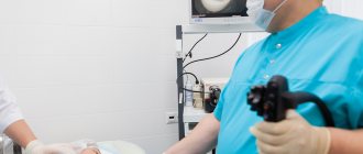
To perform FGDS, the patient is placed lying on his left side. The endoscope is inserted into the oral cavity through a mouthpiece after preliminary anesthesia of the throat with lidocaine. After which the doctor carefully moves the device deeper into the digestive tract, conducts an examination and performs the necessary manipulations. There is also a transnasal method of administering EGD, you can read about it here.
If gastric ultrasound and endoscopy data are not enough to make a diagnosis or for some reason they cannot be performed on the patient, then gastric fluoroscopy with or without contrast can be used as an alternative method.
If ultrasound and FGDS rather complement each other, then x-ray may be an alternative to both of them. In this article, we take a closer look at the advantages and disadvantages of FGDS and x-rays in the diagnosis of gastrointestinal diseases.
FGDS and ultrasound are two wonderful methods that have long served medicine faithfully. How wonderful would it be if they could be combined? And you will be surprised, but such a procedure exists. And it is called endouzi, you can read about it in our article.
Ultrasound of the stomach and FGDS are studies that do not replace, but complement each other. Although the indications for them are not very different, the information obtained from these diagnostic procedures is very important. And if one method fails to identify the cause of poor health, then another comes to the rescue.
Before finishing the article, I would like to address the audience:
- Have you ever had a case where the conclusion of an FGDS was not enough for a specialist to make a correct diagnosis?
- Have you ever had to examine your stomach using an ultrasound? Was this research able to answer all the doctor’s questions and solve the problem?
Share with the community in the comments!
Advertising

Ultrasound of the stomach is an important part of the gastrointestinal tract examination using ultrasound.
For a long time, ultrasound diagnostics was not used in the study of the stomach. This is due to the fact that the stomach is a hollow organ, and the air does not allow full use of a conventional ultrasound sensor - special sensors are needed to examine the back walls. In addition, accumulated gases distort the displayed results. However, medicine does not stand still, and modern techniques already provide sufficient information to make an accurate diagnosis.
Sensors for studying the stomach appeared relatively recently, in the late 2000s. However, the speed and safety of scanning makes ultrasound examination of the stomach increasingly popular.
During an ultrasound examination, the doctor evaluates the organ according to the main indicators:
- Stomach volume.
It is a hollow muscular organ that resembles a pouch. The volume of an empty stomach is 0.5 liters, and when full it stretches to 2.5 liters. The height of the stomach reaches 18-20 cm, width - 7-8 cm. When filled, the stomach stretches up to 26 cm in length and up to 12 cm in width. - Structure.
Near the heart is the cardiac region, in which the esophagus passes into the stomach. On the left you can see the bottom of the organ, where air entering with food accumulates. The body of the stomach is the largest part, rich in glands that produce hydrochloric acid. The pyloric zone is the transition from the stomach to the intestine. There, partial absorption of substances received from food occurs. - Structure.
The walls of the stomach have a muscular layer that is responsible for contracting and promoting the food coma. The serosa is intermediate between the muscular and mucous layers. Lymph nodes and blood vessels accumulate in it. The mucous layer is covered with the finest villi, which secrete gastric juice produced by the glands. - Blood supply.
The circulatory system covers the entire organ. The organ is supplied with venous blood by three main vessels: the left, hepatic and splenic. The venous network runs parallel to the arterial network. Various bleeding occurs when the gastric mucosa is damaged (ulcers, tumors).
How is the preparation going?
Before conducting the analysis, a qualified specialist must explain in detail to the patient the rules for preparing for FGDS. They are as follows:
- 10-12 hours before the start of the procedure you should not eat. For this reason, FGDS is often carried out in the first half of the day;
- smoke right before the test. The fact is that nicotine can dramatically increase the volume of gastric juice secretion.
If the patient suffers from any diseases in acute or chronic form, this must be reported to the attending physician before the study. This also applies to the presence of allergic reactions to medications.
Endoscopy results
The doctor will be able to draw the first conclusions about the condition of the digestive tract organs immediately after a visual examination. Having assessed the condition of the mucous membrane, a specialist doctor immediately diagnoses pathologies:
- cardia failure;
- gastritis;
- stomach and duodenal ulcers;
- enterocolitis, colitis;
- diverticula, polyps, tumor-like formations of the gastrointestinal mucosa.
Based on the results of the endoscopic examination, the gastroenterologist establishes a complete diagnosis and agrees with the patient on a further treatment plan.
What sensations may occur during the procedure?
Many patients are afraid of FGDS, considering this type of research extremely unpleasant. Progress in the field of medical equipment has reduced the level of human discomfort during analysis to almost zero.
A modern endoscope has a minimal size and therefore creates a minimum of unpleasant sensations for a person.
The medical procedure itself using an endoscope takes an average of 6 minutes. However, if any serious problems are detected, it can be increased to a quarter of an hour.
In most cases, the person does not feel any serious pain or particular discomfort. However, many patients state that they experienced unpleasant sensations during FGDS.
The following consequences of FGDS are considered the norm:
- belching;
- nausea;
- impulses to cough and sneeze;
- the appearance of tears;
- nasal discharge.
All these phenomena are considered completely normal, and in some patients they do not occur at all.
Is it possible to perform the procedure for examining the stomach or intestines under anesthesia? In fact, FGDS can be performed under both local and general anesthesia. This greatly simplifies the manipulation itself, since the patient does not feel any pain, which often complicates the study and even distorts its results.
In most cases, anesthesia is prescribed by a specialist when, in addition to the examination itself, long-term medical procedures are planned.
It is worth noting that the use of anesthesia is possible only if the patient does not suffer from allergic reactions to the components of the drug. In this case, anesthesia must be administered to the patient exclusively by a qualified anesthesiologist. And if a person has even the slightest contraindications, it is not worth performing FGDS under anesthesia.
+