Salivary glands are glands in the oral cavity that are responsible for the secretion of saliva. This is a transparent, colorless liquid (liquid biological environment of the body), which moistens the oral cavity, helps a person perceive taste, and promotes the process of swallowing. Moreover, saliva has a bactericidal effect and protects teeth from multiple damage from pathogens. Ultrasound is used to assess the functionality of the salivary glands or a comprehensive examination of the oral cavity. For what diseases is ultrasound diagnostics prescribed, how exactly does the procedure take place and how safe is it for the human body?
Briefly about the salivary glands
The salivary glands are divided into 2 groups - large (submandibular, parotid, sublingual) and small (lingual, labial, buccal, molar, palatine).
The glands are connected to the oral cavity by ducts through which liquid enters the mouth to participate in digestion. About 2 liters of saliva are secreted per day, which moisturizes the mucous membrane and breaks down complex carbohydrates (therefore, it is important to chew food thoroughly before swallowing). It also has a barrier function and protects the oral cavity from microorganisms.
The largest percentage of pathologies are inflammatory diseases, and the most vulnerable are the paired parotid organs.
Ultrasound of the submandibular salivary gland
The norm for an ultrasound examination of the salivary submandibular gland corresponds to the following parameters:
- Fine-grained homogeneous structure.
- The edges of the glands are smooth.
- The excretory duct is well visualized, small in size, without signs of blockage.
Determining the normal size of the submandibular glands is not particularly difficult precisely because of its clear contours. During inflammation (sialoadenitis), the organ increases in size and its edges become blurred.
Symptoms for diseases of the salivary glands
If the symptoms listed below appear, you can immediately do an ultrasound of the salivary glands and contact a specialist with the results. Symptoms are specific and most likely indicate problems:
- feeling of pressure in areas where organs are located;
- pain and swelling in the mouth and throat;
- irradiation of pain to the occipital and/or temporal part of the head;
- increase in size;
- dysphagia;
- local increase in body temperature at the location;
- decreased amount of saliva production (dry mouth).
How is the procedure done?
Ultrasound of the salivary glands is non-invasive and does not cause pain or discomfort to the patient. The procedure does not harm health and is not associated with receiving even a minimal dose of ionizing radiation. Therefore, ultrasound of the salivary glands is considered a harmless and safe procedure.
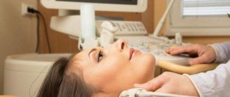
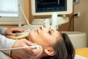
During the examination, the specialist attaches sensors to the outside of the oral cavity - their specific location depends on the gland being examined. After this, the patient takes a horizontal position. During an ultrasound of the salivary glands, the patient needs to put his head on the pillow, throw it back and turn it to the right or left.
The movement of the transducer during the examination depends on the area being examined. During ultrasound of the submandibular or sublingual salivary gland, the sensor is placed in the oral cavity; during ultrasound of the parotid salivary gland, the sensor is moved in the area of the ear.
How the research is carried out
There is no preparation for the study, other than hygiene. The last meal is planned 4 hours before the test, after which you should thoroughly brush your teeth and rinse your mouth. The examination takes 10-20 minutes - it all depends on the individual characteristics of the anatomical structure. The parotid glands are examined externally, ultrasound of the sublingual and submandibular glands is performed from the inside of the oral cavity, or from the chin with a transverse position of the sensor.
The procedure is carried out as follows:
- the patient is positioned on the couch lying down, the head is tilted back, a wedge-shaped support or pillow is placed under the head;
- A universal gel for ultrasound examination is applied externally to the skin, the specialist evaluates the parotid organs on both sides, in the longitudinal and transverse plane;
- the sensor is placed in the oral cavity to examine the mandibular glands;
- the sensor is lowered to the floor of the mouth to examine the sublingual glands on the left and right.
The basic principles of ultrasound of the submandibular and other large glands are sequential examination of the neck and face areas symmetrically from the center and polypositional scanning of the area that is of interest to the researcher. With normal indicators, homogeneous echogenicity is observed without very light and dark spots on the images, the glands themselves have a flattened round shape, the contours are clear and even.
What do you need to know about ultrasound diagnostics?

Ultrasound is sound vibrations whose frequency exceeds 20 kHz. It should be noted that the human ear is capable of perceiving sound with a frequency of 16 to 20 kHz. Ultrasound is outside the zone of human acoustic perception, but bats, dolphins and, for example, whales communicate freely with its help.
One of the properties of high-frequency sound vibrations is free propagation in the soft tissues of living organisms. Once the wave penetrates the skin, it encounters a huge number of barriers. These can be various fluids, bones, soft tissues and other heterogeneous internal structures. All of them block the path of ultrasound and prevent it from spreading further. This process is called acoustic impedance. The intensity of acoustic impedance differs depending on the density/elasticity of the barrier. As soon as a sound wave reaches, for example, soft tissue, one part of it is absorbed by the medium, and the second is reflected from it. This point is key for ultrasound examination.
What is the principle of diagnosis? A special liquid gel is applied to the patient's skin. It acts as a transition medium and allows ultrasound to penetrate inside our body. Without liquid gel, penetration is impossible, since human skin reflects close to 100% of sound vibrations. The sensor is applied to the prepared area. The sensor creates ultrasonic waves, records their propagation and transmits information to the computer. The system evaluates the intensity of sound vibrations and generates a three-dimensional image of the internal cavity. All this happens so quickly that the doctor and patient can observe what is happening in real time. The picture is displayed on the screen immediately after the sensor is placed against the skin.
To create the final image, 64 different shades of black and white are used. The intensity of white or black shades depends on the strength of the ultrasonic wave. The minimum signal is recorded in black (most often these are bones and compacted areas of the body), and the maximum is recorded in white (liquids, soft tissues, air).
Possible deviations in the functioning of the organ
The occurrence of pathologies in the glands, as a rule, is facilitated by various infectious diseases. They, among other things, can provoke cell proliferation, which is expressed in benign (polymorphic adenoma) and malignant (carcinoma, sarcoma) tumors. The formation of a benign tumor does not have pronounced symptoms, so it is detected in late stages, while malignant tumors have certain symptoms.
Symptoms of malignant tumors:
- ulcers in places of occurrence;
- pain in the glands;
- damage to the facial nerve.
Of all tumors, the majority are benign, and the minority are malignant. Metastases from other organs penetrate these tissues quite rarely. Ultrasound of the parotid glands and other small and large glands helps identify the tumor, but cannot reliably determine its quality. If neoplasms are detected, the use of Doppler ultrasound examination of the vessels of the neck is recommended.
The main difficulty in diagnosis is that the nature of the symptoms resembles colds or inflammation of the lymph nodes - patients do not consult a doctor in a timely manner.
General characteristics of the study area
Salivary glands are glands that are located in the oral cavity and are responsible for the secretion of saliva. In the human body there are multiple small glands (localized on the mucous membrane of the palate, cheeks, tongue, lips) and 3 pairs of large salivary glands - sublingual, submandibular, parotid. Small glands are also classified according to the nature of the secretion - serous, mucous and mixed. Most of the serous secretory glands are located on the tongue, and their saliva is rich in protein. The palatine and part of the lingual glands are called mucous because their saliva is rich in mucus. The mixed category includes the molar, buccal, labial, and part of the lingual glands, the secretion of which contains saliva mixed in composition.
The parotid secretory glands produce thin saliva that is high in sodium/potassium salt and amylase (a digestive enzyme). The acidity of the liquid is 5.81 pH, which is the highest among other glands. During the day, these cells produce from 0.2 to 0.7 liters of fluid - this is a third of the total production of saliva. The submandibular glands produce serous and mucous secretions. Its acidity is relatively low and amounts to 6.39 pH. The sublingual salivary glands are responsible for the production of protein secretions. It is rich in mucin, has a pronounced alkaline reaction and high phosphatase activity.
Content:
- General characteristics of the study area
- What do you need to know about ultrasound diagnostics?
- Indications for ultrasound diagnostics
- Ultrasound technique
The functionality of the glands includes the secretion of hormone-like substances, protein/mucosal components of saliva and the filtration of its composition, the release of metabolic end products.
What diseases are the salivary glands susceptible to?
In most cases, the salivary glands are susceptible to inflammatory pathological processes. One of them is sialadenitis. This is an inflammation of the secretory glands, which is fraught with swelling of the gland (most often the submandibular) and the formation of stones in the salivary ducts. The patient feels dry mouth, swelling of the oral cavity, pain, decreased secretion of saliva, and possible discharge of pus. Another common problem is mumps. This is an inflammation of the parotid gland, which makes itself felt by sharp pain, severe swelling of the cheeks and entire face, loss of appetite, apathy, headache and increased body temperature.
The proliferation of salivary gland cells can lead to the development of neoplasms of various types. Among malignant tumors, doctors most often diagnose sarcoma/carcinoma/mucoepidermoid tumor. The most common form of benign tumor is polymorphic adenoma. The main problem is that the tumor develops without pronounced symptoms. Pathology is most often diagnosed in the later stages, when precious time is lost, and therapy will require much more time, effort, material and emotional resources.
If you notice even minor deviations (edema/swelling/pain/discomfort), consult your doctor immediately. The doctor will assess the nature and nature of the symptoms and prescribe a therapeutic or preventive course. Do not ignore your body's signals and always tell your doctor about suspicious changes.
How is diagnostics made based on the results?
The result of the research is a printout of an image of the gland, or a CD with images recorded on it. The doctor evaluates the various characteristics of the glands one by one and takes measurements. The first characteristic is the size (reduced, enlarged), the second contours are clear and even or blurred, torn, deformed.
What does an ultrasound show:
- echogenicity (the ability of tissues to transmit ultrasonic waves);
- position;
- structure;
- parenchyma condition;
- type of pathological changes, if any (ectasia, strictures, stones, foreign bodies).
When neoplasms are detected, the ultrasound doctor describes them in detail to facilitate the work of the attending physician.
Differential diagnosis of inflammation, cysts, and formations of vascular origin is complicated by the lack of specificity of the clinical picture of diseases characteristic of tissues in the mouth. The clinical manifestations of different processes are very similar, this requires the ultrasound doctor to have extensive experience in examining the organ.
Decoding the result
The interpretation of the ultrasound of the salivary glands is carried out by a specialist who compares many parameters, deviations in which, alone or in combination with other factors, can serve as the basis for making a diagnosis. As a rule, the specialist pays attention to the size of the glands, their contours and position, and the condition of the lymph nodes. In addition, echogenicity (the ability of tissues to reflect sound) is important on ultrasound of the ear glands. The rate of echogenicity varies from organ to organ. However, the indicator is extremely important for making a diagnosis, since reduced echogenicity of the ear glands detected by ultrasound may indicate swelling or inflammation, and an increased indicator may be a sign of a neoplasm.
Sign up for an ultrasound
The information content of the study depends on the quality of the equipment and the professionalism of the ultrasound doctor. On mid-range devices, some structures may not be visualized, for example, tubular duct structures in which problems may exist.
When choosing where to have the procedure done, always find out information about the doctors and the brand of equipment. All branches of the NEARMEDIC clinic network are equipped with high-quality ultrasound machines, and information about doctors and their professional biographies can be found on the website.
Ultrasound of the sublingual salivary gland
Normally, the sublingual salivary gland has smooth, not very clear contours. They have a homogeneous structure, but echogenicity (the ability to reflect ultrasound) may be slightly increased. In the absence of any diseases, the device will not show the ducts of this organ.
The shape of the sublingual salivary glands displayed on the device screen changes depending on where the sensor is placed. When located in the chin area, the glands have oval contours. If the device is applied parallel to the body of the lower jaw, the glands will be slightly elongated.
Which doctor gives a referral for an ultrasound?
You can get a referral for an ultrasound from a pediatrician (if the child has a problem), a dentist, or a therapist. This manipulation is provided in those dentists where the appropriate equipment is available.
Standard sizes of salivary glands determined by ultrasound
During the examination, the doctor determines the dimensions of a given organ (length, width and thickness) using ultrasound and compares them with acceptable values.
Standard indicators of the salivary glands look like this:
- Sublingual gland: width – 10-16 mm, length – 18-22 mm.
- Parotid gland: width 30-40 mm, length 40-50 mm.
- Submandibular gland: width 11-18 mm, length 30-40 mm.
Standard indicators of parotid SF according to ultrasound parameters
A healthy parotid gland has the following criteria:
- the glands should be presented in the form of symmetrically placed capsules;
- During an ultrasound, it is possible to see only the closest surface of the SG; distant (deep layers) do not provide a clear picture;
- the back wall of the capsules has vague contours;
- The capsule structure is fine-grained and homogeneous.
When examining the parotid sebaceous glands in a healthy state, they are not detected. But if a pathological process is observed in them, then you will be able to see them. SF are visible when they are compressed by a cyst or when there is blockage from a calculus.
Standard indicators of submandibular SF according to ultrasound parameters
In a healthy state, the submandibular gland is characterized by the following parameters:
- the structure of the gland is fine-grained;
- The small excretory duct is clearly visible, there are no blockages;
- the glands have smooth edges.
Since viewing the gland through ultrasound gives a clear picture, it is quite easy to establish the normal state of the submandibular glands by their outline. So, in the presence of an inflammatory process (sialoadenitis), the size of the organ increases significantly, and the edges become blurred.
Normative indicators of the sublingual gland according to ultrasound parameters
In a healthy state, the sublingual gland, according to ultrasound results, shows smooth, but slightly blurred contours. The echogenicity parameter (the ability to repel ultrasound) is slightly too high, the structure of the glands is homogeneous. In a normal state, in the absence of disease, ducts are not detected in the ultrasound picture.
If we talk about the shape of the SG, then on the monitor of the device it will be different, depending on the place where they are viewed. For example, if the sensor is pressed against the body in the chin area, the oval shape of the glands will be recorded. If the device is pressed against the lower jaw (direction along the body), then the shape of the gland will have an elongated appearance.
Methodological rules for performing ultrasound: protocol
A protocol is usually called a document in which all the data obtained from scanning the salivary glands is entered.
For example, the parotid SF is examined according to the following scheme:
1. First, a transverse image is studied; it is obtained when the sensor is moved, starting from the angle of the jaw and ending with the tragus;
2. Then the longitudinal picture is studied. It is obtained by moving the sensor along the cervical area.
This is followed by a bilateral examination of the organ. That is, the condition of the gland symmetrical to it is studied. The need to examine the opposite organ makes it possible to compare them and determine the extent of damage by the disease.
The submandibular gland is examined according to the following plan:
- A cross-sectional image is examined. The midline of the neck area enters this incision, starting from the hyoid bone and moving towards the lower jaw along a horizontal line. Bilateral examination is used.
- A detailed examination of the diseased organ is performed, the device is installed slightly to the side along the line of the lower jaw.
The plan for studying the sublingual gland is similar to the protocol for studying the submandibular gland. The device moves along the center line of the cervical area, located under the lower jaw. This place allows you to clearly see both symmetrical organs.
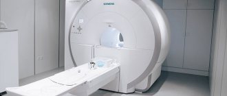
Ultrasound of the salivary glands is a fairly informative method for diagnosing various diseases of these organs. Thus, diagnostics makes it possible to detect the disease at an early stage and prevent the development of the pathological process. But, despite this, doctors do not always limit themselves to ultrasound examination, additionally turning to such techniques as:
- X-ray;
- CT scan;
- Magnetic resonance imaging.
In cases where the doctor suspects the presence of neoplastic processes, the patient is prescribed a diagnostic puncture to carry out a cytological study of the material. It should be noted that ultrasound cannot replace histological examination and biopsy, which can determine the degree of malignancy. Therefore, in certain cases, a comprehensive diagnosis is required to obtain a detailed picture of the disease.
How is preparation for an ultrasound done?
The study of the salivary glands does not involve any preparatory steps. The procedure itself is safe and painless. Patients, regardless of age category, tolerate the manipulations well. At all stages, from beginning to end, patients do not show any anxiety. Premedication (that is, taking sedatives) is not required, even for small patients.
Before conducting the study, doctors advise not to eat for 4 hours, and before the procedure itself, thoroughly rinse the mouth to remove any leftover food.
Decoding and diagnosis
The decoding and interpretation of the results should be carried out by the doctor who referred for the study. Normally, the salivary glands have a homogeneous structure, are surrounded by a capsule, and the ducts are freely visualized. Each gland, from a large group of large salivary glands, has its own standard sizes:
- sublingual gland 17-21 mm long and 10-15 mm wide;
- the submandibular gland can normally be up to 40 mm long and up to 18 mm wide;
- parotid salivary gland up to 45 mm in length and up to 40 mm in width.
The ultrasound diagnostician enters the ultrasound results into the examination protocol. The decision on further treatment tactics is made by the attending physician, taking into account the medical history and other results.
Back to list of articles
Treatment of pathologies of the salivary glands
If the corresponding symptoms appear, it is very important to do an ultrasound in a timely manner. The fact is that if diseases of the salivary glands are neglected, they can become chronic. This is fraught not only with regular exacerbations, but also with the resistance of pathologies to drug treatment. In such cases, surgery may be required.
But if measures are taken in a timely manner, conservative treatment will be sufficient. In many cases, salivary gland diseases in the early stages can be treated on an outpatient basis.
Preparation for the procedure and progress of the study
Before ultrasound of the salivary glands, the patient must carefully clean the oral cavity. You should not eat food 3-4 hours before the procedure. As a rule, it is enough to clean the dentition and the oral cavity as a whole by brushing your teeth using a toothbrush and special floss.
How is an ultrasound of the salivary glands done? During the diagnosis, the patient lies on the couch, the diagnostician applies a special gel to the outer surface of the cheek, which improves the conductivity of the ultrasound signal. Then the inner surface of the cheek is examined. So, the specialist analyzes the data on the monitor and draws a conclusion about the structure of the parotid, sublingual, and submandibular glands. The procedure does not bring any discomfort.
You can sign up for an ultrasound scan of the salivary glands in Moscow at the Alpha Health Center medical clinic using the phone numbers listed on the website. Our consultants will be happy to answer all your questions; together with a staff member, you will choose a convenient day and time for the study.