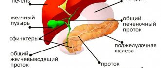
The liver is the largest gland responsible for digestion. The liver produces bile and healthy cholesterol, where proteins are broken down into amino acids, excess glucose is converted into glycogen, fat metabolism occurs, and various hormones are synthesized. It filters the blood and protects the body from toxins.
The liver tissue, parenchyma, consists of lobules that resemble a honeycomb in appearance. Each lobule is penetrated by blood vessels and ducts, it is separated from neighboring connective tissue.
Liver cells have the ability to renew, but when exposed to toxins, the liver cannot cope with the load, and changes occur in it. Alcohol, an abundance of fatty foods, toxic substances, and viruses have a particularly negative effect on liver function.
Liver cells (hepatocytes) die or change their structure, leading to disruption of the organ. Today there are about 50 liver diseases; in Russia, 40% of pathologies are caused by excessive alcohol consumption.
According to WHO, 30% of the adult population of the planet suffers from liver diseases, of which 2.2% are liver cancer caused by hepatitis B or C. Every 3rd inhabitant of the planet is infected with the virus; in the CIS countries hepatitis C is the leader, which mainly affects the young population aged 15 to 29 years, and in 3.6% of cases it is diagnosed in children under 15 years of age.
In addition to viral liver damage, every 5th resident of Russia has stones in the bile ducts and liver.
Not everyone pays due attention to the condition of the liver, attributing it to the organ’s ability to heal itself. However, this only applies to a healthy liver that is not exposed to adverse factors. It really copes with removing toxins from food and water, but sometimes failures occur, and the liver undergoes changes that lead to the development of the disease.
What liver diseases are there?
The content of the article
Depending on the provoking factor, liver diseases are divided into 4 groups:
Caused by exposure to a foreign microorganism (viral, bacterial and parasitic).
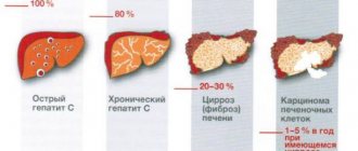
Hepatitis is the most common viral liver disease. It has 6 varieties, each of which is characteristic of a specific region of the planet.
- Hepatitis A (Botkin's disease) occurs in 28% of cases. It is transmitted through unwashed hands, drinking or bathing in contaminated waters, and through blood. Most common in Latin America, Africa and Southeast Asia.
- Hepatitis B affects 18% of the total number of infected people, and this is the most common type of hepatitis in Russia. It is transmitted through blood, as well as during childbirth from mother to child.
- Hepatitis C is transmitted during unprotected sexual contact or during childbirth. It has the most severe consequences for the liver.
The other three varieties of the virus are concomitant with the three main ones. The most dangerous are hepatitis B and hepatitis C, they lead to liver destruction and death.
Caused by metabolic disorders:
Fatty hepatosis involves the death of functional liver cells of hepatocytes due to their damage by fat cells. This occurs due to metabolic disorders when dietary fat accumulates in the liver, causing inflammation and cell necrosis. As a result, they are replaced by connective tissue, disrupting the normal functioning of the organ.
Fatty hepatosis has a different nature: in people suffering from alcohol addiction, fat cells accumulate as a result of the death of liver cells under the influence of aldehydes.
Cholestatic hepatosis occurs as a result of excess “bad” cholesterol. Non-alcoholic hepatosis occurs in 65% of people who are overweight. Also, 1% of women expecting a child develop hepatosis during pregnancy.
Caused by an inflammatory process in the liver:
Liver cirrhosis is an inflammation of the organ in which the parenchyma (spongy tissue) is replaced by stroma (connective tissue). In 70% of cases, cirrhosis is a consequence of alcohol abuse, the rest is the result of damage by viruses, parasites or infections. Cirrhosis also occurs due to severe intoxication, for example, when consuming poisonous mushrooms. However, in 10% of cases it is impossible to determine the exact cause of liver cirrhosis.
Clinical interpretation of liver ultrasound results
Ultrasound scanner HS50
Affordable efficiency.
A versatile ultrasound scanner with compact design and innovative capabilities.
Introduction
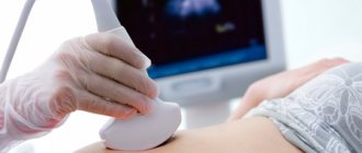
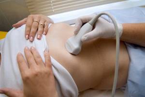
Ultrasound examinations (ultrasound) are often the first in a series of diagnostic searches for suspected liver pathology, determining the choice of other techniques. Improvements in ultrasound diagnostic technologies have led to the fact that in some cases it has become possible to abandon other radiological examinations. But this also increases the responsibility of the specialist and requires a clinical interpretation of the results obtained, without reducing the description to the recording of “echo-positive” and “echo-negative” formations. The main share of ultrasound in outpatient settings is performed on the abdominal organs. And the liver is the first thing that a specialist tries to visualize in this case. Liver diseases can be suspected based on complaints, clinical picture, laboratory data, but they are often detected by chance. The latter applies, first of all, to children, in whom the proportion of congenital pathologies is higher than in adults. The reserve capacity of the liver is incredible, so the doctor’s task is to identify pathology before it becomes clinically significant.
General provisions
Embryogenesis of the liver and its vessels, anatomy. In early ontogenesis, the liver, gallbladder and bile duct arise as a single outgrowth (hepatic diverticulum) in the caudal part of the anterior duct. This formation grows ventrally into the septum transversum, which is the mesoderm between the developing heart and the midgut. The liver bud grows very quickly, occupying most of the abdominal cavity in the first 10 weeks. During this period, the sizes of the right and left lobes of the liver are the same, but due to oxygenated blood from the hepatic vein, the right lobe quickly overtakes the left in size and weight. From the 6th week, the liver is a hematopoietic organ, and from the 12th week it synthesizes bile. A small caudal process on the hepatic diverticulum gives rise to the gallbladder, and its stalk gives rise to the cystic duct. From the cord running from the hepatic and cystic ducts to the duodenum, the common bile duct is formed. During the embryonic period, blood enters the liver through the umbilical vein, which passes along the free edge of the ligamentum falciforum into the left branch of the portal vein. This part v. portae connects to the hepatic vein through the ductus venosus, then blood enters the right atrium. Thus, the ductus venosus is a large venous shunt that bypasses the liver, thanks to which the bulk of the blood from the placenta flows directly to the heart. This is the only route through which the catheter can be passed. If the catheter, when probing the umbilical vein, enters the right branch of the portal vein, thrombosis, obliteration and portal hypertension develop. After the birth of the child, the umbilical vein is obliterated and turns into the round ligament of the liver (ligamentum teres). Ductus venosus transforms into ligamentum venosum. In the first hours after birth, these structures can be visualized as canalicular. Later, the ligamentum teres is visible as a dense structure extending from the left side of the portal vein. If intrahepatic pressure increases (in cirrhosis), the round ligament of the liver is recanalized. Portal blood begins to flow from the liver to the navel, resulting in varicose veins (caput medusae). Anomalies of the inferior vena cava are possible, but they are extremely rare. Among these rare defects, the most common is the left-located inferior vena cava, which independently flows into the right atrium through the coronary sinus. Possible underdevelopment of the hepatic segment of the inferior vena cava with blood drainage through the vv system. azygos et hemiazygos. The hepatic vein drains independently into the right atrium. It is possible for the inferior vena cava to double, with the left vein being significantly smaller in diameter than the right. Knowledge of these features is especially important in pediatric practice, since a significant part of pediatric pathology is congenital.
Location features
(The author expresses gratitude to Medison for the equipment provided. Figures 2-4 are also provided by this company)
.
When starting your workday, check the condition of your equipment. Make sure the sensor set allows you to complete all studies. Ensure that the referral outlines the indications for the study, formulates the purpose and objectives, and presents the main clinical, laboratory and instrumental results. In a conversation with parents and the child (if he can formulate his problems), clarify all unclear points. Look at the patient as a doctor, and not as a performer with a device. Critically evaluate previous data and formulate your testimony. Have a clear work plan: the period during which the child is calm and allows the procedures to be carried out is short.
For ultrasound of the liver, spleen, and upper abdomen, a convex sensor is most suitable. The viewing angle is large, and the near-field deformation is minimal. The linear sensor is suitable for visualizing small structures. Doppler sonography is necessary if vascular anomalies or hemangiomas are suspected.
Scanning is best done with the child in the supine position with maximum inhalation or exhalation (depending on the child’s characteristics). When inhaling, the lungs push the liver, gall bladder and spleen below the costal arch, displace the colon, increasing the acoustic window. In older children, examination through the intercostal spaces or in a lateral projection is sometimes required. Intercostal access is optimal when the liver is located deep in the hypochondrium. To avoid false conclusions with non-standard approaches, the researcher must remember the changing topography of the organ, violations of the usual relationship with it of the kidneys, inferior vena cava, and intestines. If during the examination of the liver doubts arise about the condition of the gallbladder or biliary tract, and the child is not sufficiently prepared, it is better to complete the examination and repeat it after preparation. The most important advantage of ultrasound over other non-invasive imaging methods (radiography, magnetic resonance imaging - MRI or computed tomography - CT, retrograde cholangiopancreatography - RP) is the ability to repeat the study as often as dictated by the clinic.
Sonographic picture of a normal liver.
Normally, a child's liver is homogeneous. The nature of signal processing is chosen depending on the purpose of the study (Fig. 1a, 1b). The liver capsule is thin and echogenic. The echogenicity of the liver parenchyma is higher than that of the renal cortex parenchyma. The medullary tissue of the kidneys is darker than the liver parenchyma. The ventral edge of the liver is thin and sharp. As the size of the liver increases, it becomes rounded. A frequent indication for ultrasound is a suspicion of an increase in liver size. There are many formulas for calculating liver size, including volumetric ones. The latter are used mainly for scientific purposes or in specialized clinics. The size and configuration of the liver are quite variable. With routine studies, liver size can be assessed qualitatively. The right lobe of the liver extends to the lower pole of the kidney, but if there is a Riedel lobe (a normal variant), then it is slightly lower. In children 1 year of age, the left lobe extends beyond the midline during transverse scanning; in older children, it extends to the aorta. With hepatomegaly, the left lobe extends all the way to the spleen, moving it down. Displacement of the spleen may cause an erroneous conclusion about splenomegaly. Thus, the liver consists of right and left lobes. In addition, there is a caudate lobe. The liver lobes are divided into segments. Simplified - the right lobe has anterior and posterior segments (the border is the right hepatic vein). The left lobe consists of medial and lateral segments (the border is the left hepatic vein). Each segment has its own blood supply. Therefore, with Budd-Chiari syndrome (obstruction of the hepatic veins), the segments increase in size and their echogenicity increases. Knowledge of liver segments is also important for accurate localization of space-occupying lesions.
Rice. 1.
Echogram of the liver in normal mode and in MRI reconstruction mode.
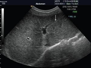
A)
Echogram of the liver of a 12-year-old teenager in normal mode. Note that even at 12 years of age, the round ligament of the liver (arrow), which arises from the left side of the portal vein, has a canalicular appearance (lumen inside).
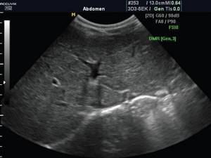
b)
Echogram of the liver of the same teenager in MRI mode. Multiple small echoes, most likely originating from connective tissue, were removed. The parenchyma and vessels themselves are clearly visualized.
Hepatic veins
carry blood from the liver to the inferior vena cava and right atrium. Three-phase Doppler curve. In diastole, the wave is directed towards the liver; in systole, two short, low waves have the opposite direction. Factors determining wave amplitude:
- increased liver density reduces amplitude;
- normal fluctuation changes with heart failure, expansion of the inferior vena cava;
- extrahepatic formations and ascites, compressing the liver and veins, change the fluctuations of the Doppler curve.
Portal vein
brings blood from the intestines and spleen. At the porta hepatis it divides into right and left branches. The total diameter of the vein varies depending on age, body position (standing or lying down), and depth of breathing. For practical purposes, we can assume that in children under 10 years of age the diameter is v. portae is about 8 mm, after 10 years - 10 mm (see table). The walls of their intrahepatic sections are highly echogenic and stand out well against the background of the parenchyma. Wall density is used as a standard for assessing the density of liver parenchyma. In chronic liver pathology, its density approaches the density of the walls of the intrahepatic branches of the portal vein. The Doppler wave is monophasic, fluctuations are determined by respiration and cardiac output. Among the anomalies of the portal vein, its agenesis, duplication and preduodenal portal vein are known. The latter is often combined with the reverse arrangement of internal organs, annular pancreas, duodenal stenosis or other anomalies.
table 2
. The diameter of the portal vein in children of different age groups.
| Age, years | 1 | 3 | 5 | 7 | 9 | 11 | 13 | 15 |
| Median, mm | 3,3 | 4,5 | 5,8 | 7,7 | 8,0 | 8,2 | 8,7 | 8,9 |
| Oscillation limits, mm | 2,9-5,7 | 3,5-6,9 | 4,3-7,6 | 4,5-8,5 | 4,9-9,4 | 5,1-10,0 | 5,6-10,1 | 5,7-10,6 |
Hepatic artery
carries oxygenated blood from the aorta. The pulsation of the Doppler curve corresponds to the cardiac cycle.
Private problems
Liver changes in newborns and children of the 1st year of life
are similar to its changes in older children, but their spectrum is somewhat different. Hepatomegaly is the most common reason for ultrasound examinations in newborns.
Causes and echographic picture.
- Metabolic liver diseases
(glycogenosis, etc.). The liver is enlarged in size and its echogenicity is increased. - Congestive heart failure.
The echogenicity of the liver is increased. The veins of the liver and the inferior vena cava are dilated. - Congenital hepatitis.
By the time of birth, the liver usually has increased echogenicity. - Hemangiomas and hemangioendotheliomas
are the most common local formations in the liver in children 1 year of age; in 80% of cases they manifest themselves in the first 6 months of life. Formations can be single, multiple or diffuse. Multiple small hemangiomas are hypoechoic; solitary hemangiomas are usually hyperechoic, as in adults. At the same time, lesions can be detected in the spleen. Calcification is observed in 13-17% of cases. Cavernous hemangiomas may appear as rough, irregularly shaped cystic lesions. A thorough Doppler analysis is required to identify feeding and draining vessels and arteriovenous shunts. - Neuroblastoma
(4S neurobastoma). It accounts for up to 50% of all cases of neonatal tumors. Median age at diagnosis was 22 months. Up to 80% of all cases are detected during the period of distant metastases. Echographically, the enlarged liver looks like it is infiltrated with metastases, and the primary focus in the adrenal glands is detected in only 40-50% of cases.
Diffuse liver changes in children
can be caused by both systemic and local pathological processes.
The echographic picture in most cases is not specific, combined with hepatomegaly and often jaundice. Diffuse changes in the liver usually occur in combination with hepatomegaly. The exception is cirrhosis of the liver, which in advanced stages leads to a decrease in liver size. In addition, diffuse changes in the liver are usually combined with its hyperechogenicity. Exceptions include acute liver edema and acute hepatitis
. Against the background of the hypoechoic liver parenchyma, the walls of the veins of the portal system are especially clearly visible.
In case of diffuse changes in the liver, it is necessary to determine its size, the nature of the surface (smooth, bumpy) and edges (sharp, rounded), the condition of the spleen, pancreas, kidneys, liver vessels, and lymph nodes.
Fatty liver
- the result of fat accumulation in hepatocytes. The echogenicity of the parenchyma increases, the pattern of the veins of the portal system is erased. The contrast between the liver parenchyma and the kidney is sharp. The severity of the changes depends on the degree of fatty infiltration. Fatty infiltration is observed as a result of chemotherapy and steroid therapy, with malnutrition, obesity, cystic fibrosis, tyrosinemia, Wilson-Konovalov disease, glycogenosis, etc. Fatty infiltration can also be focal. The focus of fatty infiltration can be of any size and shape, which simulates a tumor.
Cirrhosis
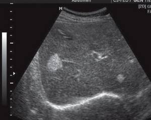
- not an independent disease, but the outcome of many liver diseases, against which the development of hepatocellular carcinoma is possible. In an acute process, fatty infiltration is first recorded. Later, granularity of the parenchyma appears. The process can be micronodular, in which the entire liver parenchyma changes, and macronodular, in which nodes with a diameter of 1 cm or more are visible in the altered parenchyma. The parenchyma is heterogeneous due to hypoechoic foci of regeneration. Fibrosis forms as strands of connective tissue, so the attenuation of the beam is less than with fatty infiltration. The liver decreases in size, and the left and caudate lobes increase compensatoryly. With the macronodular type of cirrhosis, the edge of the liver becomes uneven. Increased density of the liver leads to its rigidity and impaired blood flow. The usual three-phase wave along the hepatic veins can turn into fluctuating or monophasic. As an additional sign of cirrhosis, ascites is indicated (Fig. 2).
Cystic fibrosis
is transmitted in an autosomal recessive manner and is registered among Caucasians with a frequency of 1:3000-1:2500 live births. The cause of the disease is a disruption in the transport of ions from the epithelial cells of the exocrine glands with the formation of thick secretions, obstruction of hollow organs, infection and the development of sclerosis. Liver damage is characterized by macrofocal fibrosis.
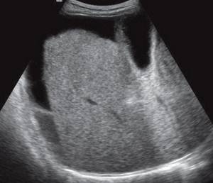
Rice. 2.
Echogram of the liver with cirrhosis.
Portal hypertension
- inability of the portal system to freely transport blood to or from the liver. The resulting excess pressure leads to an enlargement of the spleen and the appearance of varicose veins of the portal system and the opening of collaterals. The basis of diagnosis is the identification of splenomegaly, varicose veins, and collaterals. There are three types of portal hypertension:
- subhepatic - the most common variant in childhood. May be idiopathic, the result of catheterization of the umbilical vein, ascending infection (omphalitis; appendicitis with phlebitis of the superior mesenteric artery flowing into v. portae, and thrombosis of the latter);
- intrahepatic. Cirrhosis with obstruction of intrahepatic branches v. portae;
- suprahepatic. Veno-occlusive disease is a partial or complete occlusion of the small hepatic veins, usually reserve for the larger veins, with blockage of blood flow through the liver. In children, it usually occurs after hematopoietic stem cell (bone marrow) transplantation, radiotherapy or chemotherapy. The term “Budd-Chiari syndrome” is often used as a synonym, although this is not entirely true. The term “Budd-Chiari syndrome” is commonly understood as a blockade of blood flow through the hepatic veins during hypercoagulation. Therefore, the term veno-occlusive disease is more universal. Vascular occlusion usually begins with intrahepatic venules. The veins flowing into the inferior vena cava may still be preserved for some time, which can cause a false negative diagnosis. Sonographic diagnosis is difficult. Ascites, thickened gallbladder walls, and atypical blood flow through the portal vein are detected. In severe veno-occlusive disease, the hepatic veins have a small diameter and are poorly visualized.
The echographic picture depends on the level of obstruction. With subhepatic portal hypertension v. portae is practically not visualized, but its cavernous transformation (portal cavernoma) is clearly visible. Cavernous transformation - many small collaterals bypassing the thrombosed v. portae. It develops in the next 1-3 weeks after thrombosis. According to Doppler ultrasound, portal blood flow is depressed and may be reversed. Collateral channels open. In total, at least 20 such channels have been described, but the following are of clinical significance and are easily identified by ultrasound.
- Esophageal and gastric veins
are identified easily and most often. On longitudinal scanning, the gastroesophageal junction is located immediately to the left of the aorta. Color Doppler scanning reveals varicose veins around the lower esophagus and along the undersurface of the liver. Some veins are located in the wall of the esophagus, others along its surface. The latter serve as a source of bleeding and are clearly detected by esophagogastroscopy. - Splenorenal collaterals
form from the hilum of the spleen and conduct blood into the left renal vein. Echographically they look like an additional vessel between the spleen and the left kidney in combination with an enlarged renal vein. This is an optimal shunt: there is no bleeding from it. - Paraumbilical collaterals
are rarely found in children and only in cases of cirrhosis. They originate from the left branch of the portal vein and extend to the navel. Blood is drained through the epigastric veins into the external iliac. It is these veins that form the caput medusae. Another sign of opening of paraumbilical collaterals is thickening of the lesser omentum. To identify this phenomenon, a longitudinal scan is performed at the level of the celiac artery. Normally, the distance from the celiac artery to the lower surface of the liver (the thickness of the lesser omentum) is 1-1.5 times the diameter of the aorta. If there are collaterals in the lesser omentum, its thickness is 1.7 or more times the diameter of the aorta. - An arterioportal shunt
connects the hepatic artery and the portal venous system. Its hemodynamic significance varies greatly. With a large discharge, portal hypertension and heart failure occur due to the large stroke volume of the left ventricle (a sharp reduction in the time of blood return to the heart). Occurs with tumors, vascular anomalies, as a result of injury due to unsuccessful liver puncture.
Focal changes in the liver
can be benign or malignant. Benign focal liver lesions include cysts, abscesses (purulent, fungal), hydatid disease (echino-, alveococcosis), hematomas, mesenchial hamartoma, hemangioma.
True cysts
are rare in childhood. They can be present in polycystic disease, therefore, if a cyst is detected in the liver, and even more so multiple cysts, it is necessary to examine all parenchymal organs. A true cyst is anechoic, its wall is not visualized, and the phenomenon of increased sound behind the cyst is clearly visible. Cysts are usually detected by chance; they are asymptomatic until they reach large sizes and do not displace or compress the liver structures. Small cysts are located along the periphery of the liver and are much better detected by CT than by ultrasound. Secondary cysts form as a result of a hematoma or abscess. Such cysts have heterogeneous contents and a thick wall.
Abscesses are pyogenic, fungal, amoebic
have common echographic manifestations. Pyogenic and fungal abscesses are typical for children with primary immunodeficiency (especially chronic granulomatous disease), HIV infection, and chemotherapy. Abscesses are usually localized in the posterior part of the right lobe and can be multiple or single. Echographically they may look like:
- small echo-negative formations;
- the bull's eye phenomenon, i.e. round echo-negative formation with an echo-dense center;
- target liver. Because abscesses can be very small, high-frequency probes are required.
Hydatid liver disease
- echinoid alveococcosis. The incidence among children increased by 2.9 times (“Distribution of parasitic incidence across the territory of the Russian Federation” on the website www.rusmedserv.com). Echographically, echinococcosis reveals a cyst with a distinct wall and a very thin echo-negative rim around it. The daughter hydatids are internally defined as small echo-positive floating formations. The capsule of an old hydatid cyst may become calcified. (They try to explain the most common localization of echinococcus in the liver by the discrepancy between the size of the parasite and the diameter of the vessels, presenting the liver as a sieve. In fact, echinococcus is selectively fixed in the liver because its capsule is built of five-membered sugars, and the pentose cycle is most intense in the liver, i.e. e. Echinococcus is fixed where there are optimal conditions for its development. And the fact that the pentose cycle occurs most intensely in the liver also has great biological meaning: the liver, being perhaps the main depot of glycogen (built from six-membered sugars, competes with it only muscles), provides its energy metabolism with five-membered sugars, without wasting carbohydrates consumed by other organs. In the same way, the erythrocyte, which carries oxygen, itself lives due to anaerobic metabolism. The platelet, whose task is to die at the right moment, is not a living cell, but only a fragment of a cell. There are many such examples. And if after all this seemingly narrow medical knowledge we do not come to the idea of a higher expediency, then how will we hear the voice if we do not want to hear it.)
Liver hematoma
should be excluded for any injury, especially abdominal and automobile trauma. In cases of recent trauma, the hematoma appears as a solid echogenic formation. Gradually, due to necrosis, a pseudocyst is formed.
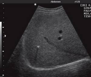
Liver hemangioma
- the most common benign tumor-like formation of the liver. A small hemangioma, due to the many small cysts and septa that create the effect of sound reflection, looks like an echogenic formation (Fig. 3). With large hemangiomas, episodes of thrombosis are possible with a deficiency of platelets in the periphery and a tendency to hemorrhage.
Rice. 3.
Hemangioma of the liver.
Mesenchymal hamartoma
usually detected in children during the first 2 years of life and debuts as a painless formation in the abdominal cavity. It accounts for 7-8% of all liver tumors in children. Hamartoma arises from the mesenchyme of the periportal tract; inside the tumor, hepatocytes, components of the bile ducts, blood vessels, and mesenchyme are found, but without lymphatic tissue. Due to the accumulation of fluid in the mesenchyme, cysts arise. The different ratio of tissue and cysts led to the identification of two types of hamartoma: predominantly stromal and predominantly cystic. The echographic picture of mesenchymal hamartoma is extremely variable. The hamartoma is well demarcated from the liver tissue, usually located in the right lobe, large in size, and contains many septal cysts.
Liver adenomas
are rare in children. They occur in children with glycogenosis (especially type I), glucose-6-phosphatase deficiency, galactosemia, Fanconi anemia, aplastic anemia (in the latter case, treated with androgens). Echographically, a solitary hypoechoic formation is recorded, in some cases it is isoechoic.
Focal nodular hypertrophy
- primary liver tumor without capsule. It is a hyperplastically regenerating node of hepatocytes, Kupffer cells, separated by fibrous cords. Characteristic of focal nodular hypertrophy is the presence of a central stellate field (scar) with proliferating bile ducts and blood vessels radiating from it. Focal nodular hypertrophy is more often detected in women, but can also be found in children of any age. Estrogens accelerate the growth of education. Echographically, the tumor is detected accidentally, looks like a well-circumscribed formation, which can be hyperechoic or isoechoic to varying degrees. In 50% of cases a halo is recorded. The formation may be located on a stalk at the edge of the liver.
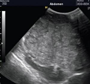
Intrahepatic calcifications
are rare (Fig. 4). They are typical for an infectious process, especially granulomatous (tuberculosis, brucellosis, histoplasmosis, coccidioidomycosis), cytomegaloviruses, toxoplasma, pneumocystis, hydatid disease, hemangioma, hemangioendothelioma, malignant tumors (calcifications of the radial ray type are characteristic of hepatoblastoma), trauma, vascular calcification, disease Caroli.
Rice. 4.
Calcification in the liver parenchyma with a typical acoustic shadow phenomenon.
Malignant liver tumors
occupy 3rd place in prevalence after Wilms tumor and neuroblastoma. They account for 5% of all childhood malignant tumors. 65-70% of liver tumors in children are malignant. All liver tumors manifest as mass formations in the abdominal cavity. The role of echography is to clarify the localization of the formation, provide data for a preliminary diagnosis and create conditions for the next stages of diagnosis (MRI, angiography, etc.).
Hepatoblastoma
- the most common liver tumor. It accounts for 47-50% of all childhood malignant liver tumors. Originates from fetal hepatocytes. The average age of patients at the time of diagnosis is 18 months (range of fluctuations is from 0 to 3 years). Girls get sick more often. It manifests with an enlarged abdomen, hepatomegaly, and often with symptoms of an acute abdomen. Possible osteopenia with bone fractures, hypoglycemia, hypercholesterolemia. In 90% of cases, the level of α-fetoprotein is elevated. Echographically, the tumor looks like a fairly clearly limited formation, often massive. Inside the tumor there may be foci of calcification and necrosis. According to Doppler ultrasound, the tumor is well vascularized. Hepatoblastoma quickly grows into the hepatic artery, inferior vena cava or covers them in the form of a dense coupling (Fig. 5, a, b).
Rice. 5.
Echogram of a liver tumor in normal mode and in MRI reconstruction mode.
A)
Echogram of a liver tumor in normal mode. The tumor is not visualized clearly enough.
b)
Echogram of a liver tumor in MRI mode. The structure of the tumor and its boundaries are visible quite clearly.
Hepatocellular carcinoma
in children, histologically it is completely similar to that in adults. Tumor cells are histologically very similar to normal hepatocytes, which makes diagnosis difficult. Affects children aged 10-12 years. It accounts for 20% of all malignant liver tumors in children. Predisposing factors are biliary atresia, familial cholestasis, hepatitis B, obesity, glycogenosis type I, hereditary tyrosinemia. Manifests with hepatomegaly, jaundice, anorexia, and a significant increase in the level of α-fetoprotein. Echographically, the tumor can be hypo- or isoechoic, multifocal, solitary (giant in size), diffuse (affecting the entire liver). Sometimes a thin echo-negative rim-halo is recorded around the tumor, which is an echographic reflection of the capsule. Central tumor necrosis with the formation of false cysts is possible.
Undifferentiated embryonal sarcoma (malignant mesenchymoma)
is a rare tumor consisting of undifferentiated spindle cells and mainly affects adolescents. It accounts for 5% of all malignant liver tumors in children. It manifests itself, like all liver tumors, with a mass formation in the abdominal cavity, fever, exhaustion, while the concentration of α-fetoprotein does not change. Echographically, the tumor can be presented as either solid masses with dense calcifications and the phenomenon of acoustic shadowing, or cystic formations with multiple internal septa. These pseudocystic cavities are formed due to hemorrhages and necrosis. Angiography confirms hypervascularization of the tumor.
Metastases.
The liver, as a result of the complexities of its blood supply, is more predisposed to the appearance of metastases in it than any other organ. Tumor cells are usually introduced with the bloodstream through the portal system (for example, from intestinal tumors), through the hepatic artery (tumors of the lungs or breast), and through the lymphatic system. Less commonly, metastasis occurs along the peritoneal surface (ovarian cancer). In the latter case, the initial metastasis is localized subserosally. In contrast, with hematogenous metastasis, the foci are located centrally. Small peripheral lesions are difficult to detect echographically, as they may be hidden by a rib shadow or near-field artifact. Metastases can be hyper-, hypoechoic (lymphoma metastases are the most hypoechoic, they are multiple, very tender), isogenic, heterogeneous (due to necrosis or hemorrhages). But it is impossible to talk about the origin of metastases based on their acoustic properties according to ultrasound data. The stage of the disease should be clarified using MRI or CT.
Metastases on ultrasound are usually solid, the edges are not clearly defined. Large metastases may contain fluid (necrosis) or mucin in the center (ovarian cancer metastasis). As a result of chemotherapy, calcifications may appear in the metastasis. In cases of changes in the texture of the liver (expansion of intrahepatic bile ducts, consequences of chemotherapy, cirrhosis as an outcome of hepatitis), identifying metastases is very difficult. According to Doppler ultrasound of the vessels, there are no metastases inside. In large metastases, rare small vessels may appear along the periphery. In the presence of ascites in advanced stages of cancer, it is possible to detect metastases on the surface of the omentum or peritoneum.
Conclusion
Ultrasound is used to diagnose and monitor liver diseases, as well as in treatment using invasive ultrasound-guided procedures. At the same time, it should be remembered that the liver changes with the pathology of many other organs, for example, with kidney diseases, and it is impossible to establish the causes of changes in the morphology and function of the liver without a thorough systemic analysis. To do this, along with examining the gallbladder and pancreas, do not forget to scan the retroperitoneal, subdiaphragmatic spaces and the small pelvis. If you do not work next to the device as a doctor, then even with the most golden stars on your shoulder straps, you will not rise above a paramedic.
Ultrasound scanner HS50
Affordable efficiency.
A versatile ultrasound scanner with compact design and innovative capabilities.
Benign and malignant tumors
Most often, liver cancer is diagnosed in men over 40 years of age and less often in women over 55 years of age. The most common is secondary liver cancer as a result of metastases in the organ. The risk of developing primary liver cancer increases in people who smoke and drink alcohol, as well as those who work in hazardous industries, suffer from diabetes and obesity.
Hemangioma is a tumor of a plexus of blood vessels that tends to increase in size without developing into a malignant neoplasm. It occurs in women 5 times more often than in men. It is believed that the formation of a tumor occurs during the intrauterine period of development, when the baby’s mother is adversely affected.
The trigger for tumor growth is hormonal changes associated with pregnancy, menopause, and taking contraceptive medications. The tumor develops asymptomatically and makes itself felt only when it reaches 4 cm, when the patient experiences pain in the right side. However, in most cases, hemangioma does not manifest itself in any way and is discovered by chance during a medical examination.
Biochemical and general blood tests do not reveal any abnormalities; only when the tumor is very large, bilirubin increases. Hemangioma itself is not dangerous, but as the tumor grows, the hepatic ducts become blocked, making it difficult to remove bile.
There is also a high probability of blocking the main feeding artery, which provokes a liver infarction and disruption of the functioning of neighboring organs. Complications of hemangioma occur in 15% of patients; if the tumor ruptures, internal bleeding occurs in 85%, leading to the death of the patient.
Signs of the disease
The main symptoms of diffuse liver changes include:
- Increased sweating and fatigue. If the liver is malfunctioning, sweat has a pungent and unpleasant odor. The body's performance is reduced: weakness, drowsiness, severe fatigue with minor physical exertion.
- Pain and discomfort in the area of the right hypochondrium in front or from the back indicate serious problems. Severe pain occurs when an organ is injured, malignant tumors, or purulent inflammatory processes.
- Poor digestion of food. Nausea and heartburn, bitterness in the mouth, frequent and loose stools, change in color of stool.
- Increased body temperature. Thermometer values may not exceed 37.2°. The so-called low-grade fever can last for several weeks.
- Deterioration of skin condition. Yellowing, acne, various lumps and papillomas, severe itching, swelling and peeling.
- Unpleasant coating and cracks on the tongue.
- Bleeding, hematoma formation.
- Unmotivated aggression, irritability, mood swings, headaches.

How to detect liver disease at an early stage
For a long time, liver diseases develop without any significant symptoms. Hepatosis has no manifestations, and only ultrasound shows structural changes in the organ.
Hepatitis has a relatively long incubation period, so immediately after infection it can only be detected by an increase in ALT and AST in a biochemical blood test. Cirrhosis is similar in symptoms to acute respiratory viral infections or ordinary food poisoning (flatulence, vomiting, weakness), and cancer becomes noticeable already when an enlarged abdomen forms on the side of the liver.
Also a common symptom for all liver diseases is jaundice of the skin. But all these manifestations are characteristic of a late stage, when the disease has already developed and affects the patient’s quality of life.
The only way to early diagnose liver diseases is an examination, including a series of tests and ultrasound diagnostics.
Diagnostics
All patients are required to undergo liver ultrasound, regardless of health status. Conditionally healthy people - as a preventive examination once every 6 months or a year, for sick people - to clarify the diagnosis, assess the condition of organ tissues. Using echography, you can detect cirrhosis, hepatitis, various seals and cysts.
In addition to an ultrasound of the liver, the doctor prescribes several more studies, with the help of which you can accurately establish the correct diagnosis:
- Radionuclide scanning. A number of special radioactive drugs are injected into the patient’s circulatory system, which, together with the bloodstream, enter the gland tissue. Then, using the device, a scan of the organ is performed. This technique is used when serious liver damage is suspected after injury or the presence of metastases is detected.
- CT. Computed tomography helps to see foci of parenchymal bleeding, small malignant and benign tumors and other organ pathologies.
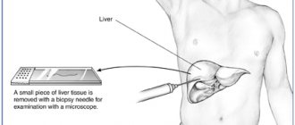
- Tissue biopsy with fine needles. Tissue samples taken for analysis are subjected to histological examination. The method is necessary if preliminary tests indicate serious problems. Histology will either confirm or refute the preliminary diagnostic conclusion.
- Biochemistry of venous blood. A detailed blood test with liver tests helps the doctor create an overall picture of pathological disorders in the organ, make an accurate diagnosis and select adequate therapy. If the patient is already taking a course of drugs, an interim biochemical blood test will show how effective such treatment is.
- Antibodies to the hepatitis virus. This analysis helps determine the type of pathological changes in liver tissue and the degree of virus activity.
Laboratory tests to detect liver diseases
A biochemical blood test helps assess liver function. The sampling is done on an empty stomach in the clinic, and after a few days a specialist will decipher the results. The main indicators include:
Bilirubin. This is the main antioxidant, a bile pigment formed during the breakdown of hemoglobin. Contained in the general, free and bound fractions. The first is 96% in complex with albumin, 4% with glucuronic acid. When the hepatic ducts are blocked or the liver is inflamed, bilirubin increases. Free bilirubin is destroyed in the liver, and bound bilirubin is eliminated through the kidneys. When an organ malfunctions, the concentration of bilirubin in the blood increases. This pigment is toxic, so its increase causes deterioration in health.
The level of bilirubin in the blood indicates a violation of the liver and kidneys. AST and ALT. Enzymes synthesized by the liver. In a healthy person, it is found in the blood in small quantities, but with the breakdown of liver cells, the levels increase significantly, and if the heart functions are disrupted, AST rises.
Alkaline phosphatase. It is a component of the cell membrane and is involved in phosphorus-calcium metabolism. Its concentration increases during various processes in the liver and other organs, and decreases with the destruction of bone tissue, decreased functionality of the thyroid gland, and anemia.
Cholesterol . This is a low-density lipid produced by the liver and is used for the synthesis of bile, cell membranes, etc. The liver produces about 1 gram of cholesterol per day, and this is enough for the body’s needs. In the hepatic ducts, bile acid is formed from low-density cholesterol molecules, which is absorbed into the blood after combining with proteins. Excess lipids are excreted from the body through feces.
When liver function is impaired, cholesterol concentrations increase. This leads to the development of cardiovascular diseases and obesity. The most dangerous consequence of excess low-density cholesterol is fatty hepatosis.
Over time, with constant high cholesterol, atherosclerosis develops, affecting blood vessels and arteries and leading to heart attack or stroke.
Only a doctor can interpret the tests, because different laboratories have different units of measurement. With fatty liver hepatosis, the level of bilirubin increases and albumin in the blood decreases.
Diet
In case of diffuse changes in the liver, it is necessary to follow a special diet. In fact, healthy eating rules are not difficult to follow. It is more difficult to overcome psychological dependence on harmful products. Willpower, a positive attitude and a healthy, varied menu help with this.
The following foods, drinks and dishes must be excluded from the patient’s menu:
- black tea, coffee;
- tomatoes and juice from them;
- any alcoholic and low-alcohol drinks;
- sweet soda;
- fat meat;
- any strong broths;
- barley, millet, pearl barley;
- mayonnaise and other fatty sauces;
- sausage and any smoked meats;
- fatty fish;
- white bread and baked goods;
- fatty fermented milk products;
- marinades, salted and spicy vegetables;
- mushrooms;
- legumes;
- fresh berries and fruits;
- chocolates and other confectionery products;
- spices.
In case of diffuse changes in the liver, it is necessary to eat such dishes and drinks as:
- weak green tea, dried fruit compote, rosehip decoction;
- rye and bran bread, crackers from it, unsweetened biscuits;
- dietary meat: chicken, turkey, rabbit, veal;
- lean fish - pike, pike perch, cod;
- vegetable oil and butter in small quantities;
- skim milk and dairy products;
- eggs;
- boiled and stewed vegetables;
- buckwheat, oatmeal, rice porridge;
- leaf salad;
- fresh bell pepper;
- pasta;
- marmalade, honey, fruit jam.
You need to eat small portions 5-6 times a day. Preference should be given to steamed, boiled and baked products. Soups and aspic are prepared from lean meat and fish. You can eat a small amount of squash caviar, vinaigrette, and sauerkraut. Sugar is replaced with xylitol. The amount of salt should not exceed 3 grams per day.

Cirrhosis of the liver
Liver cirrhosis can be seen on ultrasound only at stages 2 and 3, when echo signs of organ damage are noticeable. The indicators of a healthy organ are taken as guidelines: uniform, smooth edges, the left lobe is up to 7 cm in length, the right lobe is up to 13 cm, the diameter of the bile duct is up to 7-8 mm, the diameter of the portal vein is 12 mm. Deviation from the norm requires a more detailed diagnosis.
Liver cirrhosis has 3 stages:
- The disease at stage 1 can be detected only after abnormalities in the biochemical blood test. The changes are so minor that it is almost impossible to see them on an ultrasound. The specialist sees some blurring of the contours, but it is minimal. There is also an increase in one of the lobes, but the echostructure remains homogeneous. The blood vessels of the liver may be slightly dilated.
- At stage 2, the organ undergoes significant changes. The surface of the liver is covered with hyperechoic scars, which can be detected by an experienced specialist. The vascular pattern is poorly visible, the outlines of the organ are blurred. The echogenicity of the liver is heterogeneous; there are foci of hyper- and hypoechogenicity.
- At stage 3, the liver significantly increases in size, the portal vein swells, and the surface is covered with connective tissue scars. Also, the spleen enlarges beyond normal (over 12 cm in length and 6 cm in width). The vascular pattern is displaced, the caudate lobe of the liver is 0.65-0.75 times larger than the right lobe. The accumulation of fluid is so large that the abdomen bulges forward.
What causes increased echogenicity of the liver?
Reasons for which a diffuse (spread) increase in the echogenicity of the liver tissue is detected:
- Acute hepatitis of various nature (viral, toxic and others).
- Metabolic liver diseases (eg fatty liver disease).
Advice to a sick person from gastroenterologist Daniela Purgina, an expert on the website Pokhmelye.rf.
If you have been diagnosed with fatty hepatosis (and this is one of the most common causes of diffuse changes in the liver), then the main treatment is weight loss. This is the simplest and most difficult at the same time. Alas, no drugs will remove fat from the liver, so you will have to make efforts for the sake of your own health.
The increase in echogenicity can be either diffuse, diffuse, covering the tissue completely, or local, that is, limited. Moreover, limited areas (individual formations of increased or decreased echogenicity) can have smooth or uneven contours. What does it mean?
Hyperechoic (that is, denser) areas can mask:
- benign tumors (usually hemangioma, vascular tumor);
- malignant tumors;
- metastases from other organs;
- stones;
- calcifications;
- areas of fatty degeneration of liver tissue.
Hypoechoic areas (with reduced density) are usually:
- cysts with liquid contents,
- hydatid cysts,
- abscesses.
When the doctor sees a separate area with a changed shade in the image, then we are talking about a lesion. During ultrasound diagnostics, there are a lot of subtleties that suggest the nature of the formation, for example:
- Benign tumors most often have smooth, clear edges.
- In a malignant tumor, growth most often occurs expansively: in all directions and uncontrollably, so that the contours are uneven and they grow without a clear boundary.
This division is conditional. There are features characteristic of each volumetric formation that a specialist looks at and describes. To clarify the diagnosis, the doctor may prescribe a computed tomography or MRI. In this case, layer-by-layer images with the introduction of contrast into the organ are very helpful in clarifying the nature of the formation. For information about what areas of abnormal density look like on CT and MRI in various diseases, read the article about hypodensity formations in the liver.
The liver is a very patient organ. If there are no changes in the biliary system, then it may not manifest itself clinically for a long time. However, over time, pathological processes in which the echogenicity of the liver is increased can lead to cirrhosis of the liver. Therefore, it is imperative to know exactly how the organ speaks about its malaise, so as not to miss dangerous symptoms.
Fatty liver on ultrasound
Fatty hepatosis is extremely rare in completely healthy people. It develops as a consequence of existing diseases, such as diabetes mellitus, pancreatitis, gastric ulcer, alcoholism, etc. Hepatosis is characterized by extremely rapid development of the disease. Thus, after toxic damage to the liver as a result of poisoning (mushrooms, chemicals), fatty infiltration occurs within a week. Fatty liver disease can be easily detected using ultrasound. Depending on the location of fat deposits, the disease is of three types:
- Diffuse hepatosis . Fat cells are evenly distributed throughout the organ. This type also develops in completely healthy people who violate their diet. For example, in vegetarians, a lack of protein due to the refusal of animal foods disrupts the production of lipids, which also occurs after a long-term strict diet, after eating fatty and carbohydrate foods. Metabolic disorders also occur after a course of antibiotics.
Unlike a healthy organ, with diffuse hepatosis, the freedom to conduct ultrasound is reduced, which is displayed as white spots on the screen. The deep parts of the liver are poorly visualized, and the small venous pattern is not visible. The contours of the liver remain clear, but the size of the organ increases slightly.
- Local hepatosis . Fatty formations are found only in certain areas of the liver. On ultrasound, this is reflected by hypoechoic areas against the background of healthy parenchyma. focal hepatosis. Foci affected by fatty deposits are visualized as voluminous hyperechoic areas.
Common signs of hepatosis are a “light” liver - a predominance of low-echoic tissue areas that do not conduct ultrasound well. Areas affected by fat cells prevent the organ from functioning normally, which leads to serious consequences: stagnation of bile, intoxication of the body, decreased immunity. bile does not arrive in full, food is digested worse, enters the intestines not completely digested, provoking dysbacteriosis and intestinal infections.
The liver is less able to cleanse the body of toxins, reducing the supply of nutrients and vitamins. With fatty hepatosis, the cardiovascular system suffers, hypertension, myopia, and cardiac dysfunction develop. General intoxication reduces immunity, creating favorable conditions for ARVI, influenza and other infections.
Degrees of liver fibrosis in adults
Experts distinguish 5 degrees of scarring of liver tissue.
No fibrosis.
In this case, there is no evidence of liver cell death (necrosis) or scarring, despite liver inflammation (hepatitis).
Portal (mild) fibrosis.
In this case, there are areas of necrosis and scarring affecting the small and medium branches of the portal vein, which carries blood from the small intestine. The structure and function of the liver remain normal.
Periportal (moderate) fibrosis.
This is an option with an increase in foci of necrosis, scarring and impaired liver function.
Bridging (severe) fibrosis.
At this stage, scarring has disrupted normal blood flow to the liver and function is further impaired.
Cirrhosis.
This is permanent scarring and irreversible loss of liver function.
Hepatitis and liver on ultrasound
Hepatitis is a viral infection that leads to inflammation of the organ. In the acute form of inflammation, there is an accumulation of fluid in the tissues, and it conducts ultrasound well, so inflammatory processes are characterized by hyperechogenicity.
The size of the liver also increases. Its surface becomes too smooth and even, but the contours are blurred.
For hepatitis C, Doppler scanning is used to identify areas with increased blood flow and places where it is impaired or absent. With hepatitis B and C, you can see the affected areas of the parenchyma replaced by hyperechoic connective tissue.
What is liver fibrosis
Liver fibrosis is scarring of the tissue.
The liver is capable of healing itself, however, this system does not work at full capacity if the liver is damaged or the injury is long-term and very serious. All attempts at regeneration cause an accumulation of connective tissue (these are tough, inert fibers) instead of functioning liver cells. Liver fibrosis is not a specific disease, but rather a symptom of another liver problem. The most common conditions that lead to liver fibrosis are alcoholism, chronic hepatitis C and non-alcoholic fatty liver disease (NAFLD).
Liver fibrosis itself does not cause any symptoms. Doctors can detect signs of liver fibrosis using blood tests and an ultrasound, CT scan, or MRI. If it is detected early, it can be treated. But if it goes undetected and damage continues, fibrosis can progress to cirrhosis.
Liver cysts on ultrasound
Liver cysts are diagnosed in 0.8% of the population, and they are detected more often in women than in men. Usually accompanied by diseases such as polycystic ovary syndrome, liver cirrhosis, cholelithiasis.
When the liver ducts narrow, bile cannot pass through, it clogs the channel, and a cavity is formed in it, resembling a capsule in appearance. Over time, it fills with anechoic fluid, and new cysts with a diameter of up to 25 cm appear. There is no blood flow in a simple cyst.
Echinococcal cysts are formed as a result of the deposition of parasite larvae in the liver. They have the appearance of a capsule with dense walls, which gradually expand and put pressure on the parenchyma, blocking ducts and blood vessels.
A growing cyst can compress the hepatic vein, causing myocardial infarction. The danger of an hydatid cyst is that the capsule may burst and its contents may spread throughout the body.
Echinococcal damage reaches the brain and lungs. Also, if the capsule breaks, anaphylactic shock and death occur. An empty capsule has the property of calcification, the liver parenchyma loses functionality, leading to disruption of the organ.
If the disease is not treated, alveococcosis develops, in which parasitic nodules spread throughout the liver. If signs of an hydatid cyst are detected, the patient is referred for a more detailed examination.
Prognosis and prevention
If the cause of pathological changes in the liver is an unhealthy lifestyle or alcohol abuse, then if treatment is started in a timely manner, recovery occurs in almost all patients. If you follow the doctor's recommendations, liver function is restored almost completely.
Liver cirrhosis is considered life-threatening for patients. With this disease, the prognosis for recovery is only 50%. The remaining patients die within 5 years. The reason for this outcome is irreversible pathological processes in the gland. To prevent this, you need to consult a doctor in a timely manner, lead a healthy lifestyle, undergo regular medical examinations, and, if necessary, get vaccinated against hepatitis.
Liver hemangioma on ultrasound
Hemangioma is a vascular formation inside the liver, consisting of a plexus of blood vessels. It is benign in nature, but tends to increase in size. The cavernous tumor reaches a diameter of up to 20 cm, and if left untreated it turns into an atypical hemangioma covered with keratinized tissue.
The tumor is easily diagnosed by ultrasound; it has clear tuberous contours and hyperechogenicity. Increased blood flow is confirmed using Doppler scanning. However, ultrasound only confirms the very fact of the presence of a tumor, but MRI provides more reliable information.
Preparing for an ultrasound
The purpose of preparation for the procedure is to increase the information content of the study by reducing gas formation in the intestines. 3 days before an ultrasound scan of the liver, it is necessary to completely exclude fresh vegetables and fruits, baked goods, legumes, fermented milk products, alcohol and carbonated drinks from the menu. Food should be taken in small portions. For digestive disorders, enzyme preparations (festal, pancreatin, enzistal) are indicated. If you are prone to constipation on the eve of a liver ultrasound, you need to cleanse the intestines with an enema or a mild laxative.
Hyperechoic formations on ultrasound
Almost always, dense hyperechoic formations in the liver are metastases. In colon cancer they become calcified; in breast cancer they have a hypoechoic shadow. Also, with metastases, the lymph nodes are enlarged, which causes dilation of the ducts. But even if all the echo signs of cancer are present, the patient is not given a final diagnosis. He is sent for further, more detailed examination.
What is not visible on an ultrasound:
stones in the liver
Hepatolithasis is the formation of stones in the liver from clots of bile, which for some reason cannot pass through the ducts, thickens and stagnates. Due to the fact that the liver does not have nerve endings, stones do not make themselves felt for a long time.
Ultrasound diagnostics is not very informative due to the specific location of the organ.
The liver is blocked by the costal arch, so the sensor does not visualize stones in the ducts. The only diagnostic method remains MRI and biochemical blood test. With stones, bilirubin increases significantly, which indicates stagnation of bile. An MRI shows the exact location and size of the stones.
What does increased echogenicity of the liver mean?
Echogenicity is the ability of tissues and organs to absorb or transmit a signal (wave). An ultrasound machine is both a receiver and a transmitter at the same time. It sends out ultrasonic waves and then catches what is reflected back.
Echogenicity is the density of an organ during ultrasound, the ratio of absorption and reflection of a wave from an organ or tissue. The level of difference between the values of the sent and returned signal displays the picture on the screen. The degree of wave absorption depends on the density, elasticity, and humidity of the medium through which the ultrasonic waves pass.
Increased echogenicity of the liver on ultrasound can be due to various reasons. Contact your doctor to find out.
Ultrasound diagnostics allows you to see many indicators needed to assess the condition of the liver and surrounding organs:
- overall size of the organ
- ratio of the sizes of the right and left lobes,
- the condition of the gallbladder, the density of its walls,
- inclusions in the gallbladder,
- homogeneity of bile in the gallbladder,
- density of liver tissue - parenchyma,
- structure of large vessels of the liver,
- structure of large bile ducts,
- the presence of various formations in the liver.
If space-occupying formations are detected in the liver tissue, then from the image one can guess their nature, structure, and state of the cells. It is in assessing the condition of the tissues, that is, the structure of the liver, that the assessment of echogenicity helps. In the picture, the echogenicity levels, when compared with the video image, represent a difference in contrast. The ultrasound picture is drawn with dark and light shadows. And the difference in colors means different echogenicity.
Any conclusion requires a standard. During the study, the standard of average echogenicity of the ultrasound machine is the renal cortex. The density (and, accordingly, echogenicity) of a healthy liver is close to the standard. The liver parenchyma is represented by hepatocytes; they provide an almost equal ratio between absorption and reflection of ultrasonic waves. In the ultrasound picture it looks like a smooth gray mass. But the organ has vessels, bile ducts, membranes of lobes and lobules - and they all differ in echogenicity from the liver parenchyma (tissue) in the picture.
For example: the bone reflects almost 100% of the signal that was sent. In liquids, on the contrary, the signal is completely absorbed. Parenchymal organs (that is, organs made of cellular tissue, including the liver) typically absorb and reflect 50% to 50% ultrasonic waves.
An expert on the website Pokhmelye.rf, gastroenterologist Daniela Purgina, warns against making a common mistake.
Do not self-medicate. It's ineffective. And sometimes, trying to treat yourself, the patient may waste precious time without achieving the desired result.
conclusions
Our ultrasound clinic offers a complete liver examination, including consultation with a highly qualified doctor, a full range of blood and urine tests, as well as ultrasound diagnostics.
It is recommended to undergo a liver examination for men over 40 years of age and women over 50, as well as younger people working in heavy industry, residents of megacities, as well as patients with a family history of liver pathologies.
ONLINE REGISTRATION at the DIANA clinic
You can sign up by calling the toll-free phone number 8-800-707-15-60 or filling out the contact form. In this case, we will contact you ourselves.
If you find an error, please select a piece of text and press Ctrl+Enter
What symptoms should you be wary of?
When you still feel well, but the following signs appear, do not ignore them. By contacting a doctor right now and starting treatment on time, you can avoid a lot of problems. Here are the most common early symptoms of liver damage:
- Pain or heaviness in the right hypochondrium is the most common first sign of liver problems.
- Itchy skin.
- Decreased appetite.
Next, consider the symptoms that develop when the liver is already severely damaged:
- jaundice;
- an increase in abdominal volume with a decrease in muscles;
- gynecomastia, or an increase in the volume of the mammary glands in men;
- menstrual irregularities, infertility;
- red palms. In the medical community they are called “liver”;
- the presence of dilated veins on the anterior abdominal wall;
- bleeding gums, bruising on the skin;
- change of size. The doctor can detect hepatomegaly even during the initial examination, during palpation (feeling with hands);
- the presence of a large number of spider veins - telangiectasias.
A case from the practice of gastroenterologist Daniela Purgina, an expert on the website Pokhmelye.rf.
There is nothing special in this case, but I remember it precisely because of the reaction of the patient herself. A 54-year-old woman came to the appointment and cried incessantly. As it turned out, the day before she went to have an ultrasound about dull pain in her right side. The ultrasound revealed diffuse changes in the liver, and the ultrasound diagnostic doctor told her that these were signs of liver cirrhosis. In fact, the woman did not have any liver cirrhosis: all her blood counts were normal, viral hepatitis markers were normal, and she had no signs of cirrhosis. The only thing the woman had was excess body weight and increased cholesterol levels. And diffuse changes in the liver were nothing more than fatty liver disease. The patient was recommended to lose weight and take medications to normalize cholesterol levels.