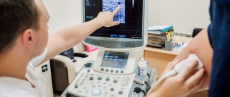
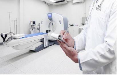
Arteriography of the lower extremities is a method of visualizing the lumen of the arteries by introducing a radiopaque substance into them through a catheter and simultaneous fluoroscopy (video recording of x-rays), with recording and processing of the resulting image using special equipment. In this case, the specialist receives objective information about the anatomical structure of the vascular bed of the area under study, the speed of blood flow, the presence of stenoses (narrowings) and occlusions (complete blockage), the degree of development of collateral circulation in the limb under study.
Based on this information, a council of doctors sets indications for surgery and determines the stages of treatment; taking into account the entire complex of preoperative examination, the choice of revascularization method is made (open bypass, endovascular or hybrid intervention).
Treatment under the compulsory medical insurance policy is free!
Submit your application
Follow the news, subscribe to our social networks
Details
Advantages of angiography at the Innovative Vascular Center
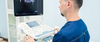
Arteriography of the lower extremities is performed on a stationary Philips Allura Xper FD20 angiographic complex, or on a Veradius mobile angiograph if performed intraoperatively. The use of modern angiographic systems makes it possible to conduct research at any required frame rate, which gives an objective picture of the nature of blood circulation in the limb.
Our clinic uses CO2 angiography without the use of iodine contrast. This method is well suited for patients with iodine intolerance and allows any operation to be performed using the endovascular method.
To reduce the risk of complications, we perform angiography most often through a peripheral radial approach on the arm, which allows the patient to begin walking immediately after the procedure.
What does a CT scan of the vessels of the lower extremities show?
Computed tomography of the vessels of the lower extremities helps to detect many different diseases at an early stage, some of which are fatal. Therefore, this method allows you to identify the problem in time and begin treatment in order to avoid serious consequences.
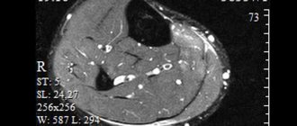
CT scan of the vessels of the lower extremities allows you to obtain detailed three-dimensional images of the venous, arterial systems and bone tissue. Thanks to this, it is possible to study the changes occurring in the legs, to examine in detail the network of blood vessels and certain parts of it. The images show where the disease is spreading, as well as the approximate size of the outbreaks.
CT angiography of the vessels of the extremities helps in diagnosing the following pathologies and conditions:
- Takayasu's disease;
- atherosclerosis;
- thrombosis;
- venous insufficiency;
- the condition of soft tissues and organs adjacent to the arteries;
- the size of the lumen of the arteries;
- compression by tumor or scar tissue;
- neoplasms.
How is angiography of the lower extremities performed?
Angiography is performed by a highly qualified x-ray endovascular surgeon in a sterile cath lab and with sterile consumables. Anesthesiological monitoring is carried out throughout the study.
Under local anesthesia, a puncture of the artery of the arm or leg is performed, after which a thin catheter is inserted into its lumen, through which a radiopaque substance is injected. During the examination, the patient may feel warmth as the contrast is injected, and very rarely there may be pain. The duration of the angiography itself is 10-15 minutes.
When performing angiography, the specialist receives objective and accurate information about the structure and damage to the arterial bed of the area under study. The resulting images are saved electronically and can subsequently be written to disk and given to the patient.
CT scan of the vessels of the lower extremities: how is it performed?
CT scan of vessels of the lower extremities
lasts no more than 25 minutes, during which the patient is positioned on a special tomograph table. This examination is always carried out using special contrast.
Contrast enhancement
Contrast enhancement is used to obtain a resonance image, in which the picture of the condition of the veins and arteries will be clearer. Puncture, or invasive penetration, is carried out using a special catheter with a mandrel, a rod that closes the lumen.
Possible complications of angiography
Complications after angiography are very rare. However, due to the fact that this study is invasive (penetrating through the natural external barriers of the body) and is accompanied by the introduction of a radiocontrast agent into the systemic bloodstream, associated complications may still develop: contrast-induced nephropathy,
- bleeding
- hematomas and false aneurysms at arterial access sites
- local infectious complications
- allergic reactions to contrast
Our specialists do everything possible to prevent these complications, and also know the necessary treatment methods in case of development.
CT angiography of vessels of the lower extremities
Tomography of the vessels of the lower extremities is currently the most effective examination method, the analogue of which can only be MRI. However, the price of CT is much lower, which makes this study more accessible. This diagnosis allows you to study in detail the problems associated with impaired leg function, identify pathologies even at an early stage, and also establish the causes of pain. Most often, a vascular surgeon gives a referral for a computed tomography scan of the capillaries of the vascular system of the legs after the patient has been examined using Doppler ultrasound.
.
When is a CT scan of the lower extremities prescribed?
CT angiography of the vessels of the lower extremities is usually prescribed for obliterating lesions of the arteries, as a result of which problems with blood circulation appear. The doctor may prescribe a tomography of the blood vessels of the legs when the patient complains of swelling of the legs, pain, numbness, loss of skin sensitivity, and varicose veins. In addition, indications for examination are persistent tissue hematomas, various injuries to the vascular system, thrombosis, abnormal development of blood vessels, rheumatic complications with blood vessels, pain in the leg muscles during long walking, vascular tumors and many other diseases. CT angiography of the vessels of the extremities
It is also indispensable in preparation for surgery to reduce the risk of complications, and in the postoperative period, as it makes it possible to monitor the recovery process.
Decoding the results
To decipher the results of angiography of leg vessels, the following principles are used:
- thrombosis, thromboembolism or thrombophlebitis is indicated by a picture that shows that the injected contrast agent has not spread throughout the vascular bed;
- an image showing that the lumen of the vessel has decreased by 30-90% may be evidence of the following pathologies: atherosclerosis, acute and chronic ischemic disease, tumor or hematoma in the vessel, thrombosis, thrombophlebitis, arteritis, phlebitis, endarteritis, congenital vascular pathology;
- if the images show zones of dilation of blood vessels, protrusion of their walls, then the diagnoses may be as follows: varicose veins, aneurysm, congenital vascular pathology;
- atypical distribution of the contrast agent, its flow into other arteries, too much branching of the vascular system, visualized on X-ray images, indicate a congenital vascular anomaly.
As you can see, the same situation in the image can indicate several diseases simultaneously. Therefore, in order for the decoding to be the most reliable and informative, several specialized specialists take part in it: a vascular surgeon, a radiologist, a phlebologist surgeon and others. In addition to angiographic images, previously taken blood tests, results of ultrasound, plain radiography and other studies are taken into account.
What will need to be done in the ward
- You should drink plenty of fluids. This is necessary to compensate for the loss of water that you had during preparation for the study and to quickly remove the contrast agent from your body. For most people, this means drinking at least 6 glasses of water, juice or tea. Check the required volume with your attending physician or x-ray surgeon after the study.
- You must strictly adhere to bed rest for 6-8 hours after the examination and keep your leg straight on the side of the puncture site (when using the femoral approach). If using the radial carpal artery approach, you should remain in bed for 1.5 to 2.0 hours, but limit use of your right arm for the next 24 hours.
The puncture site may be slightly numb for a few days.
The next day the pressure bandage will be removed and you can return to your normal lifestyle. You will receive a conclusion about the study and will be able to discuss the results with your attending physician or x-ray surgeon.
Advantages and diagnostic capabilities of CT
The accuracy of the data obtained is one of the main advantages of CT examination. Even minor violations are visible in layer-by-layer images. Tomography shows:
- inflammatory processes;
- blood clot formation;
- phlebeurysm;
- developmental abnormalities and vascular obstruction;
- tumor formations of various origins.
The procedure takes little time (10–30 minutes), is absolutely painless and does not require invasive intervention. A CT scan is carried out using x-rays, but this is not something to be afraid of. The radiation dose is calculated taking into account the individual characteristics of the patient and does not affect health.
MSCT angiography
What is multislice vascular computed tomography (MSCT angiography, CT angiography)? Computed tomography of blood vessels (CT antiography) is a unique method that allows us to study the condition of the blood vessels of the human body. CT Angiography is a minimally invasive test that helps doctors diagnose and treat vascular diseases in the body. When performing CT Angiography, various image processing technologies are used and, as a rule, it is carried out with intravenous administration of a contrast agent. To perform MSCT angiography, a computed tomograph is used, which allows you to obtain a detailed image of blood vessels and tissues in various parts of the human body. An iodinated contrast agent is administered through an intravenous catheter. The contrast agent passes through the large and small vessels of the body, allowing them to be clearly imaged during scanning. After scanning, the image is processed using special computer programs that allow you to visualize the vessels in different planes and projections. Indications for vascular computed tomography (MSCT angiography).CT angiography is performed to study various organs and anatomical areas of the human body: brain, neck, heart, chest, abdominal cavity, retroperitoneal space (kidney and ureter area), pelvis, lower limbs, upper limbs, etc. CT (MSCT) angiography is prescribed for suspected:
Used to assess the condition of blood vessels before and after various surgical interventions:
CT angiography is also performed to identify features of the structure of blood vessels and assess blood flow to organs; The characteristics of the blood supply to areas of organs affected by the tumor are often assessed with the aim of conducting selective chemoembolization or selective internal radiation therapy. |
How is the research going?
Angiography is performed according to the following scheme:
- treating the area of skin where the puncture will be performed with an antiseptic;
- puncture of an artery or vein;
- insertion into an artery/vein of an introducer - a device that does not allow blood to flow in the opposite direction;
- insertion of a catheter into a vein/artery;
- supply of contrast agent;
- performing x-rays.
Indications and contraindications
CT angiography has its indications and contraindications
CT angiography of the lower extremities is justified if the results of other diagnostic methods cannot be interpreted unambiguously. The study can be performed if there are the following complaints:
- pain in the lower extremities;
- fatigue in the legs after minor physical activity;
- swelling in the ankle area;
- paresthesia in the feet and legs;
- cramps in the calf muscles (especially at night);
- changes in the skin (pattern wear, atrophy of hair follicles, dryness and flaking, the appearance of hyperpigmentation and areas of ulceration);
- visible deformation of blood vessels, compaction, etc.
With claudication, there is a possibility of occlusive diseases of the peripheral arteries, which are associated with the abdominal aorta (its bed is additionally assessed and an aneurysm is excluded).
During pregnancy and childhood, other examination methods are preferable, since X-ray radiation and contrast are contraindicated for this category of people. In this case, MRI and ultrasound of the lower extremities with Doppler (duplex scanning) are performed. For health reasons, it is possible to perform MSCT without enhancement. The administration of contrast is unacceptable to patients with a burdened allergic history (hypersensitivity reactions to iodine), chronic renal failure and hyperfunction of the thyroid gland.
Which is better: CT angiography of the lower extremities with or without contrast?
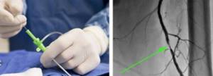
Contrast in CT angiography provides better visualization: on the left is the process of vessel catheterization, on the right is an angiogram with an arrow indicating the pathological zone
To obtain the most detailed images, contrast is necessary. There is no need to be afraid of administering contrast in the absence of contraindications; complications in the form of anaphylactic reactions are extremely rare. Doctors have a range of drugs in their arsenal to provide emergency medical care in urgent situations. The price of CT angiography of the lower extremities varies in different clinics; at the Magnit diagnostic center, the cost of diagnostics is affordable for most patients, as there is a flexible system of discounts.
How is computed tomography performed?
- The patient lies down on a table that will slide into the tomograph ring;
- To inject contrast into a vein, a special cannula is installed, which is connected to a machine for automatically administering a contrast solution.
- The CT machine is controlled by a specialist from the next room. There is a special intercom to communicate with the patient.
- During the tomography, you must stand still so that there is no movement. Any movement distorts not only the currently scanned image, but also all subsequent images. It is necessary to strictly follow the commands of the doctor performing the research. The patient holds his breath every time the specialist gives the appropriate command.
- The examination lasts 3-5 minutes, after which the cannula is removed from the vein. The received data enters the apparatus for computer processing. After studying the materials, the doctor gives a conclusion on the study.
Angiography of the arteries of the lower extremities
Angiography of the arteries of the lower extremities (other names: peripheral angiogram; angiography of the legs)
What is angiography of the arteries of the lower extremities (LAN)
Angiography of the arteries of the lower extremities (LAN) is a minimally invasive study that is performed in a specially equipped cath lab. ANC is performed using ultra-low doses of X-ray and a special contrast agent, which is injected into the arteries of the lower extremities through the lumen of the catheter.
ANC allows you to determine the state of the blood supply to the legs and will help to develop the most optimal and effective further treatment tactics for the patient.
ANC will identify areas of narrowing of the leg arteries and/or areas of completely closed (occluded) arteries.
Why is an ANC necessary?
This study is prescribed if there is a suspicion of disruption of the lumen of the arteries supplying the legs with blood. An angiogram obtained after ANC will help decide what type of surgical treatment is necessary to restore the lumen of the arteries of the legs. There are two main types of treatment for leg artery disease: bypass surgery and peripheral artery stenting. Bypass surgery is a major surgical operation in which a shunt (bypass vessel) is sutured around a narrowed or closed section of an artery, which restores blood flow in the affected area of the vessel. Currently, either pieces of the patient's own superficial vein or artificial synthetic shunts are used.
Peripheral stenting is a type of minimally invasive operation, similar in technique to ANC. A special device (stent) is inserted into the site of narrowing of the artery and straightened, which allows thrombotic and atherosclerotic masses to be pressed against the artery wall and the lumen of the artery is restored.
A stent is a metal mesh that is expanded with an air balloon at the site of maximum narrowing. The stent is made of metal (usually stainless steel, nitinol or chromium-cobalt alloys) and within 6 months grows into the vessel wall (endothelialization.)
Endothelialization is the growth of the stent with connective tissue and, in fact, its ingrowth into the arterial wall. This is a normal and desirable process.
Possible complications during ANC
The risk of complications during ANC is very small, but the following complications may occur:
- Damage to an artery by a thin and flexible tube (catheter), with which an x-ray surgeon visualizes the arteries. In rare cases, emergency surgery is necessary.
- The development of an allergic reaction in a patient to a contrast agent containing iodine. It is imperative to inform your doctor if you are allergic to iodine, strawberries or seafood.
Preparing for the ANC
- As a rule, it is recommended to stop eating and drinking large amounts of liquids 6-8 hours before the test.
- You must tell your doctor about all medications/vitamins/herbs, etc. that you are taking. You may need to stop taking some or all of your medications until the study is done. We strongly recommend that you do not stop taking any medications yourself without the recommendation of your attending physician or x-ray surgeon.
- Tell your doctor if you have any allergies, including to iodine, seafood, latex or rubber, or to medications such as novocaine, lidocaine, heparin, nitroglycerin, etc.
- It is recommended to remove all jewelry and jewelry to avoid interfering with arterial imaging.
- On the eve of the examination or in the morning on the day of the examination, it is advisable to take a shower and change your underwear.
- It is necessary to shave the artery puncture site in the groin area or the wrist of the right hand (back side). Details can be obtained from your attending physician or x-ray surgeon.
- Take with you 1.0-1.5 liters of drinking water (with or without gas as you wish). Water is needed for hydration after the examination to quickly remove the contrast agent and minimize possible complications.
- You must have a CD or flash card with you in order to receive a record of the examination performed on you and the x-ray surgeon’s report in electronic form.
What happens during the study
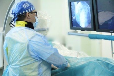
The x-ray surgeon performs the examination only in a hospital setting in a special x-ray operating room. 30-40 minutes before the examination, the patient is given an intramuscular injection in the department. This is a premedication, which consists of a mild sedative and antiallergic drug. The patient will be slightly relaxed but alert and will be able to communicate with the x-ray surgeon throughout the procedure.
The study is performed on a special angiographic table in the supine position. Metal electrodes will be placed on the arms and legs, which will transmit information about each heartbeat and its frequency - to record an ECG. The area of the artery puncture will be treated with special and aseptic solutions. The patient is covered with a disposable, sterile surgical linen and local anesthesia is performed, during which a slight prick and numbness of the skin in the area of arterial puncture is felt. Then the x-ray surgeon will puncture the artery with a thin needle (less than 1.0 mm) and then through a thin and long tube (catheter) will introduce contrast into the lumen of the leg arteries being examined (see Figure).
This will allow you to see blocked or narrowed areas of the artery. When contrast is administered, the patient may feel heat in the groin for 10-15 seconds. On average, the study lasts no more than 30-40 minutes.
After completing the study, the x-ray surgeon will stop the bleeding at the site of arterial puncture within 10-15 minutes and apply a pressure, aseptic bandage, after which the patient is taken to the ward on a lying gurney.
What will need to be done in the ward
- You should drink plenty of fluids. This is necessary to compensate for the loss of water that you had during preparation for the study and to quickly remove the contrast agent from your body. For most people, this means drinking at least 6 glasses of water, juice or tea. Check the required volume with your attending physician or x-ray surgeon after the study.
- You must strictly adhere to bed rest for 6-8 hours after the examination and keep your leg straight on the side of the puncture site (when using the femoral approach). If using the radial carpal artery approach, you should remain in bed for 1.5 to 2.0 hours, but limit use of your right arm for the next 24 hours.
The puncture site may be slightly numb for a few days.
The next day the pressure bandage will be removed and you can return to your normal lifestyle. You will receive a conclusion about the study and will be able to discuss the results with your attending physician or x-ray surgeon.
What you should pay close attention to
There may be a slight hemorrhage (subcutaneous hematoma) at the puncture site. If bleeding starts from the puncture site, immediately call the attending physician or the doctor on duty.
Also, be sure to inform your attending or duty doctor about the following cases:
- If your leg feels numb or tingling, or if your foot turns blue or cold.
- If the area around the puncture area appears blue.
- If the puncture area swells or fluid is released from it.
What can you do to improve your condition if damage to the arteries of the legs is detected?
The most important steps are:
- Quit smoking. Try not to be a passive smoker.
- Get physically active. Try to follow the level of physical activity recommended by your doctor. Ideally (unless contraindicated), walk, ride a bike, or do other types of cardio exercise 5 times a week for at least 30 minutes.
- Monitor your blood pressure. Consult your doctor to help you achieve your blood pressure goals.
- Try to reduce your blood cholesterol levels. Eat a healthy diet (high in fiber and low in saturated fat and trans fat) and take cholesterol-lowering medications if necessary.
- If you have diabetes, monitor your blood sugar levels. And aim to achieve and maintain an HbA1c level of less than 7%. HbA1c (hemoglobin A1c) is a blood test that shows the average blood sugar level over the previous 2-3 months.
- If you are obese, then set a goal to lose 5-10 kg. If you need to lose more, it is recommended to lose about 1 kg per week until you reach the desired weight.
How to learn more about leg angiography and what to ask your doctor
The necessary consultation can be obtained from your attending physician and/or x-ray surgeon.
Most frequently asked questions:
- Why do I need a leg angiogram?
- Can I take my medications on the day of the test?
- How can the angiogram results affect my treatment?
- Do I need surgery to open or improve the function of my arteries?
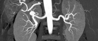
Example of an angiogram of the lower extremities, narrowing of the common iliac artery by 90% with signs of mural thrombosis
COMPUTED TOMOGRAPHY (CT)
What is CT?
CT (computed tomography) is an X-ray method of obtaining layer-by-layer images of organs. CT devices of the latest generations - multislice CT (MSCT), - which, thanks to more modern equipment, also provide the ability to construct 3D images.
Why is intravenous administration of contrast agent necessary for CT?
The contrast agent accumulates differently in normal and altered organs and tissues of the body, this primarily applies to tumor formations. Using contrast, you can detect even a small tumor and predict the degree of its malignancy. Intravenous contrast also helps determine the condition of blood vessels (arteries and veins). Such studies include CT angiography (of the abdominal aorta, arteries and veins of the lower extremities). CT coronary angiography (study of the heart vessels).
It makes no sense to perform a CT scan of the abdominal cavity without using a contrast agent, because the doctor will not receive complete information and the study will need to be repeated.
Can intravenous administration of a contrast agent lead to any complications?
No, the latest generation of contrast agents are generally well tolerated by patients. Sometimes, at the time of administration, a feeling of heat is noted, which quickly passes. Only some patients with a tendency to allergies may develop a more pronounced reaction, which will require additional therapeutic measures. All CT rooms are equipped with the appropriate set of necessary drugs. Also at the State Budgetary Healthcare Institution MKSC named after. A.S. Loginova DZM has intensive care specialists working around the clock, who, in extreme cases, will provide the necessary highly qualified assistance in a timely manner.
Is there a contraindication to CT scanning?
The only contraindication is pregnancy, especially in the first trimester, since the method is associated with radiation exposure.
The study of which organs is possible in the conditions of the State Budgetary Healthcare Institution MKSC named after. A.S. Loginova DZM?
Almost all organs and systems of the body:
- Head and brain examination
- Necks
- Chest
- Retroperitoneal space
- Urinary system
- Small pelvis
- Vessels, including heart vessels
- Skeletal system
- A separate type of research at the State Budgetary Healthcare Institution MKSC named after. A.S. Loginova DZM is CT-Enterography and virtual colonoscopy, which allow you to detect diseases of the small and large intestine without endoscopy
What is the procedure for performing a CT scan?
The examination is carried out by appointment to avoid long waits.
During the study, the patient is alone in the room, but at the same time he has the possibility of audio communication with the specialist performing the procedure.
The table on which the patient lies is moved into the tunnel during the examination to obtain more complete organ scanning results. The depth of this tunnel is very small, so there is no fear of confined space. During the procedure, the patient must lie still. In some cases, the laboratory assistant may ask you to hold your breath. The time for a CT scan is about 15 minutes, but if intravenous contrast is necessary, the time increases to 40 minutes.
It is necessary to bring with you previous photographs and descriptions of ultrasound, CT, MRI.
How long will a CT scan take?
Today, CT is of great importance in determining treatment tactics for the vast majority of diseases. The improvement of diagnostic systems has made it possible to obtain a large amount of information in a very short time - each scan lasts no longer than 10-15 minutes.
Analysis of research results is an extremely responsible process, requiring multifaceted differential diagnosis, and often collegial discussion. In this regard, at the Moscow Scientific and Research Center named after A.S. Loginov established the following standards for the time spent on describing the research:
| Name of the study | Duration of description from the end of the study |
| CT without intravenous contrast: brain, paranasal sinuses, skeletal bones of 1-2 regions, chest organs (except for oncological and interstitial diseases), abdominal organs for assessing urinary tract stones | no more than 1 hour |
| CT scan of the chest if interstitial lung diseases are suspected, as well as CT scan of skeletal bones in 3 or more areas | no more than 2 hours |
| CT with intravenous contrast: angiopulmonography, CT angiography (including CT coronary angiography), CT enterography | no more than 2 hours |
| CT with intravenous contrast: areas of the head and neck, chest, abdominal cavity and retroperitoneal space, as well as examination of several areas in case of suspicion or presence of cancer with assessment of the prevalence and dynamics of the development of the process | no more than 3 hours |
The time spent describing studies performed in other health care settings depends on the area being studied, according to the accompanying table.
With increased complexity of the study and the need to involve different specialists, the duration of the description can be increased by agreement with the patient. In addition, the conclusion can be sent to the patient’s email, which will eliminate the long wait for the result.