Diseases of the stomach and intestines - the organs that make up the gastrointestinal tract - occupy 1st place among all diseases occurring in people of different ages. These pathologies bring patients many unpleasant moments - from an uncomfortable state to excruciating pain. But the most dangerous thing is that it is gastrointestinal diseases that cause a huge number of complications - perforated ulcers, severe inflammation and cancerous tumors, leading to disability and even death. That is why it is recommended for every person to undergo a gastrointestinal ultrasound periodically, even if nothing hurts yet.
And your gastrointestinal tract is healthy: stomach health is a matter of time
The content of the article
Let's look at medical statistics on diseases of the stomach and intestines. Alas, it is frightening, even without taking into account hidden patients who have not been examined and residents of the poorest countries where there is no access to medical services.
According to statistics:
- Almost 90% of the population of developed countries suffers from gastritis of varying degrees of neglect.
- 60% of the world's inhabitants are infected with Helicobacter pylori, a bacterium that causes inflammation of the mucous membrane of the stomach and intestines, and is the cause of gastritis and stomach ulcers.
- In Western countries, up to 81% of citizens, according to statistics, periodically experience heartburn, which is a symptom of gastroesophageal reflux disease - a disease of the esophagus that leads to disruption of the gastrointestinal tract.
- About 14% of people have stomach ulcers.
At the age of over 60 years, the quality and length of life depends on the condition of the stomach and intestines, but it is possible to get rid of existing pathology only in the initial stages of the disease. That is why it is so important to be attentive to your health and not bring the problem to a chronic stage.
Prices for services at the Central Clinical Hospital of the Russian Academy of Sciences
| Primary appointment (examination, consultation) with a gastroenterologist | 2000 rub. |
| Gastroscopy | 3000 rub. |
| Colonoscopy under sedation | 13000 rub. |
| Gastroscopy under sedation | 11000 rub. |
| Colonoscopy and gastroscopy under sedation (comprehensive service) | 16,000 rub. |
| Additional services | |
| Taking material for biopsy | 750 rub. |
| Biopsy | 2500 rub. |
| ECG | 1000 rub. |
| Determination of urease activity of a biopsy of the gastric or duodenal mucosa for Helicobacter pylori (HELPIL test) | 2150 rub. |
| Appointment with a gastroenterologist | 2000 rub. |
| Appointment with a coloproctologist | 2000 rub. |
How to check the stomach and intestines quickly, cheaply and informatively?
There are several types of examination of the intestines and stomach, but only ultrasound diagnostics has a full range of advantages, which doctors consider invaluable and very effective in making a diagnosis.
- An ultrasound can be done urgently for any patient’s condition. The examination will take a maximum of 15-30 minutes.
- Ultrasound diagnostics are carried out painlessly, without causing psychological discomfort. Unfortunately, other methods of examining the gastrointestinal tract require very unpleasant procedures - swallowing tubes, inserting sharp instruments into the anus, sometimes to a considerable depth, ingesting liquids that cause vomiting, etc.
- Ultrasound is completely safe. The method is based on echolocation and does not require the use of X-ray and MRI equipment.
- This is one of the cheapest examinations. An examination of the gastrointestinal tract along with the rest of the abdominal organs will cost around 1 thousand rubles.
With all this, this technique is sometimes even more informative than other methods of examining the stomach and intestines. For example, unlike the endoscopic diagnostic method (using probes that are inserted inside), ultrasound reveals intestinal inflammation, thickening and protrusion of the walls, stenosis (expansion of the lumen), abscesses, fistulas, congenital anomalies (Crohn's disease), neoplasms in the early stages of development diseases.
Preparation for gastroscopy under anesthesia (in a dream)
Gastroscopy is prescribed by the patient's attending physician, usually a gastroenterologist. The procedure may be recommended after an MRI, in case of certain patient complaints, or in case of negative test results.
The study is usually carried out in the morning and is performed on an empty stomach. Eating is possible no later than 7 hours before the examination. You must refrain from smoking for at least 2 hours: it irritates the gastrointestinal mucosa, which negatively affects the accuracy of the study. During the examination, the patient lies on his left side, with a cushion placed under his head. When performing gastroscopy without anesthesia, immediately before the procedure, the patient receives local anesthesia: the oral cavity and pharynx are treated with lidocaine in the form of an aerosol. When performing gastroscopy under general anesthesia, intravenous anesthesia is used. With this type of examination, the patient is asleep, so he does not experience any discomfort when the doctor inserts a probe into the oral cavity, esophagus and stomach. To perform endoscopy under anesthesia, hospitalization for several hours in a day hospital is required. The duration of an endoscopic examination depends on its type and purpose. As a rule, the procedure takes from 5–7 minutes in the absence of serious pathological changes to 15–30 minutes in the presence of multiple disorders or the need to perform any therapeutic manipulations.
Specifics of the gastrointestinal tract examination: why the stomach and intestines need to be examined in detail
Despite the close relationship between the stomach and intestines, the doctor examines both organs in detail, since they not only have similar diseases. For example, ulcers can be localized in any part of the gastrointestinal tract or form in all parts at once. The same applies to oncological tumors, inflammation and other processes.
Depending on the patient’s complaints, the specialist examines the intestines and stomach separately. Having received data indicating dangerous processes, the doctor refers the patient for additional diagnostics.
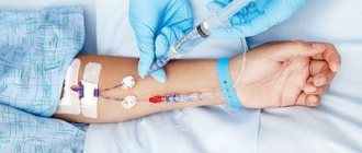
Along with an ultrasound, it is recommended to simultaneously take a breath test for Helicobacter pylori. This analysis is also not traumatic - the patient will only need to exhale air a few times. A complex ultrasound plus analysis for Helicobacter will allow literally in 15-20 minutes to identify the cause of heartburn, pain and cramps in the abdomen, diarrhea or constipation, bloating and other symptoms, establish the extent of the processes and prescribe treatment without resorting to unpleasant diagnostic methods.
How to examine the intestines: Ultrasound plus additional techniques
The intestine has three sections: the large, small intestine and rectum, and the study of each of them has its own characteristics and nuances.
- Ultrasound of the colon
helps detect cancer at a very early stage. To make sure, the patient is prescribed a contrast X-ray and colonoscopy. Irrigoscopy, an X-ray examination using contrast fluid, will also be very effective. The method allows you to “see” areas that are invisible to colonoscopy and difficult to distinguish with ultrasound, for example, areas of bends or accumulations of mucus. - Ultrasound of the small intestine
is hampered by tortuosity and deep burial, as well as the accumulation of gases that distort the image on the monitor. A special curved sensor and the latest high-precision equipment help to examine the small intestine. Ultrasound evaluates wall thickness, visualization of layers, patency, wall expansion, and peristalsis. - An ultrasound of the duodenum
is performed together with an examination of the stomach. Allows 100% diagnosis of stomach ulcers, cancer, gastroduodenitis.
Depending on the area being examined, the doctor uses a sensor with certain characteristics.
Gastroscopy at the Central Clinical Hospital of the Russian Academy of Sciences (in a dream)
At the Central Clinical Hospital of the Russian Academy of Sciences, modern equipment is used to perform gastroscopy; the examination is carried out by a highly qualified endoscopist. When performing gastroscopy under anesthesia, the patient's condition is monitored throughout the entire study by an experienced anesthesiologist. After completing the procedure, the patient spends several hours in a comfortable day hospital at the clinic - this is necessary to monitor his condition while recovering from anesthesia.
Advantages of visiting the RAS clinic:
- reliable protection of personal data;
- highly qualified endoscopists;
- consultations with doctors of the highest qualification category;
- attentive and caring nursing staff;
- modern equipment;
- quick receipt of examination results;
- the possibility of performing gastroscopy in a dream.
After undergoing gastroscopy, a consultation with a gastroenterologist is necessary. At the Central Clinical Hospital of the Russian Academy of Sciences, consultations are conducted by doctors with extensive experience in clinical practice - professors, doctors of medical sciences. The study of modern therapeutic and diagnostic methods allows our specialists to detect diseases of the stomach and other digestive organs at the earliest stages and begin treatment on time. This allows us to achieve positive results in the treatment of most gastrointestinal pathologies: both common and rare.
Appointment with a gastroenterologist – 2000 rubles.
+7 (499) 400-47-33
Ultrasound machines for intestinal examination
The intestines are examined using two types of sensors: transabdominal (through the abdominal wall) and endorectal. To study the colon, a 2D device is sufficient, which produces a flat two-dimensional image. Such an examination already provides reliable information about the patient’s health status. The endorectal method is more informative because the sensor is inserted into the anus and examines the organ from the inside.
The doctor decides which sensor to choose depending on the patient’s complaints. In special cases, both methods are used.
- In 15% of cases, the transabdominal sensor “does not see” the rectum, as well as the area of the anal canal. The endorectal method is not possible with stenosis of the terminal gastrointestinal tract (abnormal narrowing).
- The endorectal probe usually examines the distal parts of the rectum. A rectal examination requires preparation.
Is it possible to examine the stomach without gastroscopy?
For gastrointestinal diseases, doctors often prescribe various tests. Only with the help of procedures such as FGDS and colonoscopy can an accurate diagnosis be made. Sometimes the examination causes fear and anxiety in a patient who has not previously encountered such a diagnosis.
What is FGDS
During such a study, the entire upper part of the gastrointestinal tract is examined:
- duodenum;
- esophagus;
- stomach.
For examination, a special optical endoscope (gastroscope) is used, which is inserted into the patient through the mouth and then enters the esophagus. It is the need to swallow the device that often confuses patients. There are people with a particularly pronounced gag reflex who find it difficult to tolerate diagnosis. Some people experience a severe sore throat after a gastroscopy. In this case, the doctor selects alternative examination methods.
The test itself usually takes about five minutes. The procedure can be performed under local anesthesia or general anesthesia.
During the examination, the patient lies on the medical couch on his side, then the doctor inserts the device and takes pictures of the gastrointestinal tract. The results of the examination are most often known immediately. If there is a need to take tissue samples from internal organs, additional time will be required for testing.
FGDS requires special preparation on the part of the patient. Before the examination, you must observe therapeutic fasting and stop taking certain medications.
Indications
Indications for the examination include:
- gastritis;
- stomach ulcer;
- duodenitis;
- the stomach begins to ache after eating;
- frequent nausea and vomiting, regular belching occurs;
- vomiting with blood is noticed, there is a suspicion of bleeding of the digestive organs;
- there is constant heartburn, a feeling of heaviness in the stomach for no apparent reason.
The need to check the gastrointestinal tract in this way can only be determined by a doctor. The results of FGDS in themselves do not constitute a diagnosis; after the procedure, it is necessary to obtain advice from a specialized specialist.
Alternative Research Options
If there are contraindications or if the patient refuses, the doctor may recommend gastroscopy of the stomach without swallowing the probe. Using such procedures, you can obtain complete information about the condition of the organs of interest to the specialist. Alternative types of gastrointestinal examination include:
- Capsule endoscopy is an innovative method that uses a video capsule. The patient just needs to swallow a small capsule; no further action is required. This capsule naturally moves through the digestive organs and takes pictures. Up to 60,000 pictures can be taken in nine hours. All this time, the patient can live in a normal rhythm - move, communicate and work. The capsule is disposable; it leaves the human body naturally. Disadvantages of capsule endoscopy include the fact that it is quite expensive and is not fully controlled, so it may lack images of several areas.
- Electrogastrography is a method that works on the principle of an electrocardiogram. Electrodes are used to record signals produced by the digestive organs. Later, the computer processes the results of the study, and the doctor draws certain conclusions. So far, this method is rarely used and is considered experimental. Its accuracy has not been proven.
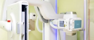
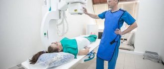
- X-ray is a painless and fairly accurate method. Before the examination begins, the patient needs to drink a contrast liquid (barium sulfate), after which the radiologist will begin the examination. During the study, images are taken that allow further diagnosis of any gastrointestinal disease. Diagnosis using this method is not recommended for people who suffer from conduction disorders of the digestive organs. The examination usually takes from twenty to forty minutes, and there are no side effects.
Any of these methods of gastroscopy of the stomach without swallowing is quite effective. Conclusions about the advisability of using it in a particular case can only be made by the attending physician who is familiar with the patient’s symptoms and medical history. Any procedure requires certain preparation and additional tests.
Preparation and performance of intestinal ultrasound
Preparation for the procedure begins 3 days in advance, the patient refuses food that causes constipation or flatulence (legumes, sweets, flour products, smoked and spicy foods).
The day before, from 18.00, the patient completely refuses any food, having first taken a laxative (Guttalax, Regulax, Duphalac, Bisacodyl). If there are problems with peristalsis, the patient is given an enema, and in special cases, a special cleansing enema is performed using a Bobrov apparatus (a glass vessel for introducing a large amount of liquid inside).
In the morning, the patient goes for an ultrasound examination until 11.00 am. This is due to the fact that the procedure is carried out only on a well-cleansed intestine and a completely empty stomach, while long breaks in food intake are contraindicated.
In the ultrasound diagnostic room, the patient lies on the couch on his side with his back to the machine, having first removed his clothes below the waist and lowered his underwear. The legs are tucked with the knees to the chest. Ultrasound begins in the direction from the lower parts to the higher ones. In parallel with this, the doctor moves the probe in such a way as to examine the intestine in the transverse, longitudinal and oblique planes. When the echogenic picture is not entirely clear, the doctor asks the patient to change position (lean on his knees and elbows, stand up).
Ultrasound of the large intestine is performed using a transabdominal probe. A contrast liquid (barium sulfate solution) is first injected into the empty intestine. Thanks to this, a clear picture is obtained on the monitor screen.
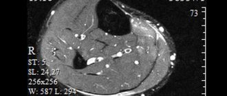
To examine the rectum, 3.5-5 MHz sensors are used. Ultrasound of a given length passes through the soft tissue of the intestine, being reflected back. The built-in receiving sensor picks up the signal and transmits it in processed form to the monitor screen. Various compactions, neoplasms and erosions are expressed in the form of white, black or mixed areas of varying echogenicity. An experienced doctor does not make a diagnosis immediately, but correlates the data obtained with the results of tests and other studies.
Gastroscopy (EGD)
Price
Diagnostic endoscopy (esophagogastroduodenoscopy without anesthesia)
RUB 4,025
Gastric biopsy using endoscopy
1250 rub.
Urease rapid test for Helicopter Pylori
575 RUR
Anesthesia
Application anesthesia
460 rub.
Medication-induced sleep (sedation)
Intravenous anesthesia for diagnostic studies up to 1 hour, 2nd degree of risk
3910 rub.
Examination (consultation) with an anesthesiologist-resuscitator
1800 rub.
Effective gastroscopy with and without anesthesia
Esophagogastroduodenoscopy (EGD, or gastroscopy of the stomach) is a modern, highly informative technique for diagnosing diseases of the esophagus, stomach and duodenum.
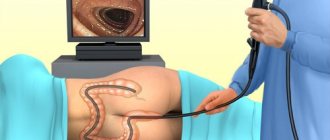
The essence of the gastroscopy procedure is that the patient swallows a special thin probe on which a small video camera is attached. After swallowing the flexible tube, the camera passes through the lumen of the patient's esophagus, stomach and duodenum. Images from the camera are transmitted in real time to the monitor screen. This allows the specialist to visualize the mucous membranes of various structures of the digestive system and diagnose conditions such as inflammation, ulcers, erosions, polyps, etc.
Carrying out gastroscopy without pain and the risk of complications is one of the areas of work of modern medical practice in Moscow.
When is EGDS performed?
Gastroscopy is recommended for patients suffering from the following pathological symptoms:
- pain, burning and discomfort in the abdomen and behind the sternum;
- nausea, vomiting, stomach upset;
- sudden change in body weight for no apparent reason;
- bleeding in the upper parts of the digestive system - vomiting with blood, black tarry stools;
- diagnosed anemia;
- cirrhosis of the liver;
- oncological diseases of the digestive system in a family history;
- long-term use of anticoagulants and antiplatelet agents;
- preparation for operations on the digestive tract.
Esophagogastroduodenoscopy is also often used as a control study to evaluate the effectiveness of treatment for diseases of the digestive system.
How is the procedure done?
Endoscopy of the stomach is an endoscopic examination that is performed only by a professional gastroenterologist. The whole procedure takes about 15 minutes. The patient is asked to lie on the couch on his left side and squeeze a special mouthpiece (mouthpiece) with his teeth. Through the hole in the mouthpiece, the doctor inserts a thin (only 8 mm thick) flexible endoscope tube into the patient’s mouth and slowly moves it along the esophagus, stomach to the duodenum. The endoscope tube is connected to a special pump, which pumps air into the lumen of the esophagus, stomach and intestines to improve the visibility of the walls.
Visualization occurs using a small camera that is located at the end of the endoscope tube along with a small lighting device. The signal from the camera is transmitted to the computer monitor. As a result, the doctor can visually monitor all movements of the endoscope in the patient’s digestive tract and assess the condition of the mucous membrane of the esophagus, stomach and intestines.
The specialist carefully examines the mucous membrane, assessing its color, structure and the presence of ulcers, damage and neoplasms. Also, during the research process, biological material is collected for biopsy. Doctors often combine gastroscopy with a test for Helicobacter Pylori as part of a comprehensive diagnosis of gastritis and stomach ulcers.
During the procedure, the patient should not experience severe pain. Discomfort may be associated with stimulation of the gag reflex when the endoscope is inserted and the appearance of belching when air is pumped into the esophagus and stomach. These manifestations are considered normal and are eliminated by preliminary irrigation of the oral cavity and entry into the esophagus with a 10% lidocaine solution. If the patient experiences severe fear and anxiety, cannot relax, or has increased sensitivity of oral receptors, gastroscopy can be done under anesthesia. In this case, the person is put into a short-term state of medicated sleep, which eliminates pain and discomfort.
You can carry out the classic procedure of endoscopy and gastroscopy with anesthesia inexpensively using. Our medical center is equipped with modern expert-class endoscopic equipment, which guarantees accuracy and high information content of diagnostics. Despite the fact that the procedure is carried out for a fee, the price in our clinic is affordable for everyone.
Share on social media networks:
DOCTORS OF THE DEPARTMENT Gastroscopy (EGD)
UrakovaYana Chingizovna
The doctor is an endoscopist. Candidate of Medical Sciences. Highest qualification category.
Sign up More details
Interpretation of intestinal ultrasound results
A healthy intestine has two membranes. The outer one is muscle tissue with low echogenicity, the inner mucous membrane is in contact with gas, and therefore is visualized as a hyperechoic layer.
During an ultrasound examination, the following parameters are assessed:
- Dimensions and shape
. The wall thickness is 3-5 mm. The picture is distorted in the event of the formation of gases that deform the ultrasound, and insufficient filling of the intestines with liquid. - The location of the intestine
relative to other organs. - Wall structure (echogenicity)
. The outer layer is hypoechoic, while the inner wall is characterized by hyperechogenicity. The contours are smooth, the intestinal lumen should not have expansions or contractions. Peristalsis is noticeable. - Length and shape of various sections.
The thermal section is 5 cm, the middle section is 6-10 cm, the middle ampullary section is 11-15 cm. - Lymph nodes.
Should not be visualized.
Deviations from the norm indicate various pathologies:
- Enteritis (inflammation of the small intestine): dilation of the intestine, increased peristalsis, accumulation of contents of varying echogenicity;
- Hirschsprung's disease (congenital pathology of an increase in certain sizes of the intestine): significant expansion of the lumen, uneven contours, heterogeneous wall thickness, noticeable places of thinning, lack of peristalsis;
- If it is impossible to determine the layers of the intestine, we can talk about acute mesenteric thrombosis - a consequence of myocardial infarction, expressed in thrombosis of the mesenteric artery;
- Uneven internal contours (which causes ulcerative lesions of the mucosal surface), weak echogenicity, thickening of the wall - all this indicates nonspecific ulcerative colitis;
- Chronic spastic colitis: areas of high echogenicity against the background of a hypoechoic surface, thickening of the walls;
- Ischemic colitis: inability to visualize layers, uneven thickening, reduced echogenicity;
- Acute appendicitis: the monitor screen shows a vermiform appendix 7 mm in diameter, the layers of the appendix do not differ from each other, the walls of the appendix are thickened asymmetrically, free fluid is visualized, increased echogenicity indicates an abscess;
- Diverticulitis (protrusion of the intestinal walls): at the site of the diverticulum, ultrasound “sees” a thickening of the wall more than 5 mm above normal, echogenicity indicates an abscess, the contours are uneven;
- Mechanical damage to the intestine: in addition to severe tension in the abdominal muscles, the patient’s echogenicity at the site of the hematoma is reduced, the walls at the site of damage are thickened;
- Oncology (cancerous or precancerous tumor): the external contours are uneven, the lumen is narrowed, peristalsis is impaired at the site of the tumor, lymph nodes of reduced echogenicity are visualized.
Of course, only a doctor can make a diagnosis. In this case, the results of other studies should be taken into account, for example, a blood test showing the degree of inflammation and the presence of parasites in the gastrointestinal tract, ultrasound of the liver and pancreas, etc.
Gastroscopy without tube insertion
01.08.2018
Currently, modern technologies make it possible to study the state of the gastrointestinal tract not only using the now common method with a gastroscope tube. Alternative methods of examination allow capsule endoscopy , virtual colonoscopy , computed tomography of the gastric , as well as electrogastrography and electrogastroenterography.
How is gastroscopy performed without inserting a probe?
Let's consider a method using a video pill, that is, capsule gastroscopy . One of the main differences from gastroscopy using a probe is the ability to obtain the most accurate information about the condition of the patient’s small intestine, which makes it possible to identify diseases in the early stages. The capsule itself is relatively small in size (about 1.5-2 cm). This makes it quite easy to swallow and wash it down with water. A sensitive video sensor is built into the capsule, which produces several thousand pictures during the period of time while the capsule is in the body, which is about eight hours. The advantages of this type of gastroscopy are obvious. Patients do not feel afraid of the procedure, and for those who are contraindicated in the classical form of the study, this procedure is the best fit. A huge advantage is the high information content of the procedure.
The disadvantages of the method cannot be ignored. The cost of conducting it is higher, and during this study it is not possible to take material for a biopsy and there is no possibility of removing polyps.
Contraindications for capsule gastroscopy
Of course, there are also contraindications for conducting this type of research. There are age restrictions: the examination is carried out for patients over 12 years of age. The study is not carried out for pregnant women, people with increased gag reflexes and patients with impaired swallowing function.
What should you do before gastroscopy using a video capsule?
To facilitate the procedure, the patient is recommended to perform a number of actions. Two days before the test, do not eat cabbage, legumes, alcohol, milk, pastries and carbonated drinks. You should take medications that reduce flatulence the day before. Stop eating 10-12 hours before the study.
The duration of the procedure is about 8 hours, the capsule must be taken on an empty stomach and washed down with plain water. During the procedure, you can behave as usual, but try not to make sudden movements or lift heavy objects. The capsule is removed naturally. During the examination, no discomfort or any deviations from the norm are observed. I would like to once again draw your attention to the fact that diagnostics using this method is highly accurate.
Timely diagnosis and an accurate diagnosis is the key to a speedy recovery!
Published in Diagnostics and examinations of Premium Clinic
What are the advantages and disadvantages of ultrasound of the intestinal gastrointestinal tract?
Ultrasound diagnostics of the intestine is used for initial examination in case of suspected pathology, as well as in cases where the endoscopic method is contraindicated due to the patient’s health condition (perforation (damage) of the intestine, inflammatory process).
Ultrasound examination of the intestines has a number of advantages:
- The patient does not experience psychological discomfort.
- The doctor receives information about the size of the organ, its structure, thickness, number of layers, without penetrating inside the organs.
- Ultrasound allows you to examine the inflamed intestines and clearly sees the upper gastrointestinal tract.
- Peristalsis is visualized in real time and intestinal obstruction is determined.
- On an ultrasound of the intestines, the specialist will see even small compactions or changes in the echostructure of tissues.
- Ultrasound allows you to do screening (endorectal method), completely confirm or refute oncology.
Despite the large number of advantages, diagnosing this organ with ultrasound has some disadvantages, the main one of which is the impossibility of making an accurate diagnosis without additional examination.
Also, the disadvantages of the method include the following:
- Only functional disorders in the functioning of the organ are detected.
- Structural changes are determined without defining the parameters of the changes.
- It is not possible to assess the condition of the internal mucous surface; if structural changes are detected, colonoscopy is prescribed - an endoscopic method
Indications and contraindications for gastroscopy under anesthesia (during sleep)
Gastroscopy
- This is one of the types of endoscopic examination. During the study, the endoscopist examines the mucous membrane of the esophagus and stomach, and, if necessary, other organs of the upper gastrointestinal tract. At the Central Clinical Hospital of the Russian Academy of Sciences, modern protocols and examination programs are used for gastroscopy, and the latest equipment is used.
As a rule, endoscopy is prescribed when the following signs of gastrointestinal diseases appear:
- burps of air;
- belching sour, bile, small amounts of food;
- sour, metallic, musty taste in the mouth;
- appetite disorders;
- nausea;
- heartburn;
- pain and other discomfort in the abdomen;
- pain when pressing on the abdomen;
- the appearance of discomfort after eating;
- cutting or aching pain on an empty stomach;
- bloating;
- difficulty swallowing;
- difficult or painful passage of food through the esophagus;
- bowel disorders;
- blood in stool;
- regular feeling of incomplete bowel movement.
Periodic EGD is necessary in the presence of chronic diseases to monitor changes in the patient's condition, assess the progress or regression of pathological changes. This type of examination is included in the dispensary program in the presence of such chronic diseases as:
- gastroptosis;
- gastritis of various nature;
- ulceration, scarring of the gastric mucosa;
- deformation of the biliary tract and other pathologies that can affect the general condition of the gastrointestinal tract.
Endoscopy allows you to identify incipient pathological changes in the tissues and mucous membranes of the gastrointestinal tract and detect in time:
- tumors;
- polyps;
- bleeding;
- changes in the gastric mucosa;
- gastroptosis;
- ulcers and perforations of the stomach;
- and other pathological changes.
There are several types of endoscopic examination. Esophagoscopy is prescribed if necessary to assess the condition of the esophagus. During gastroscopy, the stomach is examined. Esophagogastroduodenoscopy allows you to study the condition of the esophagus, stomach and duodenum.
The endoscopic method makes it possible to perform not only examinations, but also treatment procedures (if necessary) in a short time and with a minimum of complications:
- elimination of polyps;
- stopping bleeding;
- endobiliary manipulations for diseases of the pancreas and bile ducts.
To perform gastroscopy, a special flexible probe with a camera is inserted into the esophagus through the mouth, which captures the image along its path and transmits it to the monitor. Due to this, during the procedure the doctor can examine and assess the condition of the mucous membranes of the gastrointestinal tract in real time.
The procedure is very informative and is carried out quickly, that is, it allows you to obtain the most accurate data in a short time. However, gastroscopy without anesthesia has several features, due to which patients often refuse to perform this examination. This is primarily physical discomfort, due to which a person instinctively prevents the probe from moving through the esophagus, while resistance to the movement of the probe only increases the discomfort. In addition, gastroscopy without anesthesia often adversely affects the psychological state of the patient. Vomiting, stomach contractions and other factors complicate the examination, so today they try to carry out this procedure under anesthesia. The patient's condition is necessarily monitored by an anesthesiologist.
Advantages of performing gastroscopy under anesthesia:
- the patient does not experience psychological discomfort or unpleasant physical sensations;
- the physical reaction to the movement of the probe is reduced, which makes the study easier;
- the effectiveness of the examination increases (it is easier for the doctor to examine in detail all areas of the mucous membrane);
- Due to the absence of difficulties when advancing the probe, the procedure is faster.
Gastroscopy under anesthesia is to ensure the comfort and safety of the patient, as well as the accuracy of the examination results.
Using endoscopy during sleep, the doctor can conduct a painless examination of the mucous membranes:
- esophagus;
- stomach;
- duodenum.
Absolute contraindications to gastroscopy are heart attack, stroke and aortic aneurysm, bleeding disorders, mental illness, and spinal curvature. Relative contraindications are stage 3 hypertension and some other diseases of the cardiovascular system. The examination cannot be performed in case of acute respiratory tract diseases (sore throat, ARVI). As a rule, gastroscopy under anesthesia is not performed on children under 12 years of age. The final decision to prescribe an endoscopic examination is made by the gastroenterologist after reviewing the patient’s medical history.
Make an appointment
+7
Tests and studies that complement intestinal ultrasound
As mentioned above, intestinal ultrasound is not 100% confirmation of a particular diagnosis, although in many ways the method is informative and accurate. Depending on the preliminary diagnosis, in addition to ultrasound, the patient is prescribed:
- Capsule examination
. The patient swallows a capsule with a sensor inside, which conducts video surveillance and transmits the image to the monitor screen. The method allows you to see areas inaccessible to the endoscope. Significant advantages also include the absence of trauma (the intestinal walls are not scratched) and radiation (unlike X-rays).
The disadvantages of the capsule technique include the low prevalence of capsule examination, because the method was first tested in the USA in 2001, and today it is still not widespread. Its cost is very high, and this limits the circle of clients. Other disadvantages include the inability to conduct a capsule study in case of intestinal obstruction, infections, and peritonitis. The method has age restrictions associated with the peculiarities of peristalsis.
- Colonoscopy
. This is an endoscopic method that allows you to examine the internal mucous membrane for polyps, colitis, tumors, Crohn's disease, inflammation and other pathologies. The disadvantage of this method is the risk of intestinal trauma and perforation (punctures of the walls). Colonoscopy also does not see tumors between the intestinal walls. - Irrigoscopy
. This is a special method aimed at identifying hidden tumors located between the inner and outer lining of the intestine. In addition, the method, unlike colonoscopy, sees areas on the folds of the intestine and its remote areas.
Irrigoscopy involves the introduction of a liquid solution of barium sulfate through the anus, which allows a clear contrast image to be obtained upon contact with air. The advantages of irrigoscopy are the ability to examine structural changes in tissue (scars, diverticula, fistulas). The method is used for diarrhea or constipation, mucus in the intestines, pain in the anus.
Ultrasound of the stomach is an important part of the gastrointestinal tract examination using ultrasound.
For a long time, ultrasound diagnostics was not used in the study of the stomach. This is due to the fact that the stomach is a hollow organ, and the air does not allow full use of a conventional ultrasound sensor - special sensors are needed to examine the back walls. In addition, accumulated gases distort the displayed results. However, medicine does not stand still, and modern techniques already provide sufficient information to make an accurate diagnosis.
Sensors for studying the stomach appeared relatively recently, in the late 2000s. However, the speed and safety of scanning makes ultrasound examination of the stomach increasingly popular.
During an ultrasound examination, the doctor evaluates the organ according to the main indicators:
- Stomach volume.
It is a hollow muscular organ that resembles a pouch. The volume of an empty stomach is 0.5 liters, and when full it stretches to 2.5 liters. The height of the stomach reaches 18-20 cm, width - 7-8 cm. When filled, the stomach stretches up to 26 cm in length and up to 12 cm in width. - Structure.
Near the heart is the cardiac region, in which the esophagus passes into the stomach. On the left you can see the bottom of the organ, where air entering with food accumulates. The body of the stomach is the largest part, rich in glands that produce hydrochloric acid. The pyloric zone is the transition from the stomach to the intestine. There, partial absorption of substances received from food occurs. - Structure.
The walls of the stomach have a muscular layer that is responsible for contracting and promoting the food coma. The serosa is intermediate between the muscular and mucous layers. Lymph nodes and blood vessels accumulate in it. The mucous layer is covered with the finest villi, which secrete gastric juice produced by the glands. - Blood supply.
The circulatory system covers the entire organ. The organ is supplied with venous blood by three main vessels: the left, hepatic and splenic. The venous network runs parallel to the arterial network. Various bleeding occurs when the gastric mucosa is damaged (ulcers, tumors).
How is an ultrasound of the stomach performed?
Preparation for an ultrasound of the stomach is similar to an ultrasound of the intestines: the patient adheres to a strict diet for 3 days, and the night before does not eat any food from 18.00. If there is a tendency to form gas, the patient drinks 2 capsules of Espumisan before bed. In the morning, half an hour before the procedure, you should drink a liter of water so that the walls of the stomach straighten.
There is also a method of ultrasound examination with contrast. Water is an excellent conductor of ultrasound, and without it, scanning an organ is somewhat difficult.
The procedure is carried out on an empty stomach. The doctor assesses the condition and thickness of the walls on an empty stomach, looking for the presence of free fluid. Then he asks the patient to drink 0.5-1 liter of liquid, and uses an ultrasound machine to evaluate changes in the expanded stomach. A third ultrasound scan is performed 20 minutes later when the stomach begins to empty. The doctor evaluates the motility of the organ and the rate of fluid loss. Normally, a glass of water (250 ml) comes out of the stomach in 3 minutes.
The patient lies on the couch on his side, the specialist applies gel to the peritoneal area and moves the sensor over the surface. Periodically, he tells the patient to change position or change his posture slightly. The doctor pays attention to the following indicators:
- position of the stomach and its size
- Has the mucous surface of the stomach expanded?
- is there thickening or thinning of the walls
- what is the state of the circulatory system of the stomach?
- contractility of the stomach
- are there inflammations and neoplasms?
The entire examination takes a maximum of 30 minutes and does not cause discomfort or pain. Ultrasound, unlike FGDS, is much easier to tolerate for children and the elderly.
Preparation for the FGDS procedure
Any endoscopy requires maintaining the purity of the internal contents of the organ, since any substance that gets there or does not have time to be absorbed distorts the results of the study. A day or two before gastroscopy, you need to stop drinking alcohol, coffee, soda, heavy, spicy and fatty foods. On the eve of the study, food should be low-calorie and light.
Regardless of what half of the day the gastroscopy is performed, the last meal should be no later than 8 hours before the examination, it is forbidden to drink and smoke 2-3 hours before, as well as take medications, except for drugs to reduce blood pressure and treat the heart.
Advantages and disadvantages of ultrasound of the stomach when examining the gastrointestinal tract
The doctor prescribes an ultrasound examination of the stomach to the patient as a primary auxiliary diagnostic method.
The advantages of ultrasound are as follows:
- the exit section most susceptible to diseases is examined;
- ultrasound “sees” any foreign bodies in the cavity;
- Ultrasound accurately assesses the thickness of the walls of the organ;
- thanks to the method, venous blood flow is clearly visible;
- using diagnostics, benign and malignant tumors of minimal size are identified;
- stomach ulcers are well assessed;
- the degree of inflammation of the gastric mucosa varies;
- the method allows you to see reflux disease - the reflux of the contents of the lower sections back into the stomach;
- the organ is examined from different points and in different sections, which is impossible with x-rays;
- Ultrasound sees what is happening in the thickness of the stomach wall;
- thanks to the echo structure, ultrasound can easily distinguish a polyp from an oncological neoplasm;
- in addition to diagnosing the stomach, ultrasound diagnostics reveals concomitant pathologies of other organs (usually with gastritis, diseases of the biliary tract and pancreas develop);
- Ultrasound is performed on newborns and small children for whom it is impossible to undergo an FGDS or x-ray.
The main advantage of ultrasound over FGDS is the ability to detect forms of cancer developing in the thickness of the organ wall (infiltration forms), which cannot be detected using fibrogastroscopy.
Despite all the advantages, ultrasound has some disadvantages that do not allow the method to become widespread as an independent examination of the stomach.
The disadvantages include the following:
- Unlike endoscopic examination, ultrasound does not allow tissue samples to be taken for further study (for example, gastric juice;
- scraping of the mucous membrane, tissue biopsy);
- Ultrasound cannot assess the degree of changes in the mucous membrane;
- limitation of the areas studied (it is possible to examine only the outlet zone of the stomach).
What does ultrasound of the stomach reveal when examining the gastrointestinal tract?
The ultrasound method is not the most popular when examining the gastrointestinal tract, but it makes it possible to obtain very important information.
The stomach is an extension of the digestive canal in the form of a bag. It is a hollow organ whose walls have an outer muscular layer and an inner mucous layer. The mucous membrane is rich in glands that produce gastric juice and hydrochloric acid, as well as enzymes. With their help, incoming food is softened and treated with a natural antiseptic. The stomach is separated from the esophagus by the sphincter, and from the duodenum by the pylorus.
The organ is examined by ultrasound in two ways:
- Transabdominal (through the walls of the peritoneum). It is carried out with different sensors, but the results always require additional confirmation.
- Probe (sees the stomach from the inside). Used extremely rarely.
When conducting a study using a sensor, the specialist pays attention to the following:
- thickness, folding, structure of the mucous membrane (are there any neoplasms, bulges, or irregularities on it);
- thickness of the muscle layer (expansion or thinning indicates pathology);
- the integrity of the gastric wall (are there any perforations, ulcers or neoplasms);
- amount of free fluid (indicates inflammation);
- peristalsis, motility and contractility of the stomach;
- transitional sections of the stomach (sphincter and pylorus, their features
- functioning).
It is worth noting that ultrasound of the stomach and duodenum is significantly inferior in its informative value to the more popular method known as FGDS. But in some cases, other research methods are unacceptable for the patient due to health conditions or fear of a traumatic procedure.
Transabdominal examination identifies three layers of the gastric wall: hyperechoic mucosal layer (1.5 mm), hypoechoic submucosal layer (3 mm) and hyperechoic muscular layer (1 mm). With the probe method of research, 5 layers up to 20 mm thick are determined.
Ultrasound diagnostics of the stomach allows us to identify the following pathologies
| Symptoms | Possible disease |
| Swelling of the antral mucosa | Acute pancreatitis, nephrotic syndrome (kidney damage) |
| Thickening of the stomach wall, uneven rounded neoplasm, rich in blood vessels, no boundaries between layers, no peristalsis | Carcinoma (malignant tumor) with distant metastases |
| Lack of boundaries between layers, narrowing of the pyloric lumen | Pyloric stenosis (narrowing of the pylorus due to scarring caused by an ulcer) |
| Changes in the echostructure of the stomach walls, the walls are expanded, the contours are uneven | Neuroma (tumor developing from the tissues of the peripheral nervous system), leiomyoma (benign tumor of the smooth muscles of the stomach), adenomatous polyp |
| Expansion of the abdominal region (compared to the norm) after filling the stomach with water, splitting of the echo signal, the presence of hypoechoic inclusions, stagnation of fluid in the cardiac region | Gastroesophageal reflux (reflux of intestinal contents back into the esophagus) |
| Small amount of fluid, rapid release of fluid from the stomach, changes in the contour of the stomach | Diaphragmatic hernia |
| Dense hyperechoic formations with a clear structure, the boundaries between the layers are clearly visible, the echogenicity of the mucous and muscle layers is not changed | Cystic formations |
| Uncertain changes recorded by ultrasound | Affected hollow organ syndrome. This diagnosis requires mandatory confirmation by other types of research (CT, MRI, FGDS, X-ray). |
| Anechoic crater-like areas on the inner wall of the stomach | Stomach ulcer |
Ultrasound scanning of different parts of the stomach
Thanks to ultrasound, the doctor assesses the condition of the following areas of the organ:
Bulbar or duodenal bulb
. This part of the organ is located in the area where the stomach exits, and controls the flow of contents processed by gastric juice into the intestinal lumen. With intestinal diseases, ulcers and sites of inflammation form on the bulb. The main reasons for duodenal ulcers are increased acidity and the bacterium Helicobacter pylori, which begins to actively multiply under such conditions.
The study is carried out in real time with a linear or convex sensor with a frequency of 3.5-5 MHz. To detail the condition of the walls, sensors with a frequency of 7.5 MHz are used, but they are ineffective for obese patients with developed subcutaneous fat.
If a patient is diagnosed with a gastric and duodenal ulcer, then in most cases the walls of the bulb are affected. On ultrasound, this is reflected by anechoic areas, because, unlike healthy walls, the ulcer does not reflect ultrasound.
The diagnosis of “stomach and duodenal ulcer”, if areas of anechoicity are identified on ultrasound, is made conditionally. Additionally, the condition of the walls of the bulb is assessed (they have a mucous structure with longitudinal folds). The normal thickness should be no more than 5 mm, and in the antrum (the transition of the stomach into the duodenum) - up to 8 mm. With thickening, we are not talking about an ulcer, but about an oncological neoplasm. The patient will need additional research: endoscopic with sampling of material for biopsy.
Due to the fact that ultrasound is not able to establish an accurate diagnosis, the patient is given a preliminary diagnosis of “anechoic areas”, and then he is sent for fibrogastroduodenoscopy. It is this method that makes it possible to take tissue from the wall of the bulb to determine the nature of the pathology. FGDS also allows you to assess the condition of the organ’s vessels.
Pyloric canal or pylorus of the stomach.
This is a slight narrowing at the junction of the bulb and the duodenum. It consists of smooth muscle walls 1-2 cm long, located both in the annular and transverse directions. Normally, there is some curvature of the canal. Ultrasound can detect diseases such as polyps, stenosis (narrowing), ulcers, and pyloric spasm.
Sphincter (cardia)
- This is the border between the peritoneum and the esophagus. Normally, the sphincter opens only after eating, and remains closed the rest of the time. Due to its functional significance, the sphincter has a stronger muscular layer than the stomach, which allows it to open and close like a valve. When eating, the sphincter closes the exit from the stomach, allowing food to be digested. But as a result of increased acidity and other pathologies, the organ ceases to function normally, and the contents of the stomach enter the esophagus.
Pathology detected: should it be rechecked?
Ultrasound of the stomach and intestines is very informative, but it is impossible to make a diagnosis based on the data obtained. If problems are detected, the patient undergoes additional examination. The most popular methods for examining the gastrointestinal tract include:
- FGDS. This is an endoscopic method that allows you to see bleeding, tumors in the stomach and intestines.
- Probing. It involves taking the contents of the stomach for further laboratory testing.
- Gastropanel. This is an innovative method, according to which the patient is drawn from a vein, and a possible ulcer, atrophy, or cancer is detected using certain markers.
- CT scan. They take cross-sectional images in different projections and identify the location of tumors, hematomas, hemangiomas, etc.
- MRI. This is the most expensive and effective research method. Allows you to visualize not only the organ itself, but also nearby lymph nodes and blood vessels.
- Endoscopy. Used when collecting material for biopsy.
- X-ray. Reveals incorrect location of the stomach and intestines relative to other organs, pathology of shape, and various neoplasms.
- Parietography. Translucent the walls of the stomach and intestines thanks to the injected gas.
- Laboratory tests (blood, urine, stool tests).
After undergoing additional diagnostics, the doctor decides on treatment methods. It is important to understand that treatment of the gastrointestinal tract cannot be done in a “mono” mode - it is always a set of measures related to restoring health and preventing relapses and complications. You can also monitor the quality of treatment using ultrasound, comparing previous results of a gastrointestinal examination with new ones.
If you find an error, please select a piece of text and press Ctrl+Enter