Ultrasound diagnostics (sonography) is one of the most widely used additional research methods in medicine.
Due to its safety, ultrasound is performed on patients of any age, including children and pregnant women.
In addition to safety, the inherent advantages of ultrasound examination include its painlessness, high diagnostic information content and affordable price.
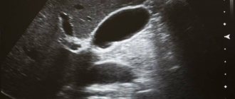
Ultrasound of female genital organs
Many diseases of the female genital organs have no symptoms and are hidden, so it is important to undergo regular ultrasound examinations. It is prescribed on different days of the cycle depending on the organ that needs to be examined:
- preventive examination is carried out in the first few days after the end of menstruation;
- if an enlarged layer of the endometrium is suspected, the study is prescribed for the second half of the cycle;
- if it is necessary to monitor the development of the disease or verify the effectiveness of treatment, then ultrasound is performed several times per cycle;
- in case of bleeding not associated with menstruation, examination is carried out urgently, regardless of the cycle.
An ultrasound can help you learn more about the condition of the uterus, fallopian tubes, ovaries and cervix.
Types of ultrasound during pregnancy

Sonography for pregnant women is the main instrumental method for diagnosing pathologies and monitoring the course of pregnancy. Ultrasound is safe for the mother and fetus, which is why it is preferred to all other diagnostic methods.
In the first months of pregnancy, when the embryo is still very tiny and difficult to see with a conventional transabdominal ultrasound, women are examined with a transvaginal sensor. It is inserted into the vagina and scans the uterus through the cervix. The signals obtained in this way are clearer, which makes it possible to examine the embryo and the walls of the uterus in detail at high magnification.
If it is not technically possible to conduct a transvaginal examination at short notice, the woman undergoes a transcutaneous ultrasound. Since anatomically the uterus in the pelvis is located behind the bladder, its muscular walls do not allow the uterus to be seen. Before a transabdominal examination, an early pregnant woman needs to drink 0.5-1 liters of water to fill her bladder. Then the ultrasonic waves pass unhindered through the liquid and scan the uterine cavity with the fertilized egg located in it.
After the 12th week of pregnancy, ultrasound is performed transabdominally (through the wall of the abdominal cavity). The pregnant uterus increases in size and rises above the bladder, so it becomes easily accessible for examination using ultrasound. The fetus at these dates is of sufficient size to be well visualized.
There are the following types of ultrasound during pregnancy:
- Fetometry (Latin "fetus" - fetus, Greek "metry" - to measure) is the taking of intrauterine measurements of the fetus. During the study, the length of the embryo, coccygeal-parietal, biparental (from temple to temple) dimensions, circumference of the head, abdomen, chest, and fetal femur length are determined. During the diagnostic process, the obtained sizes are compared with standard indicators for a certain week of pregnancy. In parallel with assessing the condition of the fetus, during fetometry the condition of the uterus and its cervix is examined.
- Doppler ultrasound is an auxiliary type of ultrasound, which is not performed on all pregnant women, but only as prescribed by the obstetrician-gynecologist managing the woman’s pregnancy. Doppler ultrasound is prescribed to assess blood flow in the vessels of the placenta and fetus. Doppler testing is indicated for gestosis, diabetes mellitus and kidney disease in pregnant women, suspected fetoplacental insufficiency, intrauterine growth retardation, umbilical cord abnormalities, fetal heart defects.
- Volumetric ultrasound (3D/4D) makes it possible to determine the general condition, assess the blood flow, breathing, swallowing of the fetus, exclude the presence of malformations, the quantity and quality of amniotic fluid, the location and condition of the placenta, and establish the correspondence of fetal development to the gestational age. 3D/4D ultrasound is often used by expectant parents to get realistic photographs of their baby before birth.
These examinations can be carried out at any stage of pregnancy, however, a certain cyclicity has been established for fetometry. According to the protocols for pregnancy management, ultrasound examinations for the purpose of screening the course of pregnancy are carried out:
- at 5-8 weeks - to confirm pregnancy, establish the viability of the embryo (by heartbeat) and determine the place of its fixation;
- at 10-12 weeks - to clarify the gestational age, determine the correspondence of the development of the embryo to the gestational period, measure the thickness of the nuchal fold (to exclude some genetic pathologies), establish the place of attachment and condition of the placenta, assess the quantity and quality of amniotic fluid;
- at 22-24 weeks - to monitor the development of the fetus, detect developmental defects in it, assess the condition of the placenta, umbilical cord, amniotic fluid);
- at 30-32 weeks - to assess the development of the fetus, the degree of its motor activity (often combined with Doppler ultrasound) and confirm the timing of delivery;
- before birth (36-38 weeks) - to determine the position of the fetus in the uterus, establish its condition, the degree of maturity of the placenta and the characteristics of the amniotic fluid.
Ultrasound during pregnancy is the main diagnostic instrumental study. Based on the findings from the ultrasound examination, other diagnostic tests may be prescribed, if necessary.
Indications for the procedure
The doctor may suggest an ultrasound of the genital organs if the following symptoms are present:
- pain in the lower abdomen not associated with the menstrual cycle;
- impossibility of pregnancy;
- bleeding outside the cycle;
- suspicion of the appearance of neoplasms;
- absence of menstruation;
- bleeding, accompanied by nausea, weakness and vomiting.
In addition, examination of the female genital organs is necessary to monitor pregnancy in the first trimester, to diagnose pregnancy or infertility, and to obtain general information about the condition of the reproductive system.
Normal indicators during examination of the uterus
Normally, the external contour of the uterus should have clear, even boundaries. Otherwise, this indicates inflammation of the surrounding tissues and the possible development of pathology. The shape of the uterus is pear-shaped. The position of the organ is forward tilt. However, tilting back does not mean pathology; in this case, complications during pregnancy and childbirth are only possible.
Sizes before menopause:
| Length | 45 – 67 mm |
| Width | 46 – 64 mm |
| Thickness | 30 – 40 mm |
The size of the uterus after menopause can change significantly; the transformation process occurs over 20 years:
| Length | 42 mm (maximum) |
| Width | 44 mm |
| Thickness | 30 mm |
The walls of the uterus should be uniform in structure. The structure and thickness of the endometrium changes throughout the cycle:
| About 4 days | 3 – 9 mm |
| 5 – 15 day | 3 – 15 mm |
| Second half of the cycle | Up to 20 mm |
Normal indicators during examination of the ovaries
| Circuit | Uneven, lumpy, clear |
| Structure | Homogeneous with inclusions up to 6 mm (follicles) and up to 25 mm in the middle of the cycle (ripe follicle) |
| Length | 20 – 37 mm |
| Width | 18 – 30 mm |
| Thickness | 14 – 22 mm |
| Volume | 20 – 80 mm3 |
Normal indicators for examining the fallopian tubes
Normally, they are not visualized on ultrasound, otherwise it is a sign of pathology.
Normal indicators for examination of the cervix
| Structure | homogeneous |
| Circuit | smooth, clear, without bending |
| Length | 30 – 40 mm |
| Cervical canal | up to 3 mm, contains mucus |
Norm of indicators when examining the retrouterine space
There may be a few milliliters of fluid in the middle of the cycle, after ovulation.
Pathological indicators: when treatment by a gynecologist is required
The doctor does not rely solely on sonography data to make a diagnosis. The specialist prescribes additional tests, with the help of which he can confirm or refute the suspicion of pathology.
| Signs on ultrasound | Possible pathologies |
| Deviation of the uterus from normal positions, fluid in the retrouterine space | Adhesions, inflammation |
| Neoplasms in the uterine cavity (the size of the organ often increases) | Cyst, fibroids, polyps |
| Thickening of the endometrium | Hyperplasia |
| Visualized fallopian tubes | Inflammation, ectopic pregnancy developing in this area |
| Blurred areas of the uterus | Inflammatory process |
| Uneven contours of the uterus | Fibroids, cancer or other type of tumor |
| Heterogeneous structure of the cervical canal or its causeless expansion | Infectious and inflammatory process |
| Distorted contour of the ovaries | Inflammation |
| Increased ovarian size | Polycystic disease, single neoplasm |
The interpretation of gynecological ultrasound necessarily contains terms for echogenicity. What echogenicity is can be found on our website in the Services - Ultrasound section.
Ultrasound methods of female genital organs
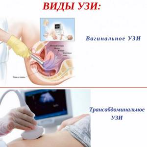
Transabdominal examination.
It is carried out through the external abdominal wall. This method helps determine the size and structure of internal organs, as well as identify pathological formations (cysts and tumors).
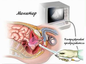
Transvaginal examination.
It is carried out using a special sensor, which is inserted into the vagina. The method allows you to study the condition of organs in more detail.
Combined study.
It combines an abdominal scan with a full bladder and a vaginal scan with an empty bladder. This is required for examining high-lying organs.
Transrectal examination.
Typically used when examining girls who are not sexually active.
How is the procedure performed?
There are several methods for examining the uterus and adjacent organs), which ultrasound method to choose is decided by the doctor:
- Transabdominal
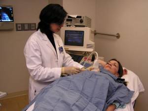
(abdominal ultrasound of the uterus, ultrasound of the cervix) is an external method of scanning through the abdominal wall. The woman lies on her back, the doctor applies a special gel to the patient’s stomach to improve the passage of ultrasound beams and conducts an examination using a sensor. The woman does not experience any discomfort during the procedure. The disadvantage of the method is its low information content compared to other methods of examining this organ. With its help, only gross pathological changes can be detected. It is usually used for general examination, for virgins and women with vaginal malformations. - Transvaginal
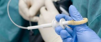
- by inserting an intracavitary sensor into the vagina. The woman also takes a lying position with her knees bent. The doctor puts a condom on the sensor and applies lubricant to it to improve gliding and minimize discomfort during insertion of the sensor. Transvaginal ultrasound of the uterus is considered the most accurate and informative, since the device is in close proximity to the internal organs. This type of ultrasound is contraindicated for virgins and for vaginal malformations. - Transrectal
- by introducing an intracavitary sensor (thinner than for TVU) into the rectum. The patient lies on her side and presses her knees to her body. The doctor puts a condom on the sensor, also applies lubricant and carefully inserts it through the anus. The patient may experience some discomfort during insertion of the sensor. This method of ultrasound is performed in some cases, when TAU turned out to be insufficiently informative, and TVU cannot be performed for objective reasons (virginity, atresia (fusion), severe stenosis (narrowing) of the vaginal opening, etc.).
Additionally, a detailed ultrasound examination of individual organs may be prescribed: ultrasound of the ovaries, ultrasound of the appendages, ultrasound of the bladder. And if there is a suspicion that other organs are affected in the process, a comprehensive pelvic ultrasound or abdominal ultrasound is performed.
Indications for ultrasound of male genital organs
A man may need to undergo an ultrasound to diagnose the following problems:
- benign and malignant tumors of the testicles and appendages;
- abnormal location of the testicle in the groin or developmental abnormalities;
- hematoma, hemorrhage, tissue damage;
- inflammatory process in the testicles and other organs (orchitis, epididymitis, tuberculosis);
- diseases of the testicles, epididymis or spermatic cord of a vascular nature: varicose veins, testicular infarction, torsion of the cord.
Ultrasound allows you to fully see the situation, assess the condition of the organs and begin treatment.
Research Alternative
Gynecological examination can be carried out in several ways: ultrasound, hysteroscopy, laparoscopy, computed tomography and magnetic resonance imaging, etc.
Alternative methods are selected for each specific gynecological disease. The universal research method is ultrasound. The value and popularity of the method, in comparison with others, is confirmed by the following provisions:
- high reliability and information content of the results obtained (85-100%);
- non-invasive;
- painlessness;
- simplicity and accessibility of the method;
- assessment of the condition of organs in “real time”, thanks to which it is possible to diagnose even an acute disease without resorting to puncture of the posterior vaginal vault and biopsy;
- harmlessness (unlike X-rays, ultrasound is not based on dangerous radiation, but on the laws of propagation of sound waves);
- relatively low cost of research.
In addition to a gynecological ultrasound, it is recommended to undergo an ultrasound of the mammary glands with a detailed examination of regional nodes. Since pathologies of the uterus and breasts in women are closely related.
Preparing for an ultrasound
The abdominal research method involves preparation, which consists of giving up gas-forming foods (legumes, brown bread, dairy products, sweets, alcoholic and carbonated drinks). In some cases, before the procedure, the doctor may prescribe an enema or the use of enterosorbents - consult a specialist. Transrectal ultrasound requires an enema before the examination, since the procedure is performed while the lower intestine has emptied.
How much does a gynecology ultrasound cost?
At Heratsi Medical Center, the cost of ultrasound (gynecology) is open on the website and you can determine the price of the visit in advance. Additionally, you can get a full consultation from a gynecologist and take the necessary tests.
If it is important for you to get a high-quality assessment of your health and it is important to understand how much it costs to do a gynecological ultrasound, then our offer will be profitable.
You will receive not just a conclusion sheet, but also all the indicators and necessary recommendations explained in detail.
Protect yourself from serious consequences, take care of your health and come to the Heratsi Medical Center, our ultrasound (gynecology) addresses are the same - Stachki Ave., 226. Sign up online.
Contraindications for ultrasound
Contraindications for transrectal examination are deep anal fissures, the presence of inflamed hemorrhoids and recent rectal surgery. Vaginal ultrasound is not performed on virgins (the transrectal method is used), with latex allergies, epilepsy, in the second and third trimesters of pregnancy, and also after operations. Transabdominal examination, which requires a full bladder, is not recommended for patients with urinary incontinence or recent bladder surgery.
Echo signs of uterine pathology
| Pathology | Features |
| Uterine cancer | Changes in the contours of the uterus, swelling |
| Myoma | Increased size of the uterine body, altered contours, “hyperechoic zone” (nodule in the muscle) |
| Polyps | Fuzzy contours of the uterus, space-occupying formations in the uterus |
| Endometriosis | “Bubbles” on the cervix, in the tubes and in the uterus itself |
| Adnexit | Thickening of the fallopian tubes, enlarged ovaries, unclear and blurry contours |
| Ovarian cancer | Increase in size with deformation of contours |
| Ovarian cyst | Accumulation of fluid with a diameter greater than 25 mm |
| Endometritis | Swelling of the endometrial layer, increased size of the uterus, thickening of the walls |
| Cervical tumor | Deformation and enlargement of the cervix |
To summarize, it should be noted that transvaginal ultrasound makes it possible to make a diagnosis in a timely and correct manner, to determine early pregnancy and its pathologies.
What kind of ultrasound should men do?
Ultrasound examination is a mandatory regular procedure for women, but in some cases, examination of the pelvic organs is also prescribed for men. So what ultrasound should I do?
to a man? In each specific situation, the examination method is selected individually; it depends on the individual characteristics of the body and the problems that led to the need for examination. In this case, two research methods are possible:
- Transabdominal.
The procedure is carried out according to general rules: the sensor scans readings through the front wall of the abdomen, and the bladder must be filled (a liter of clean water is required before the study). In the case of men, this method is not informative enough due to the location of the prostate gland; in addition, if the patient is overweight or has urinary incontinence, the diagnostic efficiency is further reduced, so it is worth choosing a different method.
- Transrectal.
The best way to check the prostate gland, because the prostate is located as close as possible to the rectum (or is in contact with its wall).
You will also be interested in our other services
Ultrasound examinations for women from 20 to 30 years old
It is between the ages of 20 and 30 that most women become sexually active and think about having a child, so the list of necessary studies is quite large:
- Examination by a gynecologist, dentist and ophthalmologist - at least once a year.
- Blood test (general, for hepatitis and HIV infection) – once a year.
- Blood pressure control - measurement at every doctor's visit.
- Fluorography – once a year.
- Checking the amount of hormones - in case of menstruation failure.
In addition, regular ultrasound examinations are necessary, since they can be used to diagnose a large number of diseases:
- Anemia (and other blood clotting problems).
- Tuberculosis.
- Ovarian cyst.
- Various oncological diseases.
- Hypertension and many others.
At the same time, the main ultrasound examination for a woman is a pelvic ultrasound, which examines the woman’s reproductive organs. This study cannot be neglected, since it is at this age that diseases develop that can cause infertility. When carrying out this procedure, you can identify:
- uterine fibroids;
- ovarian dysfunction;
- polycystic ovary syndrome;
- the occurrence of inflammation;
- infections;
- frozen pregnancy and other pathologies associated with the pelvic organs.
For women, the following types of ultrasound examination are required:
- examination of the pelvic organs (in this case, the doctor chooses the most informative method) – 2 times a year;
- ultrasound examination of the mammary glands – once a year for women over 25 years of age;
- Ultrasound of the kidneys - every third year.
In addition, if a woman has problems conceiving a child, it is necessary to undergo an examination of the uterus, ovaries and appendages.
What studies are mandatory for women aged 30 to 40 years?
At this age, it is necessary to conduct a study once a year:
- Pelvic organs (uterus, ovaries, etc.).
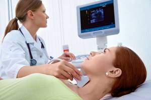
- Thyroid gland.
The normal functioning of the reproductive system, heart, and the amount of hormones depends on this organ. Any malfunction of the organ can lead to serious illnesses, especially in women of this age.
In addition, an ultrasound of the thyroid gland must be performed unscheduled if the following symptoms appear: the appearance of excessive fatigue and apathy, constant elevated body temperature, severe obesity and sudden changes in weight, tremors in the hands.
The main thing is not to delay visiting a specialist, otherwise serious health problems may arise, which will be very problematic to cure.
- Mammary glands.
At this age, the likelihood of tumor formation increases, this is due to the fact that most women after 30 years are faced with the following life events: childbirth with complications, hormonal problems due to improper use of contraceptives followed by abortion, and so on. The body's immunity weakens, which is why more and more women are experiencing the formation of cancerous tumors.

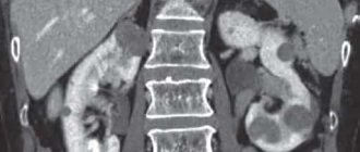
- Abdominal organs.
A study of the gallbladder and kidneys should be carried out on all women after 30 years of age, since their functions are often impaired at this age. In this case, problems can arise for several reasons: the consequences of a difficult birth, infection, frequent consumption of spicy and fried foods, cystitis.
In addition, women of this age should regularly undergo the following examinations (in addition to the list recommended for younger women):
- Blood examination (lipid profile and sugar level).
- Body mass index (hormonal processes change, so many women after 30 years are prone to obesity).
Important! The likelihood of having a stroke or heart attack increases dramatically at this age. Timely diagnosis (including using ultrasound) and competent treatment (including recommendations for proper nutrition) will help avoid these problems.