Cystoscopy is an endoscopic diagnostic method, during which the doctor examines the bladder from the inside. During the study, surgical manipulations can be performed in the organ cavity: removal of tumors, biopsy, destruction of stones, etc. To perform cystoscopy, special equipment is required - a cystoscope. Depending on the purpose of the study, a regular viewing or catheterization cystoscope can be used.
- Indications and contraindications
- How to prepare for cystoscopy of the bladder
- Execution method
- Possible complications
- Advantages and disadvantages of the cystoscopy method
Indications and contraindications
Bladder cystoscopy is performed for both diagnostic and therapeutic purposes. In the first case, the study allows you to make an accurate diagnosis. It is prescribed in the following cases:
- Cystitis with frequent exacerbations.
- Macro- and microhematuria.
- Suspicion of a tumor.
- Overactive bladder.
- Chronic pelvic pain.
- The presence of atypical cells in the urine according to the analysis result.
- Painful, difficult urination.
For therapeutic purposes, cystoscopy is used in the following situations:
- When removing tumors.
- To stop bleeding.
- For the purpose of destruction and removal of urological stones.
- When performing a biopsy.
In addition, this procedure is used to help patients with urinary retention. During cystoscopy, it is possible to restore the patency of the urinary tract in case of prostate adenoma, strictures, or obstruction of the bladder neck.
Cystoscopy is contraindicated if there are signs of an acute inflammatory process in the organs of the genitourinary system or if there is damage to the urethra.
Book a consultation 24 hours a day
+7+7+78
Types of cystoscopes
To carry out the procedure, a cystoscope is used, which is a special design made of a thin tube with optics built into it, which transmits the resulting image to an external monitor. A cystoscope is inserted into the bladder through the urethral canal, sequentially examining all parts of the organ. To carry out the procedure, two types of structures have been developed:
- Rigid cystoscopes . The optical system of the device is designed to observe and assess the condition of the organ directly with the eyes without displaying the image on a large screen.
- Flexible cystoscopes. The image is transmitted to the monitor thanks to the special features of the optics.
Important. In modern medicine, rigid structures are extremely rarely used, since they have a number of negative features - their insertion and movement through the body causes pain, and the quality of visualization is not as accurate as that of flexible cystoscopes.
How to prepare for cystoscopy of the bladder
Before carrying out the procedure, you need to undergo simple preparation. First, the patient is prescribed a general urine test and other laboratory tests that will help eliminate contraindications. If the patient is taking anticoagulants or certain other medications, the doctor may stop them a few days before the cystoscopy.
Depending on the type of anesthesia chosen to be performed, the anesthesiologist will give recommendations on immediate preparation for the intervention. For men, due to the anatomical structure of the urethra, it is recommended that the examination be performed under spinal or general anesthesia. In this case, preparation for cystoscopy involves refusing to eat 11-12 hours and refusing to drink 3 hours before the procedure.
Cystoscopy in women is often straightforward, so general anesthesia is not required. The female urethra is straight and up to 5 cm long, unlike the male urethra, which can reach 15-20 cm. For pain relief, the doctor uses a local anesthetic. It is applied to the endoscope tube. If cystoscopy is performed using local anesthesia, no special preparation is required.
In all cases, before the study, it is necessary to empty the bladder and thoroughly clean the external genitalia. On the eve of cystoscopy, you should refrain from sexual intercourse.
Chromocystoscopy
To obtain information about kidney functionality, diagnostic cystoscopy in adults is often combined with another procedure, chromocystoscopy.
This study requires preliminary intravenous administration of a contrast agent, after which the doctor assesses from which ureter, after what time and at what speed colored urine will be released.
If the appearance of urine with dye is detected 3-5 minutes after the contrast is introduced into the vein, then this is normal. If excretion is delayed for up to 10 minutes, renal dysfunction is diagnosed.
Execution method
Cystoscopy of the bladder is performed using general or spinal anesthesia. Local anesthesia can also be used if the study is carried out for diagnostic purposes.
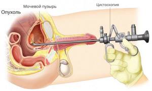
In order to facilitate insertion of the cystoscope into the urethra, it is lubricated with sterile glycerin. With gradual advancement of the instrument, during cystoscopy the inner wall of the urethra is first examined (ureteroscopy is performed). When the tip of the cystoscope is close to the pubic symphysis, it is directed downward and enters the bladder.
Procedure for examining the bladder
First of all, the urothelium of the anterior wall of the organ and its apex is examined. Next, the internal mucous membrane of the organ is examined in a circle by turning the cystoscope clockwise. Particular attention is paid to the area called the Lieto Triangle. The boundaries of this area are determined by the orifices of the ureters and the internal opening of the organ’s neck. The mucous membrane of Lieto's triangle is most often affected by various pathological processes.
For ease of orientation, the bladder is conventionally divided into four parts according to the watch dial. In this case, normally the air bubble, which is located at the top of the organ, will correspond to 12 o’clock, the mouth of the right ureter will correspond to 7 o’clock, and the left ureter will correspond to 5 o’clock.
During the study, the color of the endothelium is assessed, the presence of certain changes on the surface of the mucous membrane (ulcerations, areas of tissue degeneration, neoplasms, etc.). The specialist also pays attention to the location and shape of the ureteric orifices.
Normally, the urothelium has a pale pinkish color and a characteristic shine. A network of small vessels is determined on its surface. In the area of Lieto's triangle there are a significant number of larger vessels. The orifices of the ureters should be located symmetrically. Normally, they are slit-like, oval, or round in shape.
Book a consultation 24 hours a day
+7+7+78
Biopsy
Cystoscopy is widely used to obtain biopsy material from pathologically altered areas of the bladder wall. Histological examination is used to diagnose neoplasms and other pathological processes. Material for analysis can be obtained in the following ways:
- Cold biopsy - tissue is collected using special forceps that are inserted through an endoscope.
- Transurethral resection (TUR biopsy) is carried out as a result of cutting off a section or completely removing the formation with an electrocoagulator. Removal instruments are also guided into the bladder to the tumor through a flexible cystoscope.
Each of these methods has its own disadvantages and advantages. With a cold biopsy, the tissue sample and the inner wall of the organ are practically not damaged, but there are difficulties in assessing the depth of spread of the tumor. TUR biopsy, in turn, allows you to determine the degree of tumor growth. In addition, thanks to the use of a coagulator, there is virtually no risk of bleeding when using this technique. The disadvantage of TUR biopsy is its relatively high invasiveness.

Possible complications
During the first day after cystoscopy, the patient may notice the presence of blood in the urine, a burning sensation and discomfort along the urethra, and pain when urinating. In order for these side symptoms to resolve more quickly, the patient is recommended to increase fluid intake to 3 liters on the first day after the intervention.
Complications that may develop after cystoscopy include:
- Acute urethritis, cystitis, pyelonephritis.
- Bladder perforation.
- Development of bleeding.
- Trauma to the urethra.
Complications of an inflammatory nature may occur due to violation of the rules of asepsis and antisepsis during cystoscopy. Another reason for the development of infectious complications may be insufficient hygiene of the external genitalia before the procedure.

The bladder can also be perforated as a result of unprofessional biopsy or transurethral tumor removal. If the integrity of the organ is violated, there is severe pain in the lower abdomen and the release of a reduced amount of urine mixed with blood. The patient needs urgent surgical care.
The appearance of microtraumas of the urethra during cystoscopy is almost impossible to avoid, but they do not manifest themselves clinically, and their healing occurs quickly. But as a result of incorrect or careless actions, the urethra can be seriously injured with the formation of a false passage. In this case, the patient feels severe pain and bleeding may develop.
Study in women
Typically, the test is easier for women than for men. The reason for this is the peculiarity of the urethra, short and straight.
General anesthesia is not required; a local option is used: an anesthetic is applied to the device.
Difficulties may arise if there are significant tumors in the uterus and pregnancy in its later stages, when the bladder is compressed by the uterus.
It must be understood that the procedure in the second case is performed only for health reasons: there is a high probability of provoking a spontaneous abortion.
Advantages and disadvantages of the cystoscopy method
The undoubted advantage of this technique over many other studies in urological practice is its high information content. In particular, during the procedure, malignant tumors can be detected in the bladder in the early stages, thanks to which the patient is successfully treated. In addition, cystoscopy is used to perform a wide range of therapeutic procedures, from restoring the patency of the urinary tract to removing tumors.
The disadvantage of the study is that for its successful implementation a number of conditions must be met:
- Normal urethral patency.
- The bladder must have sufficient capacity.
- The fluid used to fill the bladder during the procedure must be clear (heavy bleeding or large amounts of pus will interfere with the examination).
- The patient’s readiness for the study, absence of acute painful sensations during the procedure. Severe discomfort or the appearance of an uncontrollable urge to urinate are indications for forced termination of cystoscopy of the bladder.
The disadvantage of cystoscopy is its technical complexity. To perform the procedure efficiently, accuracy and certain experience are required, since unprofessional actions can lead to injuries to the organs of the lower parts of the urinary system.
Book a consultation 24 hours a day
+7+7+78
A little history
Urethroscopy began to be performed in the 20th century.
Before the advent of the urethroscope, the urethra was examined using a cystoscope during cystoscopy. The cystoscope, designed by Bozzini in 1804 in Germany, was based on a hollow tube, and a candle was used as a light source.
Kelly and Johns Hopkins then improved the cystoscope by using an open, air-filled cystoscope to view the bladder. This device became very convenient for studying the bladder, but was not at all suitable for studying the urethra.
In 1973, Robertson designed a urethroscope that allowed panoramic examination of the urethra.
Compared to a cystoscope, a urethroscope is smaller in size, and the viewing angle is also different (a urethroscope has 0 degrees, a cystoscope has a lens system at an angle).
Currently, urethroscopy in men has been improved and is now performed using an endoscopic system, which allows the slightest changes in the urethra to be observed on a computer monitor screen.
Types of ureteroscopy
- dry urethroscopy (with filling the urinary tract with oxygen) helps to assess the condition of the urethra in an unchanged, unstretched form;
- irrigation urethroscopy (performed using a clear liquid that fills and stretches the bladder).