Ultrasound examination occupies a very important place in obstetric practice, therefore it is used to diagnose and clarify various conditions. Ultrasound, due to its revealing nature, is carried out as planned at least three times during pregnancy, provided that the pregnant woman registers in a timely manner.
The first screening ultrasound is performed at 11–14 weeks of pregnancy . The method allows you to adequately assess the course of pregnancy, the condition of the fetus and mother, and, therefore, choose further medical tactics. The most important characteristic of the method is its safety for both the child and the mother.
Detailed description of the study
Prenatal (antenatal) screening is a comprehensive medical study (laboratory and instrumental) aimed at identifying a group at risk for the development of chromosomal abnormalities of the fetus during pregnancy. The screening results allow you to decide on the need for a more detailed examination (invasive diagnostics, genetic consultation).
Laboratory Gemotest LLC performs prenatal screening of the first trimester of pregnancy using PRISCA (PrenatalRiskCalculation) software from the manufacturer Siemens Healthcare Global. To calculate the biochemical risk of chromosomal abnormalities, the program uses indicators of biochemical blood markers, anthropometric data of the fetus based on ultrasound results, as well as the pregnant woman’s personal data.
Prenatal screening in the first trimester:
- It is carried out during 11-13 weeks of pregnancy;
- Biochemical (serum) markers: free β-hCG and PAPP (Pregnancy Associated Protein A);
- Ultrasound data of the first trimester indicating the date of execution: coccygeal-parietal size (CTR) for calculating the gestational age, nuchal translucency thickness (NVP), visualization of the nasal bone;
- Personal details of the pregnant woman: age, weight, race, bad habits (smoking);
- History of the pregnant woman: number of previous pregnancies, presence of multiple pregnancies, in vitro fertilization, presence of diseases (insulin-dependent diabetes mellitus).
To correctly calculate the risk of developing chromosomal abnormalities, the laboratory must have accurate data on the gestational age, ultrasound data (CTR, TVP and visualization of the nasal bone for the first trimester) and complete information about all factors necessary for screening (indicated in the referral form). The gestational age can be calculated by the date of the last menstruation (LMP), the date of conception (estimated period).
For prenatal screening, it is recommended to use a more accurate and informative method - calculating the gestational age according to ultrasound data (CTR and BPR). Due to the existence of several statistical methods for determining the gestational age based on fetal ultrasound data, the calculation results performed using the PRISCA software may differ slightly from the gestational age determined by the ultrasound doctor (range up to 5 days in the first trimester).
The computer program for calculating the analysis results PRISCA allows you to:
- calculate the probability of various risks of fetal pathology
- take into account the patient’s individual data
- consider factors influencing the detection of abnormal levels of biochemical markers.
Screening parameters in the 1st trimester (11-14 weeks):
- Ultrasound measurement of CTP + TVP (coccygeal-parietal size should be in the range of 38-84 mm; measurement of the cervical fold is important when diagnosing Down Syndrome and is increased in pathology (normally about 1 mm)
- Immunochemical determination of the free beta subunit of human chorionic gonadotropin and pregnancy-associated protein A (PAPP-A).
Features of risk calculation:
- Risk calculation depends on the accuracy of the data provided for analysis
- risk calculation is the result of statistical data processing
- Results must be confirmed (or excluded) by cytogenetic studies.
1. Pregnancy associated protein A (PAPP-A) is a plasma protein produced in large quantities by placental fibroblasts during pregnancy. Detected in the mother's bloodstream. Its concentration increases throughout pregnancy. This protein ensures the full growth and development of the placenta. With a chromosomal abnormality with fetal malformations, the concentration of PAPP-A in the blood decreases significantly from the 8th to the 14th week of pregnancy. The most dramatic decrease is observed with trisomies on the 21st, 18th and 13th chromosomes. In Down syndrome, the PAPP-A indicator is an order of magnitude lower than in the norm. The level of PAPP-A in the mother's blood serum drops even more sharply if the fetus has a genetic pathology with multiple malformations - Cornelia de Lange syndrome.
2. Free beta subunit of human chorionic gonadotropin is a hormone that is produced in the fetal membrane of the human embryo. It is an important indicator of the development of pregnancy and its deviations. According to its chemical structure, it is a combination of protein and complex carbohydrates. There are several variants, or isoforms, of hCG, including free, or unbound, beta-hCG. An increase in the concentration of free beta-hCG in pregnant women in the first trimester of pregnancy may indicate a risk of developing trisomy 21 (Down syndrome) in the fetus. The risk of developing other fetal aneuploidies also increases - conditions in which the body's cells contain an altered number of chromosomes that is not a multiple of the normal haploid set. A decrease in hormone concentration may indicate the development of other chromosomal abnormalities in the fetus, in particular Edwards syndrome, trisomy 18.
In accordance with the order of the Ministry of Health of the Russian Federation dated November 1, 2012 No. 572n. The procedure for providing medical care in the field of “obstetrics and gynecology (except for the use of assisted reproductive technologies)”: it is necessary to conduct prenatal screening in the first trimester at 10-13+6 weeks of pregnancy.
Attention! Taking medications can affect the results of prenatal screening!
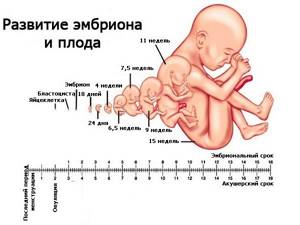
Fetal development at 13 weeks of gestation
The 13th week of pregnancy is already the beginning of the fourth month of gestation. At this time, the unborn child’s internal organs and systems are already fully developing. The length of the fetus (parietal-sacral size) reaches 8 cm, weight is about 15 - 30 grams.
central nervous system
The unborn child's brain is already differentiated into sections: there are cerebral hemispheres and the cerebellum. In the cerebral hemispheres, diencephalon and medulla oblongata, the ventricular system has already been developed; choroid plexuses have sprouted in them, which are involved in the production of brain fluid - cerebrospinal fluid. However, the large brain does not yet have grooves and convolutions, and its temporal lobes are poorly formed.
Auditory and visual analyzers
The membranes of the eye, its light-refracting structures, and the optic nerve are already formed and continue to improve. The eyelids are also already formed, but they will cover the eye until the 36th week.
The auditory analyzer continues to form at the 13th week, this process continues until the 20th week. Around 12–13 weeks, the ears begin to form.
Face, oral cavity
An ultrasound at the 13th week of pregnancy allows you to examine the face and oral cavity, which are gradually taking on their shape. They are still basic: the mouth, tongue, palate are forming, paired formations (jaws, palate) begin to fuse together. The lower part of the face will be formed by 16–17 weeks. Eyebrows - only by 28 weeks. The formation of teeth occurs.
Respiratory system
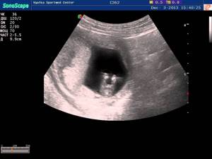
Ultrasound 13 weeks
The respiratory system is formed in the fetus quite late. This is due to the fact that the embryo developing in the womb will need air only after birth, but for now it receives oxygen from the mother’s blood. Also, the child’s pulmonary circulation is not yet functioning, since until he is born, there is very high peripheral vascular pressure, and there is also a special shunt - the ductus botallus, due to which blood can rush from the beginning of the pulmonary circulation to the beginning of the large one, not passing through the heart.
Thus, at week 13, the diaphragm, upper respiratory tract and septum, which separates the trachea and esophagus, are developed. The lower respiratory tract will begin to form only from the 16th week, and the alveoli even later. Due to the fact that the lungs are still very small, the diaphragm is still higher than in a fully mature, full-term baby.
The cardiovascular system
Since the cardiovascular system is important for intrauterine development, unlike the respiratory system, it begins to form from the moment of implantation (the first 2 weeks of pregnancy) and at 8 weeks the child already has a functioning heart with developed valves and large vessels. Further, the system continues to improve, the internal organs sprout with vessels of a smaller caliber. As for the heart, its work directly depends on the development of the lungs, therefore, unlike a child of early and even infant age, the heart is still relatively symmetrical on the right and left sides.
Digestive system
The digestive organs are formed and continue to develop. By week 12, the rotation of the internal organs ends, and the digestive system only continues to improve. As for the endocrine function of the pancreas, it is already beginning to produce insulin.
Urinary system
By the 13th week the kidney is already terminal, but the glomerular apparatus is still developing almost until birth, when they begin to function.
Reproductive system
At week 13, it is still impossible to say exactly what gender the unborn child has: female external genitalia are formed at 13–14 weeks, and male ones only at 15–16 weeks.
Musculoskeletal system
The rudiments of bones and intervertebral discs have been formed in the child since the end of the 8th week and continue to ossify throughout further development.
Skin and skin appendages
At week 13, nails begin to grow on the skin of the fingers.
The above indicates that the incomplete maturity of many organs and systems of the fetus promises high sensitivity to external and internal factors, which can lead to very severe developmental disorders.
Fertility
The best option for multiple pregnancy is bichorionic biamnionic pregnancy. In this case, ultrasound shows that each fetus has its own placenta (not communicating with others by blood flow) and there is an amniotic septum.
Ultrasound at 12 - 13 weeks of pregnancy
Objectives
At 11–14 weeks of gestation, the first screening ultrasound examination of the pregnant woman is performed. If the pregnancy has previously proceeded without problems, then this screening will be the first ultrasound examination during pregnancy. At this time, it is possible to evaluate the development of the cardiovascular system: its morphological and functional state.
It is possible to detect gross chromosomal pathologies at 13 weeks. Sometimes it is possible to determine the sex of the child, but this may not yet be accurate. It is also important that with the help of ultrasound diagnostics it is possible to determine the number of developing fetuses and their nature - by the number of chorions and amnions. It is also important to evaluate the condition of the placenta and uterus using an ultrasound picture.
How to prepare for an ultrasound?
Preparation for the study does not present any specific features. Before the examination, it is recommended not to eat food for several days that can increase gas formation in the intestines, which may interfere with the ultrasound. These foods include fast carbohydrates, cabbage, carbonated drinks, legumes, etc. On the contrary, fresh fruits and vegetables will be a good addition to your diet. It is recommended to take a towel and napkins with you. For intravaginal ultrasound you need a special condom.
How is an ultrasound done?
Ultrasound is performed in two ways: transabdominal and intravaginal.
The transabdominal method is an ultrasound through the anterior abdominal wall. The sensor is lubricated with gel (this is necessary for better sound transmission) and installed on the anterior abdominal wall. On an ultrasound scan at 13 weeks, the uterus measures approximately 12–14 cm in height, so the sensor is placed on the hypogastrium.
Intravaginal ultrasound is performed to better visualize the fetus, especially in the early stages. The sensor is inserted into the cervical canal, and a condom is first put on.
Ultrasound is reflected from denser structures and passes through less dense ones. Thanks to this, a graph or image is formed on the monitor.
Doppler sonography, as a rule, is not yet performed at this time. Only to confirm the presence of pathology.