- Brain MRI results
- Price
- doctor
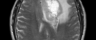
MRI of the brain is a high-tech, non-invasive method for studying the brain. Images provide information that cannot be obtained by other methods. For example: x-ray, ultrasound or computed tomograph. The magnetic field does not affect the human body, and therefore MRI of the brain is harmless to the health of patients.
You can undergo an MRI of the brain inexpensively using modern and accurate equipment. We have experienced specialists who will quickly prepare you for the procedure, conduct the scan with maximum comfort and promptly provide an interpretation.
General information about MRI
Magnetic resonance imaging (MRI) is a modern, high-precision diagnostic method that allows one to visualize pathological changes in body structures using high-quality images.
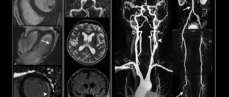
Diagnostics of this kind involve obtaining exceptionally clear and detailed images of organs and tissues using the phenomenon of nuclear magnetic resonance. Under the influence of radio signals, atomic nuclei respond in the form of various signals. The combination of excited electromagnetic waves depends on the nature of the lesion in a particular area under study. There are open-type (no tube) and closed-type MRI scans. Each option has its own advantages and disadvantages. In an open device, the procedure is carried out both for the youngest children and for elderly weakened patients. But (unlike a device with a pipe) diagnostics are contraindicated for people whose weight exceeds 120 kg. Also, the open type of tomograph is not able to evaluate moving organs - the heart, lungs. The advantages of closed-type MRI include the high accuracy of the resulting images (regardless of the area being examined). The disadvantage of the procedure is the high noise level. It also requires complete immobility to obtain high-quality images, which is impossible to achieve, for example, with the smallest patients.
Medical in Moscow has modern equipment for MRI. The examination is a universal method: it is carried out both for young children and elderly people with severe concomitant pathology. Moreover, it will not take much of your time. Medscan implements a large number of multi-directional complex programs that solve the problems of diagnosing a wide range of diseases.
Indications
An MRI of the brain with contrast is necessary to diagnose diseases:
- Tumor formations and metastases in brain structures.
- Strokes.
- Diseases of the central nervous system of inflammatory and infectious types - sclerosis, meningitis, encephalitis.
- Pathologies of cerebral vessels.
The cost of MRI of the brain with contrast is slightly more expensive than standard tomography, but it is justified. It becomes easier to distinguish between healthy and diseased tissues because the contrast penetrates and accumulates in them differently. This method is used as a final method for making a diagnosis, even if the results of other studies turned out to be uninformative. It is possible to identify small metastatic foci (1-3 mm in size) with topometric MRI of the brain with contrast with a smaller scanning step.
Symptoms of the disease for which an MRI of the brain with contrast is performed are:
- Severe headaches.
- Convulsions, epileptic attacks.
- A sharp decline in hearing and visual acuity.
- Disturbances in the threshold of pain and tactile sensitivity.
- Feelings of pins and needles, numbness in different parts of the body.
What does an MRI show?
Doctors recommend doing an MRI to confirm diseases of the brain and spinal cord, internal organs (with the exception of hollow organs), articular apparatus, spinal column, and organs of the reproductive system. Scanning is used to determine:
- spinal hernias;
- inflammatory processes;
- benign and malignant neoplasms;
- vascular pathologies (diseases of the cardiovascular system, changes in blood flow);
- presence of bone fragments;
- infectious processes of the osteoarticular apparatus;
- traumatic lesions;
- liquid in cavities.
The procedure is not relevant for identifying problems of the stomach, intestines, lungs (contain air), as well as bone structures.
The accuracy of MRI can be improved by using a contrast agent. A gadolinium-based agent is used as a contrast, administered intravenously. The drug accumulates in areas with increased blood flow (inflammation or tumor). MRI with contrast determines the pathological process in the initial stages of the disease. The substance introduced into the bloodstream is absolutely harmless to the body and is eliminated within 24 hours.
Contraindications to MRI with contrast
- Installed pacemakers and other electronic devices whose operation may be disrupted during the procedure.
- The presence of metal prostheses in the body, except titanium.
- Pregnancy. Since contrast agents can cross the placenta, their use during this period is undesirable.
- Allergic reaction to contrast.
- Severe kidney damage. Gadolinium salts are excreted through the urinary system, so the study is not carried out in case of serious renal damage. Minor abnormalities in kidney function are not contraindications.
There are also relative contraindications, in which the possibility of performing the procedure is determined by the doctor depending on the area of study and other factors:
- The presence of non-magnetic implants, braces, tattoos with metal-containing inks, permanent piercings.
- The patient’s serious condition, in which it is difficult for him to remain motionless for a long time, mental illness, claustrophobia - fear of closed spaces. In this case, the procedure is carried out after the use of sedatives or sleeping pills.
Indications for the procedure
A specialist of almost any profile can prescribe a diagnostic procedure, since the procedure helps make a diagnosis for many pathologies. There are the following indications for diagnostics using MRI:
- Brain diseases: if a stroke is suspected; neoplasms and their relapses; assessment of the condition after surgery; demyelinating disorders (multiple sclerosis); inflammatory processes; congenital features of the development of the craniovertebral zone; convulsive readiness and epilepsy; DEP (dyscirculatory encephalopathy); hypertension of unknown origin; metastatic foci; traumatic brain injury (if the injury is not recent).
- Cervical vessels and cerebral vessels: anomalies in the development of the vascular bundle; aneurysms; signs of vertebrobasilar insufficiency due to cervical osteochondrosis; vascular stenosis (narrowing); migraine pain.
- Diseases of the temporal and jaw joints, their discs; preparation for dental prosthetics.
- Problems with the eye orbits and extraocular muscles.
- Diseases and inflammation of the lacrimal glands.
- Problems with the paranasal sinuses: cysts, sinusitis, preparation for surgery.
- Diseases of the thyroid gland (including oncology).
- Spinal column and spinal cord: osteochondrosis, osteoporosis, injuries, etc.
- Osteoarticular system: rheumatoid arthritis, inflammatory, traumatic lesions, conditions after surgery.
- Organs of the thoracic cavity: lung tumors, mediastinum, myocardial infarction, coronary angiography, pleurisy.
- Organs of the abdominal cavity and retroperitoneal space: problems with the liver, pancreas, gall bladder, intestines, spleen, kidneys.
- Pelvic organs: diseases of the bladder, prostate gland, ureters, uterus, etc.
How is an MRI examination of the brain performed?
It all starts with a consultation with a doctor, checking that there are no contraindications to magnetic resonance scanning. The main reasons that will force one to abandon the procedure and replace it with another technique are the following:
- Cardiovascular devices, neurostimulators, implants, stents, pumps, braces, vascular clips, other electronic devices that are not removable or have metal components.
- Pregnancy up to twelve weeks.
- Fear of confined spaces.
- Involuntary body movements (in some brain disorders) that make it difficult to remain still.
Before the examination, the patient removes all items of clothing and accessories containing metal components. If the doctor has prescribed the use of contrast agents, then a break of 3-4 hours after the last meal is necessary.
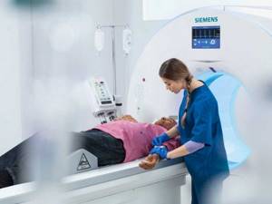
MR angiography of cerebral vessels will take from 30 to 50 minutes. The patient is placed lying on the tomograph table, the position of his body is fixed with special rollers for immobility. The doctor controls the process from the next office; communication with the patient is maintained through an intercom.
Contrast-enhanced tomography differs from conventional tomography in that before the diagnosis begins, the patient is intravenously injected with a drug that accumulates in tumor tissues and improves their visibility in the images. The substances do not pose a danger to humans and are completely eliminated from the body within a couple of days. The cost of MRI of cerebral veins will be slightly higher than simple MR angiography.
The patient will receive the result within an hour after the end of the procedure. This can be printed out the most informative images or recording on disk all the information received (the service is paid in addition to the price of MRI of the veins of the brain).
Contraindications
When choosing a clinic where it is better to have an MRI, you need to remember the main contraindications for the procedure:
- first trimester of pregnancy;
- the patient has a pacemaker;
- there are metal particles or structures in the body (including fragments after injuries).
Modern orthodontics and orthopedics (dentistry) use in their practice to install braces and dentures such materials (made of titanium and various alloys) that do not interact in any way with the magnetic field. Therefore, in this situation, performing a diagnostic procedure is not a contraindication.
How is an MRI with contrast performed?
Introduction of contrast
Before the procedure, the patient removes metal jewelry, including piercings. Otherwise, during the procedure they heat up, leading to skin burns. Another reason is that metal distorts diagnostic results.
The patient lies down on a special table where he is fixed. This is necessary because changing the body position will distort the result. After this, a control photograph is taken without contrast.
Then a contrast agent is injected intravenously by drip or jet. At this moment, unpleasant sensations may occur - dizziness, tingling, unpleasant taste in the mouth, fever or chills. This discomfort does not occur to everyone and, once it appears, goes away almost immediately.
Severe intolerance, manifested by shortness of breath, hand tremors, cardiac dysfunction, vomiting, and loss of consciousness, is extremely rare. In this case, the patient is immediately provided with qualified assistance.
After the contrast is administered, a repeat image of the same areas is taken so that the doctor can compare both results.
Because the procedure is more complex than a regular MRI, it lasts longer than a regular scan - 30-60 minutes. After receiving the images, the doctor interprets them and transmits the results to the patient. The data is provided in the form of a snapshot and can also be recorded on magnetic media.
After completing the procedure, if you feel well, you can immediately go home. If the patient does not feel well, he can stay in the clinic for some time under the supervision of doctors.
Principle and safety of the procedure
Tomography helps to determine the nuances of the location and structure of the internal organs and structures of the body. Scanning the body in thin layers helps with this. Safe contrast can be used, but there is no harmful x-ray exposure. The MRI machine recreates an extremely powerful magnetic field based on interaction with atomic nuclei. Most often we are talking about atoms of the chemical element hydrogen. Their excitation leads to a change in position, which allows the equipment to build many detailed layer-by-layer “slices” of various organs.
The magnetic field does not carry a negative radiation load on the body. There are also no other unfavorable factors that could harm human health. Therefore, the diagnostic procedure can be carried out the required number of times (according to indications).
Preparing for magnetic resonance imaging
Examination of the pelvic organs (organs of the female reproductive system, prostate gland, bladder) requires the following measures:
- the procedure is carried out with a full bladder: you should not urinate for several hours, and an hour before the scan you should drink half a liter of clean water;
- It is recommended to cleanse the intestines with an enema or laxatives;
- half an hour before the study, it is advisable for the patient to take an antispasmodic drug (“No-spa”);
- women of childbearing age indicate the day of the menstrual cycle.
Magnetic resonance imaging of the abdominal organs is performed on an empty stomach or after a light snack if the procedure is scheduled for daytime or evening hours. A few days before diagnosis, you should exclude from your usual diet foods and dishes that contribute to increased gas formation. These include legumes, whole milk, and sweets. Also avoid foods that cause constipation (flour, rice, chocolate). It is advisable to take enzymes and sorbents (activated carbon) during this period. On the eve of the study, the patient is recommended to take an antispasmodic.
Examination of other parts of the body does not require special preparation. Immediately before scanning, you must remove keys and other metal items from your pockets and remove jewelry.
Advantages of MRI at Medscan
The diagnostic clinic in Moscow performs many MRI options at quite affordable prices: joints, internal organs, soft tissues, peripheral nerves, spine, etc.
Diagnostic testing is carried out as prescribed by a doctor. Screening programs are also being implemented that involve analyzing the state of certain body systems. Some of the most relevant are cancer screening and screening of the cardiovascular system.
The leading advantages of our clinic:
- High information content of diagnostic images. The contrast agent increases the information content of the images.
- The radiology department is headed by one of the best radiologists in the entire post-Soviet space, Kirill Sergeevich Petrov.
- There is a medical center. It helps to understand controversial or complex clinical cases and clarify the diagnosis.
- The diagnostic center operates under international standards. The scanning is consistent with the recommendations of European professional societies.
- Diagnosis is carried out by highly qualified doctors with sufficient practical experience.
- Our professionals work with expert-level equipment. The equipment meets modern international standards.
- The results of the scanning are recognized by leading specialists of the Russian Federation and foreign countries.
- An MRI in Moscow can be done in one of several branches of our clinic, the addresses of which are listed on the website.
Medscan doctors will help identify health problems at the initial stages and prevent their development.
Sign up
Sources and literature
- Akberov R. F., Yaminov I. Kh., Safiullin R. R., Puzakin E. V. The effectiveness of magnetic resonance imaging in the diagnosis of brain tumors. [Electronic resource] // Kazan Medical Journal. 2011
- Kremneva E. I., Konovalov R. N., Krotenkova M. V. Functional magnetic resonance imaging. [Electronic resource] // Annals of Clinical and Experimental Neurology. 2011
- Kotlyarov P. M., Lagkueva I. D., Sergeev N. I., Solodkiy V. A. Magnetic resonance imaging in the diagnosis of lungs. [Electronic resource] // Pulmonology. 2021
Benefits of MRI
The advantages of magnetic resonance imaging are especially visible when compared with ultrasound, computed tomography, and x-rays. So ultrasound is not able to show in detail the areas hidden behind the skull. Computed tomography is performed with the introduction of iodine-containing contrast agents, which are not always well accepted by the body. X-rays are suitable for studying bone tissue, but are of little information regarding other structures and are harmful due to radiation exposure.
Advantages of MR imaging:
- No harm, even when repeated many times in a short period of time. Diagnostics can be performed on children from birth and on older people.
- The result is a three-dimensional image of the examined area with the ability to enlarge. It shows even small anomalies of various tissues (nerves, blood vessels, bone tissue, etc.) in the initial stages.
- The price of an MRI of the brain with contrast is affordable, although it is higher than the cost of ultrasound and x-rays.
- Minimal preparation - urgent MRI of the brain with contrast can be performed on the day of the patient's visit.
- MR tomography is an express technique. The results and conclusion will be ready a couple of hours after the end of the procedure.
- The scan is completely painless.