How is an ultrasound examination of the veins of the lower extremities performed?
The leading role in the diagnosis of varicose veins of the lower extremities today is occupied by ultrasound (ultrasound examination of the veins of the lower extremities). And there are many reasons for this: firstly , this procedure does not require any special preparation of the patient, secondly, it is absolutely painless, therefore does not require anesthesia, thirdly , the speed of the result obtained, and finally , fourthly , efficiency and accuracy this result!
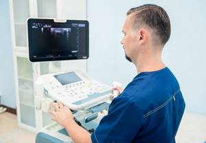
Ultrasound diagnosis of varicose veins is carried out by a phlebologist, ultrasound doctor, Ph.D. Raskin V.V.
Dopplerography, vascular doppler
Dopplerography (vascular Doppler) is a modern imaging technique that is used to detect pathological changes in large vessels before the onset of clinical manifestations or in the early stages of diseases.

Dopplerography (vascular Doppler) is a modern imaging technique that is used to detect pathological changes in large vessels before the onset of clinical manifestations or in the early stages of diseases. Doppler is most often used to diagnose vascular diseases, which are manifested by disturbances in coronary and cerebral blood flow.
"Personal health plan"
is a comprehensive examination based on evidence-based recommendations

Indications for vascular Dopplerography
:
- atherosclerotic changes in large vessels of the heart, head and neck, legs, as well as in the coronary and vertebral arteries;
- vasculitis of various etiologies;
- spasms and paralysis of blood vessels in the legs, kidneys and heart;
- thrombophlebitis and varicose veins;
- traumatic damage to blood vessels;
- if you suspect the presence of aneurysms, anastomoses or congenital anomalies of arteries and veins.
Studies of the veins of the lower extremities
Doppler of the veins of the lower extremities is the most informative method for diagnosing thrombophlebitis and varicose veins and reveals:
- lesions of the deep and superficial veins of the legs in the early stages;
- condition of the walls of veins and arteries;
- blood flow in the vessels of the upper and lower extremities.
The main symptoms of this pathology include:
- numbness and pain in the muscles of the limbs;
- intermittent claudication;
- post-thrombophlephic disorders with trophic changes in tissues;
- chronic venous insufficiency and observation of the condition of the veins with an established diagnosis of varicose veins.
Indications for Dopplerography of head and neck vessels
This method of visualizing pathological processes is very informative when examining circulatory disorders in the vessels of the neck and head in the presence of the following complaints:
- persistent and frequent headaches, migraines;
- for dizziness and repeated fainting;
- with noise in the ears or head;
- for various types of coordination disorders;
- with changes in visual fields, sudden loss or decrease in vision;
- after traumatic brain injury (to determine the integrity of blood vessels immediately after injury and the condition of brain vessels with signs of cerebrovascular accident in different periods after traumatic brain injury);
- for injuries of the cervical spine;
- with malignant hypertension and with severe hypotension;
- after a stroke;
- in the presence of signs of initial, transient or chronic disorders of cerebral circulation and dyscirculatory encephalopathy;
- if congenital or acquired vascular anomalies of the brain and large vessels of the neck are suspected;
- with osteochondrosis of the cervical spine.
The main advantages of this method are the ability to observe measurements over time, as well as perform functional tests.
Indications for examination of the abdominal aorta and its branches
Doppler of abdominal vessels (aorta and its branches, as well as visceral vessels) is widely used for diagnostics:
- if there is a suspicion of congenital pathology of the aorta and its branches;
- in the presence of signs of aneurysms and atherosclerotic lesions;
- for diagnosis and clarification of the localization of voluminous pathological formations.
Our clinic’s specialists widely use methods for diagnosing diseases using color mapping (vascular Dopplerography).
To make an appointment, call us by phone or sign up online.
How to understand the terminology of the procedure for ultrasound examination of the veins of the lower extremities
But before visiting a phlebologist, patients have a number of questions related to the terminology of this procedure. What is Doppler ultrasound (USDG)? What is ultrasonic duplex scanning? What is color Doppler mapping or triplex scanning? What is the difference?
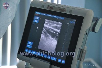
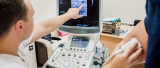
Ultrasound scanner for studying the venous system of the lower extremities
Let's answer these questions and decide what is the best way to assess the condition of the venous vessels of the lower extremities.
What is Doppler ultrasound (Doppler ultrasound)
Doppler ultrasound (Doppler ultrasound) of the veins of the lower extremities is one of the most accessible research methods, based on the Doppler effect (named after the Austrian physicist and mathematician Doppler), which consists in recording a measurement of the speed of reflected sound waves from moving blood cells. Allows you to study only one function - the patency of the vessel based on the graph of the blood flow.

Ultrasound Doppler for examining the veins of the lower extremities
It is performed blindly, the sensor is placed on the approximate projection points of the vessel. If a violation of the patency of a vessel is detected, it is impossible to clarify the cause of this violation, because there is no visualization of the vessels. Currently, it is practically not used during a consultation with a phlebologist, as it is not very informative.
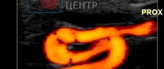
Vascular ultrasound - Dopplerography
Dopplerography
is a modern ultrasound examination method based on the Doppler effect, used to diagnose vascular disease and assess blood flow in organs. The physical basis of this method is based on the Doppler effect, in which the length and frequency of waves recorded by the sensor changes, depending on the movement of their source and/or the movement of the receiver, with the help of which the doctor can assess and determine blood flow through the main arteries and areas located 2 -5 cm from bone structures. This method is used in the best and most modern gynecology clinics, ultrasound
The technique allows us to identify the presence of narrowings in blood vessels, which can be caused by both congenital pathology, such as hypoplasia, and other functional and organic lesions, atherosclerosis, thrombosis, aneurysms, and anatomical variations. The scope of application of this technique in various branches of medicine is large and limitless. Ultrasound of the abdominal organs evaluates the condition of the abdominal aorta and inferior vena cava, determines the degree of development of atheromatous plaques and impaired blood flow, which is often observed in Leriche syndrome. Recently, Doppler ultrasound is increasingly used in obstetric practice to assess the blood flow of the fetus and placenta. During pelvic ultrasound, blood flow examination is often simply necessary for the correct interpretation of the data obtained. The main advantages of ultrasound, including Dopplerography, are its absolute safety, non-invasiveness, high information content with the possibility of high-quality visualization, as well as obtaining data in real time.
There are a number of indications in which this study will be one of the determining factors in the diagnosis and further tactics of patient management, these are primarily headaches, herniated intervertebral discs, osteochondrosis of the thoracic and cervical spine, arterial hypertension, leg fatigue when walking, varicose veins of the lower limbs, edema syndrome, excess body weight and others.
During the examination, an ultrasound transducer is placed on the skin above a blood vessel, which receives and sends sound waves reflected from solid objects, which are amplified by a microphone. If there is no blood flow, the signal is not transmitted to the microphone. Information about the reflection of low-frequency ultrasonic waves is processed by a computer, which makes it possible to see and evaluate a graphical representation of blood movement, giving a conclusion about the nature of blood flow and its speed.
There are several modifications of this method of ultrasound examination using the Doppler effect
: 1.
Color Doppler
is one of the most commonly used types of Doppler ultrasound, in which, instead of the standard ultrasound method, the image is presented in color, in which blood vessels are visible.
The Doppler sound and color signals are produced overlapping each other by a computer, allowing the doctor to evaluate the speed of blood flow through the arteries and veins. 2. Double Doppler ultrasound
is a technique based on standard ultrasound, in which a computer converts sounds, which allows you to evaluate the speed and nature of blood flow.
3. Doppler ultrasound
is a study based on changes in the transmission of ultrasonic waves.
The doctor, having a portable device, listens to sounds, the pathological sound of which is signs of a violation of the speed and nature of blood flow. The advantage of this technique is the fact that this study can be performed directly at the patient’s bedside. 4. Triplex Doppler
is the newest technology, which is also based on the Doppler effect. The sensitivity of this technique is five times higher than all previous methods. The combination of color Doppler, B-mode and pulsed wave Doppler creates the best conditions for diagnosis and visualization of the object of study. Technically, the research is quite easy to do, without any special preparation for it. In preparation for Doppler ultrasound, the patient is recommended to refrain from products such as coffee, tea, energy drinks and smoking cigarettes 2 hours before the test, since all of these products contain nicotine and caffeine, which cause vasospasm, which can give false positive results. in each specific case, which in turn will lead to incorrect interpretation of the results and, as a consequence, incorrect treatment.
Interpretation of Doppler ultrasound results requires the assessment of a highly qualified specialist with experience in diagnosing diseases of the veins and arteries using this method. It is considered very informative if Doppler sonography is carried out within a short period of time, since in this case it is possible to trace the dynamics of the disease.
Despite the fact that the “gold standard” for diagnosing diseases of the veins and arteries are angiography and venography using a contrast agent, which give definitive results, in many cases Dopplerography can become a full-fledged alternative, primarily due to its speed, non-invasiveness and low cost, and its role as an express method is generally invaluable.
DID YOU LIKE THE ARTICLE? LIKE and SHARE WITH YOUR FRIENDS!
What is ultrasonic duplex scanning?
Ultrasound duplex scanning (ultrasound duplex scanning) - this study combines two (duplex) examination modes. The usual (traditional) ultrasound scanning mode, which allows you to see the vein in a two-dimensional black and white image, and the Doppler mode, which allows you to determine the “fluidity” of the blood based on the graph. As a result, the doctor receives complete visualization of the vessel on the screen, which allows one to determine the condition of the vascular wall, the presence of thrombotic masses in the lumen, the operation of the valve apparatus, vessel tortuosity and anomalies, as well as the nature of the blood flow: its speed, turbulence.
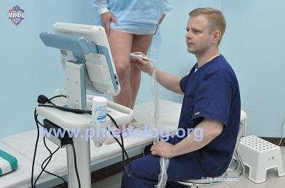
Ultrasound duplex scanning of veins is performed by phlebologist, ultrasound doctor I.I. Kalachev.
What is color Doppler mapping or triplex vein scanning?
Color Doppler mapping or triplex scanning of veins is not any other research method. This is the same ultrasonic duplex scanning device, operating in an additional third mode (three - triplex), which consists in visualizing blood vessels in a color image. Through this, a more informative examination is achieved, which makes it possible to determine with high accuracy vascular patency, the speed and direction of blood flow, and the level of thrombotic lesions.

Color Doppler mapping of veins is performed by phlebologist, ultrasound doctor D.A. Fedorov.
It is worth noting that ultrasonic waves are considered absolutely safe and do not have a negative effect on the adult body. Ultrasound can be repeated many times as necessary during treatment.
Based on the foregoing, we can conclude that today ultrasound scanning (ultrasound duplex scanning) of vessels in color mode is considered one of the best and most popular methods in the diagnosis of varicose veins of the lower extremities.
Why is it important to conduct an ultrasound examination of the veins in our veins?
In ours, all phlebologists are certified as ultrasound doctors to perform ultrasound scanning of veins, that is, all specialists have undergone multi-level professional retraining and can perform this study. In addition, for many years, working with the most modern ultrasound scanners, the doctors of our center have become experts in the field of diagnosing diseases of the veins of the lower extremities.

Ultrasound duplex scanning of veins is performed by phlebologist, ultrasound doctor Semenov A.Yu.
All this allows us to perform the most complex surgical interventions on the veins of the lower extremities under ultrasound guidance, including endovascular operations (without incisions) and complex reconstructive operations for recurrent varicose veins.
Ultrasound Dopplerography of vessels (USDG)
At the MedEx clinic you can do Doppler ultrasound (USD) of blood vessels at an affordable price. The examination allows you to assess blood flow indicators, identify diseases at an early stage of development, and establish the exact localization of the pathology.
We perform Doppler scanning of the vessels of the neck, head, upper and lower extremities, eyes, as well as intracranial structures. The procedure is performed by experienced ultrasound doctors.
What is ultrasound
Doppler ultrasound is an ultrasound method for examining cerebral vessels, which is considered a very informative method, accessible and safe for a wide range of patients. This is an ultrasound technique using the so-called Doppler effect. Ultrasound is based on the reflection of an echo signal from a static organ, that is, during the study, the doctor can only evaluate the arteries themselves, without the ability to assess the rate of blood flow to the brain. The essence of Doppler examination of blood vessels is to assess the speed of blood movement through the vessels.
Large arteries (carotid, vertebral and subclavian), as well as smaller vessels (external and internal carotid arteries), are examined. The indicators are displayed on the monitor in the form of a graph.
Mainly, the thickness and structure of the carotid artery and its branches are assessed. Through the internal carotid arteries and vertebral arteries, oxygenated blood enters the brain. Therefore, assessing the state of blood flow in the arteries of the neck and head is of great practical importance.
Types of ultrasound examination
1. Transcranial Dopplerography (TCDG) of cerebral vessels. The method is intended to study the vessels inside the skull. Due to the complexity of its implementation, TCD is a separate examination technique and since certain brain vessels are located inside the cranium, which does not transmit ultrasound waves well. TCD of the brain is done using a special technique on open areas of the skull. TCD allows you to assess blood flow through intracranial vessels. The method is often used in children to assess blood flow in case of abnormal neurological status. In adults I use it less often, often in conjunction with other studies, such as MRI and MRA of the brain, Doppler sonography of the great vessels of the head and neck.
2. Dopplerography of the great vessels of the head and neck. Ultrasound examination is prescribed to patients as part of a comprehensive diagnosis of cerebrovascular diseases or when a specific pathology is suspected, for example, to assess the degree of narrowing of an artery in patients with high cholesterol.
3. Doppler ultrasound of leg vessels. Using ultrasound, the doctor examines the state of the peripheral blood flow of the lower extremities. Doppler ultrasound data allows us to detect the presence of atherosclerotic plaques in the arteries of the lower extremities, damage to the deep and superficial veins, identify the presence of blood clots in the venous system of the lower extremities and assess the dynamics of the condition of the vessels of the legs during treatment. During the procedure, an experienced doctor can easily determine the aneurysm, venous insufficiency, the nature and location of thrombotic masses.
4. Doppler ultrasound during pregnancy. The procedure is prescribed if vascular pathology is suspected. The nature of blood flow in the area of the ovum provides information about the health of the arteries and placenta. During an ultrasound examination, the doctor evaluates the development of the circulatory system in the fetus and can detect vascular disorders at an early stage. The scan results allow doctors to prepare for the birth of a child, plan interventions and provide timely assistance to both mother and baby.
Indications for ultrasound examination
Doppler sonography is recommended for all people over 40 years of age. At this age, certain changes in the circulatory system begin in the body, and the risk of thrombosis, varicose veins, atherosclerosis, and other vascular diseases increases. The study allows you to identify them before serious complications develop.
Indications for Doppler ultrasound of the head and neck:
- arterial hypertension
- diabetes
- carbohydrate metabolism disorder
- increased levels of total cholesterol and a violation of the ratio of “good” and “bad” cholesterol fractions;
- history of cerebral vascular surgery;
- family history of cardiovascular diseases in close relatives;
Also, the doctor may refer the patient for examination of the vessels of the head and neck if he has the following complaints:
- tinnitus not associated with hypertension or hypotension;
- fainting, loss of consciousness
- frequent and severe headaches, migraine attacks that are difficult to relieve with medications
- memory impairment, inability to concentrate
- sleep disorders: insomnia or, conversely, excessive sleepiness;
- sudden speech disorder
- if there were episodes of short-term loss of consciousness
- pulsations in the neck and head area
- weather sensitivity, which reduces the quality of life;
- asymmetrical pressure or pulse on the right and left arms.
Doppler ultrasound is an effective diagnostic tool in determining the effect of cervical osteochondrosis on blood flow in the vertebral arteries.
Indications for ultrasound examination of the extremities are:
- swelling of the legs, which is especially noticeable in the evening;
- numbness or pain in the muscles of the limbs when walking;
- pulling sensation in calves;
- constant feeling of cold in the legs;
- change in skin color of the limb, the presence of lesions such as trophic ulcers;
- formation of nodules, vascular networks.
The examination helps the doctor to correctly assess the condition of the deep veins and identify the cause of impaired blood flow in the legs. This is important for choosing patient treatment tactics.
For children, manipulation is prescribed for early diagnosis of various vascular pathologies.
Indications for ultrasound scanning in a child are:
- premature birth;
- too much or too little weight;
- disturbances in speech development, memory problems;
- unusual or asymmetrical skull shape;
- difficulty falling asleep and restless sleep;
- suspicions of congenital vascular diseases, etc.
Doppler ultrasound is indicated for pregnant women after 21 weeks. Unscheduled vascular examination is carried out in the following cases:
- multiple pregnancy;
- presence of diagnosed diabetes mellitus, anemia, hypertension, late toxicosis, vascular thrombosis in the mother;
- post-maturity of the fetus;
- the age of the pregnant woman is under 18 or over 35 years;
- the appearance of bloody, dark discharge;
- the presence of miscarriages and pathologies in previous pregnancies;
- threat of interruption, placental abruption;
- suspicion of entanglement of the fetus with the umbilical cord;
- mother's immoral lifestyle;
- falls, injuries to a woman during pregnancy, etc.
Duplex ultrasound examination is also prescribed as part of the preoperative examination of the patient. During the procedure, the specialist studies the blood flow, determines the location of the defect, and plans the intervention. Under the control of a sensor, minimally invasive manipulations are performed on the vessels of the legs. The ultrasound ultrasound procedure also helps in assessing the dynamics of the disease during treatment.
What does ultrasound scan reveal?
The results of Doppler ultrasound allow the doctor to draw the following conclusions:
- identify areas of vasoconstriction and analyze their effect on blood circulation as a whole;
- detect early lesions of the vascular bed that occur without pronounced symptoms;
- examine the vertebral arteries and large veins.
- study the structure of the vascular walls, identify areas with impaired elasticity, increased or decreased tone;
- assess the structure of blood flow in the neck, head, and limbs;
- inspect the vessels for tortuosity;
- identify areas of accumulation of thrombotic masses, atherosclerotic plaques, varicose veins of varying degrees, etc.
Ultimately, the doctor who referred the patient for this study receives the following data: assessment of the state of the intima-media complex of the large arteries of the head, the degree of vascular patency - the presence or absence of stenosis (narrowing) of the arteries, the severity of stenosis, the speed of blood circulation through the vessels (to assess the intensity of supply brain blood), the degree of damage to the vessel by atherosclerosis, the presence of anatomical features (congenital developmental anomalies or acquired pathologies).
Advantages of ultrasound scanning
The main advantage of Doppler examination is its safety. Unlike X-rays and CT, the manipulation is performed without radiation exposure. Ultrasound waves pass through tissue during examination without causing harm to cells. The procedure can be prescribed for diagnostic and control purposes several times a year, even for the youngest patients and expectant mothers.
Other advantages of duplex ultrasound:
1. High information content regarding the assessment of the condition of blood vessels and tissues that are inaccessible for other types of examination.
2. Carrying out the procedure in real time - the doctor effectively examines the blood flow in dynamics and has the ability to compare the data of the diagnostic tool with external clinical manifestations.
3. No drug burden on the patient.
4. Painlessness, manipulation without the use of anesthesia or contrast on an outpatient basis.
5. Obtaining a broad clinical picture in a short time - the procedure for ultrasound examination of blood vessels takes about 20 minutes.
6. Affordable prices for Doppler ultrasound (USD).
How is ultrasound examination of blood vessels performed?
At the MedEx clinic, diagnostics are carried out by appointment. We will invite the patient to the doctor's office at the appointed time. No preliminary preparation for ultrasound examination of blood vessels is required. The method is quite simple to implement, but its effectiveness largely depends on the experience of the doctor and the quality of the equipment.
Sign up for an ultrasound Dopplerography (USDG) procedure in Moscow
We examine patients using a modern high-resolution ultrasound scanner. The procedure takes place in a comfortable office, in a calm environment. After the scan, the specialist will prepare a written report. You can learn more about the features of vascular ultrasound and interpret the results at an additional consultation with your doctor. Our receptionists will guide you to the appointment time.
To make an appointment for ultrasound diagnostics of blood vessels, call us at the phone number listed on the website.
Cost of ultrasound (USDG, ultrasonography) of the veins of the lower extremities in Moscow
The cost of ultrasound examination of veins in Moscow, as for many high-tech modern services, does not always correlate with quality. You can undergo an examination at a very high cost, but with a fairly low information content. This is possible in some commercial medical centers where there is no modern phlebology. This approach can be called “ultrasound examination for the sake of ultrasound examination.” Things are far from being the best in the public sector. Patients often wait months in line for an ultrasound examination at a public clinic. And very often the results of such diagnostics leave much to be desired. The thing is that in both of these cases, the ultrasound examination is carried out by an ultrasound doctor who does not himself treat venous pathology. The ultrasound examination, which is carried out by the phlebologist himself, is performed in strict accordance with accepted European standards. The results of such an examination may differ radically from those obtained by an ultrasound specialist. The most paradoxical thing in this situation is that ultrasound examination of veins by a phlebologist in Moscow will almost always cost less in relation to commercial medical organizations. At the Moscow Innovative Phlebological Center, we do not logistically separate specialist consultation from ultrasound scanning. An ultrasound of the veins is performed during a consultation with a phlebologist. This is the only way to quickly and accurately obtain comprehensive information about the patient’s venous system.
| Service | Treatment category | Price |
| Appointment with an expert phlebologist with ultrasound scanning of the veins of the lower extremities | 2900₽ 3500₽ | |
| Appointment with an expert phlebologist without ultrasound scanning of the veins of the lower extremities (based on ultrasound results from another clinic) | 2000₽ 2400₽ | |
| Appointment with an expert phlebologist with ultrasound examination based on the results of treatment for one year | for free |
For the convenience of our patients, we work with banks that offer credit lines. Also in our center there is a flexible system of discounts and installments. Payment by Visa and Mastercard is possible. At the end of the course of treatment, the patient is given a package of documents to submit to the tax office for tax deduction.
The price at the Moscow Innovative Phlebological Center for such a specialist consultation with expert-level ultrasound scanning is 2,900 rubles. It should be noted that the price of this examination, which meets the best European standards, has been maintained in our Phlebology Medical Center since 2015.
Dopplerography is performed to diagnose blood vessels:
- Necks
- Heads
- Spine
- Upper limbs
- Lower limbs
Dopplerography of blood vessels is carried out using ultrasound machines equipped with a special sensor that sends waves through the skin into the tissues of the patient’s body. The device receives data on the movement of blood through the vessels, combines it with data obtained from conventional ultrasound, and displays an image in which doctors can see the structure of the vessels and make a conclusion about the presence or absence of blood flow disorders.
Using Dopplerography of blood vessels, you can determine:
- Presence of vessel blockage
- The size of the vessel blockage
- Vessel diameter
- Presence or absence of blood flow disorders
Doppler ultrasound is a completely safe and painless procedure. Vascular Dopplerography has no age restrictions, it has no contraindications or any side effects. The duration of the procedure is 10-30 minutes.
Attentive diagnostic and treatment specialists will professionally perform Doppler sonography for you and make the correct diagnosis. Our medical center uses only modern high-tech equipment, thanks to which Doppler ultrasound of blood vessels is performed as accurately as possible.
Frequently asked questions from our patients on the Internet about the vein ultrasound procedure
Where is the best place to undergo an ultrasound examination of veins in Moscow?
In Moscow, the best option would be to perform an ultrasound examination of the veins as part of a consultation with a phlebologist. In our clinic you will not only undergo modern research, but also be advised by an experienced phlebologist. You can make an appointment for a consultation at any time convenient for you by phone: +7 (499) 450-71-16.
Ultrasound examination of the veins of the lower extremities, what is it for?
Ultrasound examination of the veins of the lower extremities is a very good method for identifying venous pathology, the gold standard for diagnosis in modern phlebology. The prevalence of venous pathology in modern society is so great, and the complications are serious, that the study of the venous system before many surgical interventions has become a necessary condition. It is worth noting that ultrasound of the veins of the lower extremities also has its disadvantages. The technique has a pronounced operator dependence. That is, the information content of the method largely correlates with the professionalism of the specialist conducting the research. Therefore, it is better to perform an ultrasound examination from a competent phlebologist.
Good modern ultrasound diagnostics of the venous vessels of the lower extremities, where is it done and how much does it cost?
Ultrasound examination of the veins of the lower extremities is best performed in a good phlebological center. Obvious advantages of this solution:
- High level of diagnostic reliability.
- Ultrasound of veins in a modern phlebology clinic is usually cheaper than in other medical organizations.
The cost of an ultrasound scan of the veins of the lower extremities with a consultation with a phlebologist in our phlebology center is 2900 rubles.
Where can I get a free medical ultrasound examination of veins?
Medical ultrasound examination of veins can be done at a public clinic on a first-come, first-served basis. But, there are two nuances:
- The wait for your turn may last more than one month.
- The information content of such research often leaves much to be desired.