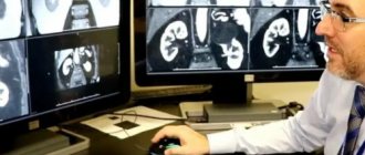
Computed tomography is a modern method of painless and highly accurate diagnostics. A three-dimensional image of internal organs allows you to evaluate their shape and structure, which allows the doctor to determine with maximum accuracy possible pathologies, variants of the course of the disease and choose the most effective treatment methods.
Specialists from the medical center tell you more about why and how an abdominal CT scan is performed, as well as the specifics of preparing for the procedure.
In what cases is a CT scan of the abdominal organs prescribed?
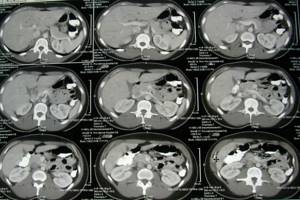
Unlike X-rays, computed tomography creates a three-dimensional image of the organs and tissues of the abdominal cavity: they are not superimposed on each other, but are visible in section. CT allows you to identify disturbances in the functioning of the gastrointestinal tract: liver, pancreas, stomach, large and small intestines, gall bladder and spleen; and retroperitoneal space: vessels, lymphoid tissue, kidneys, adrenal glands and urinary system.
Computed tomography of the abdominal cavity with contrast is performed according to doctor’s indications in a number of cases:
- inflammatory processes;
- pancreatitis;
- suspicion of appendicitis;
- damage to abdominal organs due to injuries and accidents;
- pathologies of the liver, spleen, adrenal glands;
- suspicion of cysts and abscesses;
- search for malignant and benign formations.
Why take creatinine before MRI and CT with contrast?
Before CT with contrast it is necessary to donate blood for creatinine
A creatinine test is needed to undergo a computed tomography scan with contrast. Once in the blood vessels, enhancing drugs affect the composition of the blood. These are temporary changes, and for most people they go away without a trace. But some patients may experience impaired kidney function (contrast-induced nephropathy):
- the amount of urine produced decreases (oligo- or anuria);
- renal edema appears;
- feeling weak and drowsy;
- blood pressure rises sharply;
- loss of appetite;
- a secondary infection with high fever quickly follows.
If a person suffers from acute renal failure or chronic end-stage renal failure, further progression of contrast-induced nephropathy leads to serious complications, including problems with the heart and lungs. To avoid the unpleasant consequences of iodine contrast, check the level of creatinine in the blood. This chemical compound is the end product of a chain of transformations of the amino acid creatine. Exceeding the norm is a contraindication to CT with contrast.
Unlike iodine, the nephrotoxicity of gadolinium drugs is extremely low. Therefore, if a person does not suffer from kidney disease, creatinine analysis is not required for MRI with contrast.
How to prepare for the procedure?
To conduct a high-quality study, it is necessary to empty the gastrointestinal tract as much as possible; the procedure is carried out on an empty stomach. Before contrast-enhanced CT examination of the abdominal cavity and retroperitoneal space, it is necessary to:
- take a blood creatinine test;
- for 2 days, adhere to a diet that reduces gas formation;
- when drinking liquid, give preference to still water;
- do not eat 6-8 hours before the test;
- cleanse the intestines naturally or with an enema.
What should you tell your doctor about?
- about the presence of a pacemaker, this is not a contraindication as in the case of MRI, but it is necessary to inform the medical staff;
- about a possible allergic reaction to iodine-containing drugs and allergies to medications;
- about bronchial asthma and renal dysfunction, if any in the anamnesis;
- about taking any medications;
- about the presence of fixed prostheses, pins, splinters and other metal-containing products, they can affect the image quality.
The patient must have a doctor's referral and the results of previous diagnostic examinations (ultrasound of the abdominal cavity, FGDS, X-ray of the stomach, colonoscopy - if available). When performing a CT scan with contrast, the medical laboratory technician will ask you to sign a consent to perform a medical procedure - the introduction of a contrast agent.
Blood test for MRI with contrast
Intravascular contrast is used for different purposes:
- increased visual distinction between healthy and pathologically altered tissues;
- identification of foci of vascular growths;
- examination of the circulatory system (angiography, venography, etc.);
- perfusion diagnostics;
- differentiation of anatomical structures.
Indications for MRI with contrast are:
- tumor recognition;
- assessment of local and distant prevalence of neoplasms;
- postoperative control;
- observation of dynamics during therapy.
When contrast agents enter the body, reactions are possible in the form of nausea, vomiting, itching in the eyes and skin, in severe cases - swelling of the face and larynx, convulsions, arrhythmias and respiratory arrest. Acute conditions can develop within a few minutes, delayed manifestations are observed much later. At risk are patients with:
- bronchial asthma;
- intolerance to contrast components;
- severe allergies to any drugs or substances;
- cardiovascular diseases;
- acute and chronic failure of the kidneys and liver in the terminal stage of processes.
Mostly, side effects occur after using iodine-containing CT products. Gadolinium, used for magnetic resonance imaging, provokes unwanted reactions much less frequently. Therefore, there is no need to do a blood test before an MRI with contrast.
MR contrast agents
Gadolinium drugs are considered potentially dangerous for people with kidney disease, as they can provoke the development of nephrogenic systemic fibrosis. Such patients are recommended to have their blood tested before an MRI with contrast. If the glomerular filtration rate is less than 60 ml/min, gadolinium is not used. In cases of extreme necessity, contrast scanning is performed using CT scans.
What can and cannot be eaten before an abdominal CT scan with contrast?
| Can | Not recommended |
|
|

Groups at risk for developing CIN
In whom should we, in principle, expect toxic effects of CV on the kidneys? You will be surprised, but not all patients. The risk group for developing CIN according to ESUR 10 and ACR 2021 recommendations includes patients with:
- diabetes mellitus;
- any known kidney disease (condition after kidney surgery and the only remaining kidney are also included);
- protein in the urine (proteinuria);
- increased levels of uric acid in the blood (gout);
- arterial hypertension requiring drug correction;
- age >60 years.
Of course, if we are talking about patients with kidney cancer, then they are all at risk, so determining their creatinine level before contrast is justified.
For patients not at risk, according to domestic, European and American guidelines, DETERMINATION OF CREATININE BEFORE INTRAVENOUS CONTRAST ON CT is NOT REQUIRED, but this is not our case.

By the way, according to the recommendations of ESUR 10, THE EXPIRATION DATE OF THE CREATININE ANALYSIS:
- 7 days for outpatients with acute diseases or exacerbation of chronic diseases and for all inpatients;
- 3 months for all other patients.
If an urgent examination is necessary (if the life and health of the patient is at risk), it is not necessary to determine creatinine so as not to delay the diagnosis.
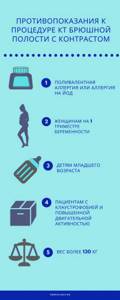
Contraindications
Computed tomography is a safe diagnostic method, since the radiation dose is significantly less than with traditional radiography of the gastrointestinal tract. However, there are contraindications for this study:
- allergy to iodine or polyvalent allergy;
- children under 14 years of age;
- pregnancy and lactation;
- severe form of claustrophobia;
- weight over 130 kg;
- renal failure.
MRI of the liver: is a blood test done before the study?
Magnetic resonance imaging of the liver with contrast is prescribed:
- if tumors and metastases are suspected;
- to assess the condition of hepatocytes (liver cells) when affected by cirrhosis and other pathological processes;
- to determine the nature of disorders caused by congenital anomalies of the organ;
- to identify areas of narrowing of the bile ducts.
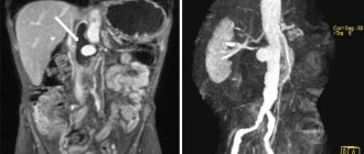
On MRI scans with and without contrast, a single metastasis in the liver is visualized
The most important function of hepatocytes is the deactivation of substances that have entered the body from outside, for their subsequent excretion by the kidneys. In case of hepatic-renal failure, this process is disrupted, and therefore, after the procedure with contrast, the patient’s health condition may deteriorate. In such cases, it is recommended to do a blood test to find out the levels of creatinine, albumin, liver enzymes, residual nitrogen and urea. If there are significant deviations from the norm, MRI with contrast is not done.
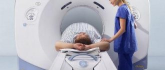
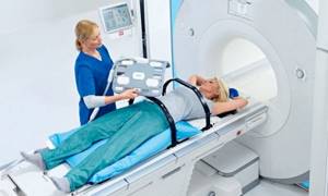
How is an abdominal CT scan with contrast performed?
The examination itself is painless and does not cause discomfort. The patient is placed on a mobile tomograph couch, and the scanner rotates around him. At the right time during the examination, a contrast agent is injected through a special catheter. The patient does not experience any unpleasant sensations, but for correct data acquisition, it is necessary to remain completely still.
The entire CT scan procedure with contrast takes up to half an hour on average. At the end of the examination, within 1.5-2 hours, the patient is given a conclusion based on the diagnostic results.
What is creatinine?
Creatinine is a breakdown product of muscle tissue that is excreted by the kidneys, and its level is a marker of their function. And since this is a product of muscle breakdown, its normal level depends on the gender and age of the patient: you must agree that a muscular 30-year-old man has significantly more muscle tissue than a dandelion grandmother, and the load on the muscles of the former is clearly higher due to a different image life (and therefore more breakdown of muscle fibers). Therefore, the creatinine level itself, not normalized for the patient’s gender and age, says little: 130 mmol/l may indicate normal in a young man and renal failure in an elderly woman.
Therefore, for a real assessment of renal excretory function in adults, the glomerular filtration rate (GFR) must be used, for the calculation of which there are several formulas. The most common, CKD-EPI, takes into account creatinine level, age and gender. Google the words “GFR calculator” or “eGFR calculator”, there are many online calculators.
All restrictions on the administration of CV are tied specifically to GFR, and not to the level of creatinine, therefore, if you are denied contrast only by looking at the creatinine numbers, this is an incorrect approach: a laboratory technician or doctor must calculate GFR, but it is impossible to do this in your head, you will have to sit down at the computer or pick up the phone and enter data to calculate using the formula.
Indications:
- detection of radiopaque stones in the projection of the kidneys, ureters and bladder.
- study assessing the structure of urinary tract stones
- renal colic, history of urolithiasis
- inflammatory damage to the kidneys and perinephric tissue
- suspected adrenal tumor
- identifying concomitant changes in the pelvic organs and upper urinary tract, complications on their part, as well as excluding various developmental anomalies
- injuries of the urinary system
List of sources
- Acute Kidney Injury After Computed Tomography: A Meta-analysis
- Meta-analysis: serum creatinine changes following contrast enhanced CT imaging
- Clinical guidelines for the prevention, diagnosis and treatment of contrast-induced nephropathy
- ESUR Guidelines on Contrast Agents
- ACR Manual on Contrast Media
Author
: Petrov Kirill Sergeevich, chief radiologist of the Medscan Group of Companies, Ph.D., member of the European Society of Radiologists (ESR), member of the North American Society of Radiologists (RSNA), member of the Russian Society of Radiologists and Radiologists (RORR) and the Moscow regional branch of the Russian Society of Radiographers and Radiologists (MRO RORR).