Lymph nodes are sentinels of the immune system and the zone of development of the first metastases, since these organs receive lymph with circulating cancer cells from the tissue and skin of the mammary gland. A tumor lesion of the lymphatic collector signals the spread of cancer in the body. The degree of involvement of the lymphatic system of the mammary gland determines the choice of treatment tactics and, ultimately, the prognosis for cure.
- The role of lymph nodes
- How do you know that the axillary lymph nodes have been damaged?
- How are lymph nodes affected when the tumor process spreads in them?
- Lymph nodes according to TNM classification
- Regional lymph nodes
The role of lymph nodes
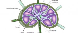
The lymph node (LN) is a kind of filter with a large number of various lymphocytes and other immune cells, into which the lymphatic fluid from the organs brings everything unnecessary and harmful. LNs form groups from several nodules to many dozens; lymphatic fluid flows to each group from a specific anatomical area.
Several lymphovascular pathways lead from the mammary gland to the lymph nodes of the axillary region and further to the subclavian and supraclavicular ones:
- The first and most voluminous, absorbing almost 95% of all waste lymph from the mammary gland, is the axillary path; fluid from the arm, half of the chest wall and the upper part of the abdomen also flows here. When cancer cells block the axillary lymph nodes, the fluid rushes to the lymph collectors of the abdominal cavity, which are connected by a network of capillaries with lymphoid formations in the mediastinum and ligament of the liver, introducing metastatic emboli into them.
- From the upper and posterior quadrants of the mammary gland, lymph immediately or in a roundabout way through small nodules between the pectoral muscles enters the subclavian collector, and from there flows into the supraclavicular lymph nodes to subsequently flow into the thoracic duct.
- The parasternal pathway collects fluid from the internal parts of the mammary gland and carries it to the nodules in the intercostal spaces and further to the subclavian and supraclavicular areas. The path anastomoses with intercostal lymph vessels, which are connected to the lymphatic network of the thoracic vertebrae, which explains the development of metastases in the spine.
- Lymph from the center and median lobes of the mammary gland is carried into the lymphatic system of the mediastinum, causing the formation of metastases in the lungs.
- Two interconnected lymphatic networks are formed in the skin of the breast, directing fluid into the vessels of the chest and the opposite breast, helping the spread of metastases in the skin of the chest wall.
Causes of enlarged axillary lymph nodes
Typically, the proliferation of lymphoid tissue is caused by inflammatory pathologies or breast cancer in women. The symptom also manifests itself in infections, lymphoproliferative processes and hematological malignancies, skin cancer localized near the axillary region. Less commonly, the cause of enlarged lymph nodes is frequent colds with chronic fatigue syndrome, which are accompanied by increased differentiation of lymphocytes. Axillary lymphadenopathy can develop with Mikulicz's disease, an autoimmune process affecting lymphoid tissue, salivary and lacrimal glands.
Mastopathy
The main symptoms of benign fibrocystic changes in the breast are pain and thickening, which most often occur in the second half of the cycle. Enlargement and tenderness of the axillary lymph nodes are typical for 10-15% of women. Adenosis is one of the forms of mastopathy, which is manifested by limited proliferation of glandular tissue in the mammary gland and discharge from the nipple. A slight increase in axillary lymph nodes is possible with a diffuse version of the lesion.
Mastitis
Inflammation of the mammary gland often begins in women after childbirth, which is caused by the penetration of a staphylococcal infection against the background of lactostasis. Enlargement of the lymph nodes during lactation mastitis is unilateral, they are painful on palpation and mobile. Women complain of intense chest pain, swelling and redness of the skin of the breast. The disease is characterized by high body temperature and symptoms of intoxication. Less commonly observed is non-lactation mastitis, which occurs as a result of hematogenous infection and trauma to the skin of the chest. A severe complication of mastitis is a breast abscess, which occurs with disturbances in the woman’s general condition, severe pain in the affected breast, and febrile fever.
Mammary cancer
The first metastases of this malignant neoplasm are localized in the axillary lymph nodes in 60-70%; subclavian and parasternal lymphoid formations can also be affected. Lymphadenopathy is caused by the proliferation of tumor cells. On palpation, the formations are dense, slightly painful, adherent to the skin and surrounding tissues. Breast cancer can occur in men and is always accompanied by enlarged regional lymph nodes.
A malignant breast tumor is asymptomatic for a long time; as neoplasia progresses, complaints of pain, serous or bloody discharge from the nipple appear. Other clinical symptoms depend on the variant of the disease: with Paget's cancer, erosions of the nipple and areola are observed, accompanied by weeping; triple-negative cancer is characterized by rapid development and metastasis. If enlarged lymph nodes are combined with other alarming symptoms, a woman should consult a mammologist as soon as possible.
Specific breast infections
In such infectious diseases, axillary lymphadenopathy is caused by both direct reproduction of the pathogen in foci of lymphoid tissue and increased stimulation of the immune system by antigens of microorganisms. Enlarged lymph nodes usually have an elastic consistency and are not fused with adjacent anatomical structures. The proliferation of lymphoid tissue is caused by:
- Syphilis of the mammary glands
. The primary affect (chancroid) is most often localized in the area of the areola and has the appearance of an ulcer with undermined edges and a bluish-red bottom, which is surrounded by a painless infiltrate. Induration and enlargement of the axillary nodes develops 2-3 weeks after the appearance of the skin defect. After one and a half to two months, the ulcer heals, the manifestations of lymphadenopathy subside, which indicates the beginning of the secondary period. - Tuberculosis of the mammary glands
. The infection is characterized by a combination of signs of general intoxication with enlargement and hardening of the affected breast, which is caused by the formation of a tuberculous node. Skin hyperemia may be detected above the formation, and later fluctuation and softening of the lesion occurs. The lymph nodes of the axillary zone are dense, painful, and often form conglomerates. With cavernous disintegration of the focus, fistula tracts can form.
Diseases of the blood system
In lymphoproliferative conditions, an increase in the nodes of the axillary region is caused by a pathological increase in the production and differentiation of white blood cells under the influence of carcinogenic factors. The category of hemoblastoses includes two large groups of diseases - lymphomas, in which the primary focus is located in peripheral lymphoid formations, and leukemias, which occur with damage to the bone marrow. The pathologies are characterized by a severe course with a pronounced impairment of the general condition. Signs of axillary lymphadenopathy include:
- Lymphoma of the lung
. The disease is typical for people after 50-60 years of age and occurs with a persistent cough, periodic hemoptysis, and chest pain. The axillary and cervical lymph nodes are enlarged and painful. Similar symptoms are observed with mediastinal lymphomas. In this case, compression of large vessels and nerve trunks occurs with the appearance of puffiness of the face and neck, and hoarseness. - Lymphogranulomatosis
. Often, the first sign of a lymphoproliferative process is an increase in peripheral lymph nodes, including axillary ones, which patients discover on their own. Later, the lymphoid tissue of the mediastinum is involved in the process, and other organs may be affected: lungs, intestines and spleen, and skeletal system. In patients, body weight decreases, low-grade or febrile fever, and increased sweating are detected. - Leukemia
. The disease is a consequence of primary damage to the bone marrow, which leads to uncontrolled proliferation of leukocyte precursor cells with their dissemination throughout the body. The disease begins acutely with increased body temperature, arthralgia and myalgia, increased bleeding and hemorrhages in the mucous membranes. Characterized by axillary and cervical lymphadenopathy, splenomegaly, and damage to the salivary glands. - Chronic lymphocytic leukemia
. The pathology is based on the excessive proliferation of mature B-lymphocytes with their accumulation in peripheral organs - lymph nodes, liver, spleen. The peak incidence occurs at the age of 55-65 years, men are more often affected. First, the axillary nodes enlarge, then the lymphoid formations of the mediastinum, abdominal cavity, and inguinal region are involved in the process. - Autoimmune lymphoproliferative syndrome
. The lesion occurs as a result of mutation of the genetic apparatus and is accompanied by hepatosplenomegaly, lymphadenopathy, and inhibition of hematopoietic processes. Manifestation of the syndrome can occur in infants 15-20 days after birth. In the case of a somatic mutation, signs are detected in adolescence. In 20% of cases, the process is complicated by the development of lymphoma. - Sézary syndrome
. The pathology is caused by primary damage to T-lymphocytes and is manifested by a characteristic triad of signs: erythematous spots on the skin, enlargement of the axillary, inguinal or femoral lymph nodes, and the appearance in the blood of typical cells with folded nuclei. Characterized by an increase in temperature to febrile levels, chills, and weakness. Patients may complain of intense itching and burning of the skin.
Infectious diseases
Diseases accompanied by the introduction of foreign microorganisms often occur with symptoms of lymphadenopathy. This is due to accelerated division and antigen-dependent differentiation of lymphocytes. More often, enlargement of the axillary lymph nodes is observed during infection with Koch's bacillus and tuberculosis intoxication, which is associated with the route of penetration of the pathogen and the anatomical features of lymph drainage from the chest organs. The most common causes of axillary lymphadenopathy are:
- Tuberculosis of the intrathoracic lymph nodes
. This localization of the pathological process is most typical for childhood. The disease is characterized by a long asymptomatic course; sometimes patients complain of causeless weakness, increased sweating at night, and low-grade body temperature. Subsequently, the process spreads to the lymph nodes of the armpit. Due to compression of the large bronchi, a dry bitonic cough and difficulty in exhaling are bothersome. - Toxoplasmosis
. Lymphadenopathy of the axillary and groin areas is more typical for the generalized version of the disease, which occurs with general intoxication, myalgia and arthralgia, and febrile fever. A widespread maculopapular rash appears on the skin, which does not affect the scalp. With toxoplasmosis, hepatosplenomegaly, myocarditis, and meningoencephalitis are observed. - Brugioz
. The disease is caused by the penetration of nematodes into the human body and is divided into acute and chronic stages. The acute phase of the infection is characterized by a profuse urticarial rash on the skin, an increase in temperature to 39°C, and an increase in inguinal and axillary lymphatic formations. I am concerned about pain in the upper and lower extremities associated with the development of lymphangitis. In the chronic form, the infection worsens about 2-3 times a year.
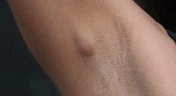
How do you know that the axillary lymph nodes have been damaged?
The size of a normal and healthy lymph node is from a few millimeters to two centimeters, it is soft and painless, and easily moves to the side when palpated. An increase in lymph nodes is a symptom of the inadequacy of the protective capabilities of the lymphoid organ to the health needs of the body, when the immune filter does not cope with the function assigned to it and zones of accumulation of cancer complexes are formed inside, which subsequently remain in the permanent residence.
Massive damage by metastases is easily determined by touch - the lymph nodes are large and dense, but painless and have not yet grown into the neurovascular bundle. Mammography and ultrasound of the mammary gland determine a tumor lymph node, although without taking cellular material for microscopy, this is only a “suspicion of cancer” that must be proven or rejected.
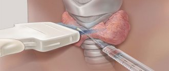
During breast surgery or before starting preoperative chemotherapy, a biopsy of the main or sentinel node, where the bulk of the lymph fluid is sent, is necessarily performed. If there are cancer cells in the sentinel lymph node, all axillary fatty tissue with everything in it is removed in one block.
Indications for ultrasound of lymph nodes
The examination is carried out to identify diseases of various etiologies, which are accompanied by activation of the lymph nodes and an increase in their size.
- Infectious pathology: viral, bacterial, fungal, chlamydial, parasitic, spirochetal, mycobacterial.
- Malignant neoplasms - hematological diseases (leukemia, lymphoma, malignant histiocytosis), metastatic lesions of the lymph nodes in melanoma, tumors of the mammary glands, lungs, gastrointestinal tract, prostate, tumors of the neck and head, Kaposi's sarcoma, seminoma.
- Endocrine diseases (hyperthyroidism).
- Pathology of fat metabolism (Neumann-Pick disease, Gosch disease).
- Mucocutaneous lymph node syndrome, familial Mediterranean fever.
- Diseases of unknown etiology: amyloidosis, sarcoidosis, sinus histiocytosis, lymphogranulomatosis, macrofollicular hyperplasia of the lymph nodes.
- Detection of acute and chronic pathology of lymph nodes and monitoring the dynamics of various diseases.
- Evaluation of the effectiveness of antitumor and anti-inflammatory treatment.
- Qualitative assessment of lymph nodes - the state of the architectonics, the intensity of the vascular pattern.
- Determination of the most effective trajectory for puncture of lymph nodes.
- Preventive examination.
How are lymph nodes affected when the tumor process spreads in them?
All removed lymph nodes, no matter how many there are in the tissue, are necessarily examined under a microscope; if there are cancer cells in them, additional studies are performed, including IHC.
The spread of cancer is believed to occur in stages: from a few cells to massive lesions, from axillary to subclavian and then to supraclavicular lymph nodes. The degrees of damage also go from minimal to maximum 3rd:
- During a morphological examination, everything pathological is detected, even cell accumulations of a fraction of a millimeter - microinvasion. The histological report will indicate рN1mic, that is, the cell complex has a size from 0.2 mm to 2 millimeters, and the letter “p” is an abbreviation for pathomorphology;
- When there is no tumor in the lymph node, the pathologist will designate the condition as “pN0”; if it is next to it (I-), then histology and IHC were performed, without immunohistochemistry - pN0 (I+);
- The next level of cancer spread through the lymphatic system is pN1 gradation, indicating a metastatic lesion larger than 2 millimeters in size, and there should be no more than three lymph nodes in the armpit;
- The pN2 lesion variant denotes a group of 4 to 9 metastatic lymph nodes in the axillary zone or a tumor in the intrathoracic lymph node;
- The final gradation pN3 suggests variations from a dozen metastatic axillary, or subclavian cancer, or involvement of supraclavicular lymph nodes.
| More information about breast cancer treatment at Euroonco: | |
| Surgeons-oncologists-mammologists | from 5100 rub. |
| Emergency oncology care | from 11000 rub. |
Preparation for the procedure
Before the examination, it is recommended to undergo an examination and consultation with a therapist and gynecologist (for women).
This is necessary, since lymph nodes can enlarge during inflammatory processes, so it is necessary to exclude the infectious nature of the symptoms. As for preparing for an ultrasound of the lymph nodes in the armpits, it is very simple. It is necessary to remove hair in the area, and immediately before diagnosis, wipe the cavities with a sanitary napkin.
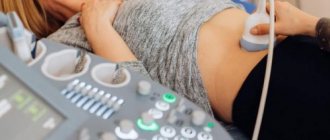
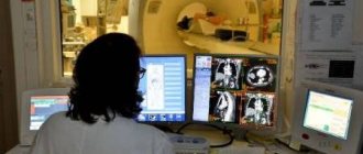
How is the diagnosis carried out?
The procedure is very simple to carry out, its duration is no more than 20-30 minutes. During the examination, the patient assumes a lying or sitting position, with his hands placed behind his head.
A special conductive gel is applied to the area and a sensor is installed, then the doctor moves it in different directions to obtain the required image. If necessary, additional examination of nodes in the groin area and neck is carried out. A conclusion about the results is issued immediately after the end of the study.
It is worth noting that the technique is completely painless and safe for the body; it can be done even for pregnant women and children.
The price for an ultrasound of the armpits in our clinic is 1000 rubles. You can find out detailed information about diagnostics and its significance from the center’s specialists - make an appointment using the online form or by phone.
We will be waiting for you at our clinic at the address: Moscow, metro station Kropotkinskaya / Arbatskaya, B. Afanasyevsky lane, building 22.
Lymph nodes according to TNM classification
In the TNM system, the letter “T” denotes the primary cancer tumor, regional lymph nodes - “N”, respectively, metastases - “M”.
Metastases formed in regional lymph nodes are always only N, while cancer in the axillary region of the opposite gland is already M, as well as the lymph collectors of the neck or groin.
Clinical classification according to the degree of damage is as follows:
- N0 - lymph nodes are healthy;
- N1 - there are already metastases under the arm, but without fixation to surrounding tissues and maintaining their mobility;
- N2 - states ingrowth or dense conglomerates in the axillary zone;
- N3 - the tumor involved the subclavian or supraclavicular, or intrathoracic.

How is the examination carried out?
Ultrasound of the lymph nodes of the neck, abdominal cavity, mammary glands, groin and any other group does not require special preparation. The doctor will tell the patient in advance what rules must be followed. The procedure is outpatient and lasts about 15-20 minutes.
After the scan, the patient is given a description of the ultrasound of the lymph nodes. To decipher the results, you must contact your general practitioner or the attending physician who referred you for the study. A diagnosis can be made using a sonogram only taking into account laboratory tests, the current condition of the patient, and other important factors. Therefore, interpretation is not included in the price of ultrasound of the lymphatic vessels and is done at a separate consultation.
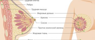
Regional lymph nodes
Lymph nodes that collect lymph fluid directly from the mammary gland are considered regional.
The first lymphatic collector is the axillary cavity, where up to 75 lymph nodes are located at five levels from the closest to the gland to the highest at the apex, on average about three dozen. The very first and largest is the Zorgius or signal node.
The subclavian lymph nodes receive lymph from the axillary lymph nodes.
Near the sternum between the ribs there is a chain of parasternal or intrathoracic lymph nodes.
Supraclavicular lymph nodes are not regional; not so long ago, metastases in them were designated as distant, and the process was considered inoperable. Today, preoperative chemotherapy makes it possible to change the situation and perform radical surgery even in such a situation.
The equipment of Euroonko clinics and specially selected and trained medical personnel allow us to quickly provide adequate treatment according to European standards.
Book a consultation 24 hours a day
+7+7+78
Where to sign up for an ultrasound of lymph nodes in Novosibirsk
To clarify information about the procedure for ultrasound of lymph nodes, you can contact the specialists of the Diagnost Center for Practical Medicine. The clinic’s doctors will answer all patients’ questions and, if necessary, select a time convenient for the appointment and examination. Instrumental research is carried out on high-quality equipment by experienced operators. As a result, the patient receives reliable information about the state of his own lymphatic system, on the basis of which specialized specialists prescribe adequate treatment.