A fracture of the radius of the wrist is a common injury that occurs primarily from a fall on the hand. Such injuries occur among people of all ages and are accompanied by pain, swelling, limitation of movement and joint deformation. The radius bone can break in the head and neck, in the lower and middle parts. A fracture of the radius just above the wrist is considered a typical hand injury. It usually occurs in old age in people with signs of osteoporosis.
If you do not contact a specialist in a timely manner for fractures of the radial bone of the wrist, deformations in the wrist joint, mid-carpal instability occur, and the radial bone shortens, which leads to uneven distribution of the load on the ligaments of the upper limb and limitations of movements. These complications cause the progression of deforming arthrosis and significantly reduce the patient’s quality of life.
A radial fracture can be isolated or combined with other injuries. But in any case, if you receive such an injury, you must go to a medical facility and undergo an appropriate examination. At the CONSTANTA Clinic in Yaroslavl you can make an appointment with a traumatologist and receive qualified medical care. Our doctors have the opportunity to use modern equipment to organize diagnostics and necessary treatment. The clinic is equipped with innovative equipment of European quality, which allows it to provide medical services of the highest possible level for many years.
Causes of beam fracture
The main cause of fractures in the radial bone of the wrist is an unfortunate fall on an outstretched arm. Damage also occurs against the background of osteoporosis, a disease that is accompanied by increased fragility and brittleness of bones. Women in the postmenopausal period are most susceptible to developing the disease, when there is a natural loss of bone tissue and the risk of developing fractures increases even after minor injuries.
Osteoporosis progresses in patients over 60 years of age. At this age, it is recommended to take special care in everyday life. Experts know of cases where even a slight fall on the hand caused dislocations and fractures of the wrist. Fractures of the radius also occur in young healthy people, especially in patients who are actively involved in sports and travel a lot.
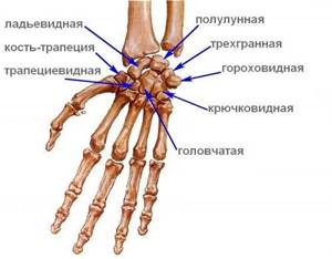
Anatomy of the radius carpal bone
The radius is one of the two bones of the forearm. It has a complex structure and is located near the ulna bone. The upper part of the beam passes into the neck and ends with the head, which connects to the bone of the shoulder, and from the side surface the beam connects to the bone of the elbow. In the lower part, the radius expands and begins to occupy almost the entire volume of the wrist joint - it is in this area that the radius passes into the wrist.
Taking into account the anatomical features of the structure of the radius described above, several types of fractures are distinguished, in which both the radius and ulna bones can be damaged.
Common types of radial carpal fractures include:
- fracture of the head, neck of the radius;
- fracture of the radius in a typical location (the injury occurs in an area located slightly above the wrist joint);
- Galeazzi damage;
- isolated fracture of the radial diaphysis.
Each of these types of fractures has its own characteristics and symptoms, which traumatologists are well familiar with. All attempts to carry out independent treatment for hand injuries lead to the development of complications, including joint deformities, infectious and inflammatory processes, chronic pain syndrome, and limited functional abilities of the limb.
Decoding the results
X-rays of the hands are interpreted by a radiologist immediately after the shooting is completed. He evaluates the relative position of the bones of the hand, their connection to each other, and integrity. Attention is also paid to assessing bone structure and density.
Normal indicators
Normally, the bones of the hands should have a uniform structure. There should be no shading in the white areas in the photographs. There should be a gap between the bones.
If only one hand is damaged, an X-ray of the healthy limb may be taken to more accurately diagnose the pathology. This will make it easier to compare and identify deviations.
Fracture of the neck and head of the radius
Fractures are more common in everyday life, and they also often occur during sports training, when an athlete falls on an extended arm. Domestic injuries occur predominantly in elderly women with signs of osteoporosis. Often, damage to the head and neck of the radius occurs in car accidents, falls from great heights, or during accidents on the street or at work. In such cases, a radial fracture is combined with injuries to the head, chest, and abdomen and requires the participation of doctors of other specializations in addition to the traumatologist.
Symptoms of a radius fracture:
- pain that intensifies with palpation and attempts to move the arm;
- tissue swelling;
- hyperemia of the skin at the site of injury;
- crunching (crepitation) of bone fragments may occur;
- deformity of the wrist joint.
Such injuries are usually treated with conservative measures. For displaced fractures, the doctor applies a cast for several weeks, after which he prescribes active rehabilitation. Displaced fractures require closed or open reduction - comparison of bone fragments. The procedure takes place under local anesthesia and does not cause any discomfort in the patient. After reposition, a control x-ray examination is required, which allows us to assess the condition of the radius bone. If unsatisfactory radiograph results are obtained, the bone head is fixed using a pin.
Fracture of the radius in a typical location
Fractures of the radius above the wrist joint are considered the most common injuries to the bones of the forearm. They are observed both in childhood and among adults. In elderly patients, injury is caused by progressive osteoporosis. A typical radial fracture occurs primarily from a fall on an outstretched limb, which may or may not involve displacement of the bone. Depending on the nature of the displacement, experts distinguish two types of such injuries - Smith fractures and Colles fractures. In a Colles fracture, the radial fragment is displaced to the back of the forearm, and in a Smith fracture, it is displaced to the palmar surface.
Typical radial fractures can be extra-articular, intra-articular, closed or open. If damaged, the patient experiences sharp pain, after which swelling, hyperemia, and hemorrhages appear. In some cases, there is a crunching sound of debris, as well as pathological mobility in the wrist joint. When examining the patient, specialists discover joint deformity. An accurate diagnosis is made after receiving the results of an x-ray examination. Complex fractures require an extensive examination, which includes computed tomography and magnetic resonance imaging.
Grade
Physical therapists should perform a thorough physical examination, including the collection of subjective and objective information.
- The subjective assessment includes any information provided by the patient, such as pain experienced, limitations in wrist range of motion, and activity limitations.
- Physical examination includes assessment of wrist and finger range of motion, grip and forearm strength, bony and soft tissue abnormalities, skin integrity, and nerve involvement. Remember that the contralateral limb may be an unreliable reference point.
Healthcare professionals should evaluate ligamentous integrity as early as possible in the presence of persistent pain associated with suspected wrist instability to avoid poor functional outcomes and prolonged recovery. Specific fracture patterns and high-energy injuries are highly suggestive of wrist ligament injury.
An X-ray examination can determine the fracture, displacement, and the number of bone fragments.
Damage to Galeazzi
This type of damage to the radius was discovered by the surgeon Galeazzi, who lived in Italy. The injury is based on a combination of a radial fracture with a dislocation of the ulna in the wrist joint. Trauma is quite rare. It manifests itself as pain, swelling of soft tissues and severe limitation of mobility. Possible formation of subcutaneous hematomas. Damage to Galeazzi is often accompanied by injury to nervous tissue, compression of nerves and blood vessels, which often requires surgical treatment. If the nerves are damaged, the patient notes a loss of sensation in the hand area.
Against the background of such fractures, compartment syndrome often develops - a complex symptom complex, which is based on an increase in tissue pressure in a closed musculoskeletal space, which leads to oxygen starvation of muscle tissue, necrosis and the formation of ischemic contracture. If the diagnosis of compartment syndrome is confirmed, surgical treatment is performed.
First aid
The victim can receive the highest quality care after contacting specialists immediately after receiving an injury. This range is considered to be a period from one day to 7 days.
Immobilization

If there are rings on your fingers, then for greater convenience it is recommended to remove all the rings from the area of the injured hand. This is done to avoid additional pressure on the fingers and the formation of edema, as well as necrosis of the finger.
Cold is immediately applied to the damaged area, and the hand is placed on an elevated surface. This is also done to prevent swelling.
The limb is immobilized using a splint. In cases where you are very far from a medical facility, the splint is made from available materials (you can use plywood, wood, plastic). The main goal of applying a splint is to avoid secondary injury to the area. If there is an open fracture, then a sterile bandage is applied to the wound, and the place is fixed with a splint.
If there are painkillers nearby and the pain is very strong, then it is recommended to give the victim 1-2 tablets.
Diagnosis of a radius fracture
If a fracture of the radius is suspected, the diagnosis is established based on the results of the examination. Most often, specialists limit themselves to radiography in two projections. Radiography is the most common and accessible research method for specialists and most patients.
But for complex fractures and associated pathologies, other research methods are also used, in particular computed tomography (CT) and magnetic resonance imaging (MRI). These two examination methods provide maximum information about the condition of soft and hard tissues, allow you to plan further treatment, including surgery, and make accurate predictions for the future.
Treatment of radius fractures
Treatment for fractures of the radius is selected by a qualified traumatologist. It is possible to involve other specialized specialists in case of detection of combined injuries and damage, as well as in case of exacerbation of certain chronic diseases. Delay often leads to complications, including the development of chronic pain syndrome, noticeable joint deformities and limitations in motor activity. For “old” fractures, treatment is predominantly surgical.
Medical tactics depend on the degree of damage and the nature of the injury. Doctors at the CONSTANTA Clinic in Yaroslavl, when choosing methods for treating radial bone fractures, focus primarily on the interests of the patient. Specialists select techniques that help restore the patient’s well-being and impaired functions in a short time.
Conservative therapy
A radial fracture in a typical location without signs of displacement is subject to conservative therapy. Specialists fix the limb with a plaster cast and prescribe analgesics for pain. If the radius is displaced, doctors return it to its anatomically correct position using reposition and securely fix it for successful bone fusion. If a patient consults a traumatologist several weeks after a fracture, the risk of developing deformation of the damaged joint and progression of arthrosis sharply increases.
The rate of fracture healing largely depends on the quality of the immobilization performed. The limb is fixed in a stationary position for 1-1.5 months. Throughout the treatment period, the patient must periodically visit a traumatologist so that the specialist monitors the patient’s condition and, if necessary, can change treatment tactics. Control X-ray examinations must be carried out on time. An x-ray allows you to timely detect secondary displacement under a plaster cast and promptly take the necessary measures to eliminate it.
The plaster cast is removed about a month after the fracture. But the treatment does not end there. Patients with hand injuries need effective rehabilitation measures that help restore range of motion in the injured hand and prevent the development of delayed complications, in particular irreversible contractures.
Final indicators
To improve prospective data collection, a CL fracture complication checklist and score sheet was developed. The checklist includes a classification of all complications of PDOLK, each of which is rated according to severity (mild = 1, moderate = 2, severe = 3). The total score is then calculated. The questionnaire categories are as follows:
- Complications of the nerves.
- Complications of bones and joints.
- Complications of the tendon side.
Self-report outcome measures
- Visual analogue scale (VAS) for pain assessment.
- Disability Assessment of the Arm, Shoulder, and Hand (DASH) Questionnaire.
- Patient Rating Wrist Evaluation (PRWE).
- The Michigan Hand Performance Questionnaire (MHQ) is a hand performance instrument divided into six scales: general hand function;
- daily activities;
- pain;
- labor productivity;
- aesthetics;
- patient satisfaction with hand function.
Physical outcome measures
- Wrist range of motion measured with a goniometer.
- Grip strength is an important indicator because it is a significant function in daily activities and can be measured using a dynamometer.
Surgical treatment of radius fractures
If conservative therapy does not give the expected result or cannot be used due to the severity of the injury, specialists use surgical methods to treat the fracture. The most common method of correcting displaced fragments is osteosynthesis (reposition), which can be either open or closed. Closed reduction involves the comparison of bone fragments without incisions of soft tissues. The technique is characterized by minimal trauma, absence of postoperative scars and rapid recovery.
If closed reduction is not possible, open osteosynthesis is used using special plates or screws. The surgeon makes a small tissue incision to gain easy access to the damaged bone. After eliminating the displacement, the bone fragments are fixed in the anatomically correct position. Titanium plates are used as fixing elements, which are biocompatible with tissues and allow the development of the joint to begin as soon as possible in order to prevent the development of contracture. The clamps reliably hold the fragments in the desired position, so the period of use of the plaster cast can be reduced or dispensed with without plaster at all.
For open fractures of the radius, external fixation devices are used. They reliably fix bone fragments, preventing secondary displacement. An open fracture of the radius requires surgery within 6-8 hours after the injury. Antiseptic treatment of the wound is mandatory. Modern antiseptics can almost completely eliminate the risk of secondary infection. External fixation devices are installed for 4-6 weeks. This period is sufficient for complete healing of the fracture.
But complete recovery takes several months, and during this period it is recommended to spare the sore arm, limit physical activity and do everything to prevent recurrent injuries.
Rehabilitation
Kay et al. found that a rehabilitation program consisting of consultation and exercises under the guidance of a physical therapist provided some additional benefits compared with no physical therapy intervention for adults following casting and/or pinning for a DCL fracture. These benefits included decreased pain compared to the control group at weeks 3 and 6, increased activity compared to the control group at week 3, and greater satisfaction with treatment compared to the control group. The two groups did not differ in recovery of active wrist range of motion or grip strength.
Michlovitz et al. published the results of a survey of physical therapists, occupational therapists, and certified hand care therapists to determine common rehabilitation practices for patients with a DCL fracture.
During immobilization
At this stage, less than 10% of patients with a DCL fracture are referred for rehabilitation treatment. Priorities during immobilization include managing finger swelling and stiffness and patient education. Home exercise programs are commonly offered to increase the range of motion of the shoulder, elbow, and fingers. Heat/cold treatments may be used to reduce pain. Compression wraps and massage can be used to relieve swelling. In 50% of cases splints were used for support and protection.
After immobilization
Ninety percent of therapists surveyed include heat/cold treatments and range of motion exercises at this stage. Eighty percent use compression wraps with massage, agility exercises, and joint and soft tissue mobilization. Nearly 90% also use strengthening exercises to improve strength and function. Static or dynamic splints (or both) can be used to relieve joint stiffness.
Smith et al. provided the following brief description of postoperative rehabilitation methods.
External fixation
- Control pain and swelling early.
- Caring for the damaged area.
- Maintaining active range of motion in non-involved joints, rotation of the forearm is difficult, active range of motion in the wrist is impossible.
- Desensitization of affected nerves, elimination of complex regional pain syndrome.
- After removal of the fixation equipment: active/active-assistive/passive range of motion of the wrist and forearm; focus on wrist flexion and extension, ulnar deviation and supination; static progressive tires; progressive activity and activities of daily living.
Dorsal plate
- Control swelling early.
- Static splint on the wrist.
- Maintaining active amplitude is not
- involved joints.
- Graduated early active range of motion at the wrist.
- After bone healing: active/active-assistive/passive range of motion of the wrist with emphasis on active wrist flexion; preload and relaxation program for the extensor tendons; static progressive tires.
- Progressive activity and activities of daily living.
Volar fixed-angle plate
- Swelling control, active range of motion of the wrist and non-involved joints.
- Static splint for the wrist in a resting position with 30° extension.
- After bone healing: progressive active/active-assistive/passive restoration of wrist amplitude; protective static splint on the wrist; Static progressive splinting is rarely indicated.
- Progressive activity and activities of daily living.
Features of recovery after a fracture of the radius and possible complications
The speed of recovery after a radial fracture depends on many factors, including the professionalism of the doctor and the general health of the patient. For several weeks after the injury, pain may persist, which can be relieved with analgesics and nonsteroidal anti-inflammatory drugs. If the pain gets worse, you should tell your doctor about it.
After applying a plaster or polymer bandage, the patient should monitor the condition of the skin near the plaster. If they change color (turn pale, blue), this may be a sign of compression of blood vessels and soft tissues. A decrease in sensitivity indicates compression of the nerves and also requires consultation with a traumatologist. In rare cases, purulent complications occur due to the addition of a secondary infection (due to late antiseptic treatment and delayed treatment).
Symptoms of a broken finger
The types of finger fractures differ from each other. There are fractures of one phalanx and multiple fractures of several fingers. Determining a fracture is not difficult. With a recent injury, symptoms include the following:
- the finger becomes bluish and swells due to internal hemorrhage and vascular injury;
- very strong and sharp pain in the finger, intensifying with movement;
- in many cases you can see the deformed area with the naked eye;
- the damaged finger is shortened;
- unusual mobility of the phalanx, which has not been noted before;
- inability to fully clench the hand into a fist or fully unclench the hand;
- the appearance of hematomas under the nails, which cause acute pain;
- when you try to move your finger, you hear an uncharacteristic crunch;
- if there is an open fracture, then the victim experiences pain shock and bleeding.