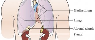
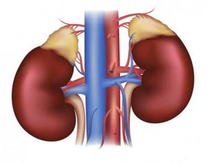
The adrenal glands, endocrine glands, are located at the upper pole of the kidneys, so their condition is often described on MRI of the kidneys. The structure of the adrenal gland consists of 2 layers - cortical and medulla. Each of them produces hormones that are very important for the functioning of the body.
The cortex synthesizes corticosteroid hormones, which have a regulatory effect on metabolic processes and the function of the reproductive system.
The medulla produces so-called “stress” hormones, which help the body cope with changes in environmental conditions, that is, adapt.
What does an MRI of the adrenal glands show?
The adrenal glands can be examined using different methods: X-ray, CT, ultrasound, laboratory (determining the level of hormones in the blood serum). But MRI occupies a special place: it has a greater resolution than ultrasound, and does not pose a danger to the patient given the lack of radiation (compared to CT). And the level of hormones does not immediately change with tumor damage to the adrenal glands.
Using MRI, it is possible to detect an adrenal tumor at an early stage, when there are no clinical manifestations of the disease. Sometimes tumors are an incidental finding on MRI of the kidneys.
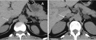
MRI clearly separates both layers of the adrenal glands and reveals tumors in them. In this case, it is possible to determine not only the location of the tumor, but also the exact size and nature (benign or malignant). The accuracy of the data obtained increases the procedure with contrast.
General information about the study
MRI of the adrenal glands in Moscow is performed to identify pathologies of internal organs in order to evaluate the structure of adjacent tissues. The kidneys ensure the removal of metabolic products from the body; their importance is difficult to overestimate.
The adrenal glands are glands that produce vital hormones. Their lack or excessive quantity leads to disruption of the endocrine system.
Using MRI of the adrenal glands and kidneys, you can determine the structure of organs, tissue structure, their condition, promptly identify the presence of new formations, identify pathologies in the circulatory system and begin treatment.
Diagnostics are often carried out to clarify the nature of diseases, determine the causes of pain symptoms, and deterioration in clinical test results. Main indications for performing the procedure:
- Painful sensations in the lumbar region;
- Problems with urination;
- Suspicion of the appearance of a neoplasm;
- The presence of congenital or acquired pathologies of the urinary system.
In the case of diagnosing oncology or other pathologies, it is necessary to carry out the event regularly in order to assess the effectiveness of treatment and identify relapses. It is recommended to sign up for an MRI if you suspect inflammatory processes, after injuries, etc.
Performing a study at a medical center allows you to determine the condition of the organs and identify any changes. An MRI of the adrenal glands can be done with or without contrast. The introduction of a special substance allows for high-quality visualization of organs and obtaining detailed and clear images. You can find out the price of MRI of the adrenal glands from our specialists.
Indications and contraindications
The main indication for MRI is suspicion of an adrenal tumor or monitoring after surgery to remove a tumor. Suspicion may arise based on clinical manifestations or if any changes are detected during another type of examination (for example, an imbalance of adrenal hormones).
Clinical manifestations of adrenal damage may include the following nonspecific symptoms:
- high or low blood pressure;
- cardiac dysfunction (increased heart rate);
- muscle weakness;
- changes in the patient’s appearance (enlarged mammary glands in men, the appearance of a rough voice in women).
MRI of the adrenal glands is prescribed for:
- Itsenko-Cushing syndrome;
- Addison's disease;
- Conn's syndrome.
Contraindications
Given the need for contrast in this study, MRI is not performed in cases of renal failure, pregnancy, or breastfeeding.
Contraindications to MRI are also:
- any metal objects present in the body (bullets, fragments, prosthetic joints, clips on blood vessels, etc.);
- implants in the body (heart pacemaker, insulin pump, hearing aid);
- patient claustrophobia (uncontrollable fear in a confined space);
- body weight more than 150 kg;
- epilepsy;
- state of decompensation, need for constant monitoring;
- mental disorders.
Pros and cons of the study
MRI of the adrenal glands has many advantages over other methods of studying these organs:
- the procedure is painless, non-invasive (except contrast) and does not require special preparation;
- does not emit harmful rays, therefore does not carry an x-ray load for the patient (unlike computed tomography - CT);
- can be carried out several times in a row;
- has a minimum number of contraindications;
- provides a clear image of adrenal tissue;
- identification of the earliest manifestations of ischemic disorders in organs.
To increase the contrast of images and carry out differential diagnosis of various neoplasms in the adrenal glands, it is possible to conduct MRI with contrast.
Despite the large number of advantages, the technique still has disadvantages that partially limit the use of MRI for diagnosing adrenal pathologies. These include:
- duration of the study (can last 15 minutes or more), which in some cases, for example, in restless children, requires sedation (administration of sedatives);
- low diagnostic ability to detect hemorrhages in the adrenal glands;
- does not reflect the hormonal activity of organs;
- impossibility of carrying out in those patients who have metal objects in their bodies (valves, plates, pacemakers);
- the difficulty of carrying out in patients on mechanical ventilation, for example, with acute adrenal insufficiency;
- distortion of results at the slightest movement of the patient;
- difficulties in examining patients with claustrophobia;
- high cost of examination.
In some cases, MRI of the adrenal glands is the only method for non-invasive rapid detection of adrenal pathology, for example, with tumors of the medulla.
Tomographs are very expensive, so they are not available for many medical institutions, especially provincial ones. Patients from the outback are often forced to undergo MRI examinations in large cities (for example, Moscow, St. Petersburg, Rostov, Yekaterinburg).
Preparation
No special preparation is required for MRI without contrast. The day before the procedure, it is recommended to exclude foods that contribute to increased gas formation. In emergency cases, MRI can be performed without preparation.
When using a contrast agent, you should stop eating and drinking 5 hours before. before the procedure.
On the eve of the study, a contrast sensitivity test is performed. To do this, the nurse injects a small amount of contrast subcutaneously and monitors the patient's condition for 30 minutes. If there is no reaction, contrast can be used.
The patient changes into disposable clothes, takes off jewelry or jewelry, watches, and leaves bank cards and a phone number with the laboratory assistant.
Where is the diagnosis carried out?
Screening is available for both private organizations and public medical institutions. Any diagnostic center containing a tomography room accepts patients in the specified direction. You can choose a clinic in any area of the city. To search, use the service of the “Unified Recording Center” of St. Petersburg, on the pages of which the data of all medical organizations registered in the Northern capital is collected.
The portal contains detailed lists of procedures, and when you click on any of them, a list of clinics opens. They are distributed by addresses, ratings, technical characteristics of equipment and prices for services. Mark the best offers and book your session immediately through the website. Each user of the service receives guaranteed discounts from the recording center for the selected type of hardware research. The telephone numbers of the consulting service are located on each page of the service.
results
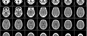
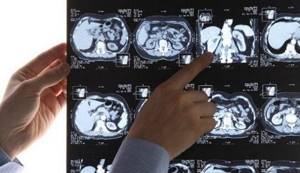
The resonance signals registered by the device, after computer processing, appear on the screen in the form of images of the glands. The adrenal glands are clearly visualized, and with the help of contrast, the medulla and cortex can be distinguished.
In the detected tumor, the doctor determines not only the exact location and origin from one of the layers of the adrenal gland, but also its nature. The malignant tumor is heterogeneous in structure and does not have clear boundaries or contours.
The doctor also evaluates the condition and size of visible lymph nodes, the condition of the perinephric (perinephric) tissue to identify tumor metastases in them. The conclusion is issued within an hour.
Treatment of any type of adrenal tumor is surgical. After tumor removal, a follow-up MRI is performed.
MRI of the kidneys: preparation for the study

During the diet before an MRI of the kidneys and adrenal glands, food is prepared in a gentle way: boiled or baked in the oven.
Preparatory measures are aimed at preventing gas formation and normalizing the functioning of the gastrointestinal tract, therefore foods and drinks that provoke flatulence are excluded from the diet for 3-4 days:
- unprocessed greens, vegetables and fruits, especially legumes and cabbage;
- yeast-containing baked goods, rye bread;
- chocolate, cocoa;
- milk and its derivatives;
- lemonade, kvass, any alcohol.
A cleansing enema and taking medications that enhance peristalsis, relieve spasms, and reduce flatulence are a separate topic that requires consultation with your doctor.
Benefits of contrast-enhanced MRI
Magnetic resonance imaging, which uses an injection with a contrast agent, has the highest information content. It is not painful at all, but can sometimes cause allergies. Therefore, before giving an injection, doctors conduct a test for the patient’s allergic reaction.
Considering MRI of the adrenal glands with contrast and other types of diagnostics, you can see the following advantages of magnetic resonance imaging:
- High-resolution images allow you to identify even the most minor changes in the structure of organs;
- the examination is safe and does not result in unnecessary exposure to radiation;
- minimum contraindications;
- The procedure is painless and quick (approximately 20 minutes).
The use of contrast provides the greatest accuracy of the study. After administering the contrast agent, the specialist immediately begins the examination. This is necessary because after 3 minutes the coloring liquid reaches the organ, then accumulates in it for 5 minutes, and after another 5 minutes is excreted.
Fifth - poor quality transcription
“How can a person find out whether he was given a quality conclusion or not? Very simple. If you were given an A4 piece of paper with a few lines, which are summarized by the conclusion “no pathologies were identified,” you spent your money in vain. The employee performing CT and MRI is not a doctor, he is a medical specialist who may or may not see certain problems, nothing more. He is obliged to describe them in detail for the doctor who will evaluate the results,” notes the doctor.
For example, if an examination of the abdominal cavity is carried out, then there may be many formations, some of them are normal, some should not be there. And the specialist conducting the research must indicate them all and describe them in detail, otherwise the procedure makes no sense, explains Serebryansky. “The correct description has quite a lot of columns and fields. There is also a reason to be wary if you are given results too quickly. If you received them less than half an hour after the study, it means that no one took care of you,” says the doctor.
It is also worth paying attention to the medium on which the information is given to you. “If these are only a few pictures, then you can assume that you haven’t actually done any research. Due to the fact that the number of sections on modern devices is quite large, many do not print pictures at all; everything is recorded on a disk or flash drive if the amount of information is too large,” emphasizes Oleg Serebryansky.