Home » Department of Orthopedics » Diseases » Chest deformity
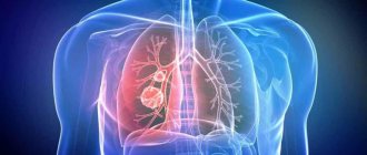
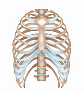
The chest is a part of the human body. It is formed in front by the sternum, behind by the spine, on the sides and partially in front and behind by the ribs. It is also formed by muscles that connect these bones to each other and to other bones. The rib cage contains the chest cavity, which contains the most important organs of the human body.
The chest contains the lungs and heart, as well as the aorta , the largest artery in the body. If these organs stop functioning, the person dies within a few minutes. That is why, during evolution, they settled in such a protected place. Due to the rigidity of the chest, the organs inside it are in a constant position, which ensures the stability of their functioning.
However, in some cases the chest changes its shape. Depending on the degree of change in shape, the position of the organs inside changes, their transformation occurs as they adapt to changing conditions. It should be noted that the chest is deformed, in most cases, in children, or is completely congenital. And with intensive growth, changes occur very quickly. This can lead to serious consequences for the entire body.
What it is
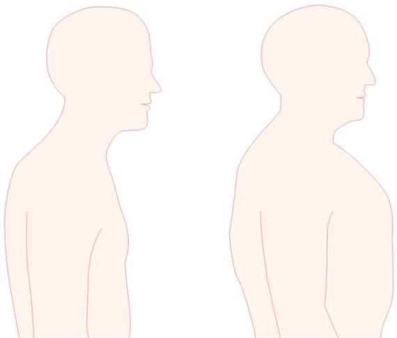
What does an emphysematous chest look like? The signs of this pathology are as follows:
- increase in transverse and anteroposterior breast size;
- large chest volume;
- protrusion of the collar bones;
- expansion of the spaces between the ribs;
- cylindrical or barrel-shaped breasts.
A wide sternum can also be observed in healthy people of dense build (hypersthenics). However, there are differences in the description of emphysematous chest and hypersthenic chest. With a stocky build, the size of the chest corresponds to the dimensions of other parts of the body. With breathing disorders, breast volume increases significantly more and looks disproportionate.
A photo of an emphysematous chest can be seen below. On the right is a barrel-shaped deformation.
Publications in the media
Congenital chest deformities are developmental defects associated with changes in the shape of the chest wall.
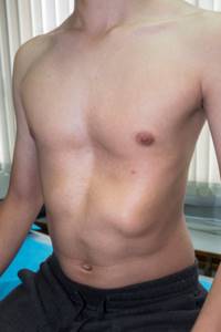
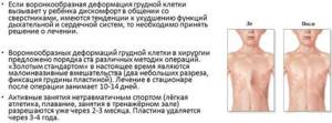
Funnel chest (pectus excavatus) is a developmental defect, which, in addition to a cosmetic defect in the form of retraction of the sternum and ribs, is accompanied by various functional disorders in the respiratory system and cardiovascular system. In boys it is observed 3 times more often. There are three stages of the disease (compensated, subcompensated and decompensated), three forms of deformities (symmetrical, asymmetrical and funnel-shaped), three degrees of deformation (I degree - deformation depth less than 2 cm, no displacement of the heart; II degree - deformation depth less than 4 cm, displacement heart by 2–3 cm; III degree - depth of deformation more than 4 cm, displacement of the heart by more than 3 cm). Treatment: general and special gymnastic exercises, massage. Surgical treatment is indicated for progressive deformities with functional impairment at the age of 4–5 years. There are many methods in the arsenal of surgical treatment, but none of them is accepted as optimal. With each, the risk of late relapses is noted • Removal of deformed costal cartilages • Osteotomy of the sternum • Correction of the sternum defect by introducing a bone wedge • Fixation with a support bar located behind the sternum • Other options: dosed traction, use of magnets, etc.
Keeled chest deformity (“chicken breast”, pectus carinatum) is a protrusion of the sternum and the costal cartilages attached to it. Damage to the costal cartilages can be unilateral or bilateral, and the sternum protrudes anteriorly in the upper or lower sections. With conservative treatment, general and special gymnastic exercises and massage are prescribed. Of the proposed surgical treatment methods, subperichondral resection of cartilage is the most acceptable. Corrected by transverse osteotomy with posterior movement of the sternum.
Poland syndrome is a combination of abnormalities including absence of the pectoralis major and minor muscles, syndactyly, brachydactyly, atelia and/or amastia, deformed or absent ribs, absence of hair in the armpit, and decreased thickness of the subcutaneous tissue. Each patient has different components of the syndrome. Deformation of the chest varies from mild hypoplasia of the ribs and costal cartilages on the affected side to aplasia of the anterior part of the ribs and all costal cartilages. Surgical treatment is necessary for only a small proportion of patients.
Sternal defects - a wide range of rare anomalies of the sternum, heart and upper abdominal wall • Thoracic ectopia cordis - the classic “naked heart”, not covered in front by any tissue, often noted is tetralogy of Fallot, pulmonary stenosis, transposition of the great vessels and VSD. Treatment of thoracic ectopia of the heart is almost always unsuccessful • Cervical ectopia of the heart. Patients differ from patients with thoracic ectopia in the distance to which the heart is moved • Thoraco-abdominal ectopia of the heart. The lower part of the sternum is split, the heart is covered with a membrane. Surgery is aimed at closing the skin defect over the heart and abdomen • Cleft or bifurcated sternum • Distal sternal defect is encountered as part of Cantrell's pentad.
Anterior scalene syndrome • Causes: accessory cervical rib, spasm and secondary shortening of the scalene muscle or traumatic deformity of the bones of the chest and shoulder girdle • Clinical picture. Pain in the neck, shoulder girdle and arm (along the ulnar nerve) and peripheral paresis of the arm caused by compression of the neurovascular bundle in the anterior interscalene space (between the anterior and middle scalene muscles). The clinical picture resembles Raynaud's disease • The diagnosis is confirmed by studying the functions of the ulnar nerve • Treatment. Decompression of the neurovascular bundle, most often by resection of the first or accessory cervical rib (if present), dissection of the anterior scalene muscle. Release of nerves from surrounding scar tissue, usually performed through a supraclavicular approach.
ICD-10 • M95.4 Acquired deformity of the chest and ribs • Q67 Congenital musculoskeletal deformities of the head, face, spine and chest • Q67.6 Sunken chest • Q67.7 Keeled chest • Q67.8 Other congenital chest deformities
Possible diseases
What diseases can cause deformation of the breast bones? Most often this is observed in cases of serious pathologies of the respiratory system:
- emphysema;
- obstructive bronchitis.
Pulmonologists classify these two diseases as chronic obstructive pulmonary disease (COPD).
In addition, emphysematous deformation of the chest is observed in patients suffering from cystic fibrosis and bronchial asthma. Curvature of the sternum bones is also observed in advanced cases of osteoarthritis.
Next, we will consider in detail each of the possible pathologies.
Chest deformity
15784 March 10
IMPORTANT!
The information in this section cannot be used for self-diagnosis and self-treatment.
In case of pain or other exacerbation of the disease, diagnostic tests should be prescribed only by the attending physician. To make a diagnosis and properly prescribe treatment, you should contact your doctor. Chest deformation: causes of occurrence, what diseases it occurs with, diagnosis and treatment methods.
Definition
Deformation of the chest is understood as changes in the shape of its bone structures of varying degrees of severity, in some cases manifesting not only as a cosmetic defect, but also leading to functional disorders of the respiratory and cardiovascular systems due to compression and displacement of the organs of the thoracic cavity. The chest is a part of the body formed by the sternum, ribs, vertebrae, and muscles connected to each other through joints.
In other words, the chest is a musculoskeletal frame that protects vital organs from external influences.
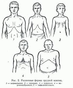
The chest changes as a person grows and develops, and in adults its shape and size depend on gender, the development of muscles and respiratory organs, type of activity, and lifestyle. The shape of the chest has several normal variants: flat, cylindrical and conical.
Types of chest deformities
All chest deformities are divided by origin
into congenital and acquired.
Congenital defects include funnel-shaped, keeled, combined chest deformities and more rare developmental defects. Funnel
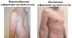
chest is characterized by recession of the sternum and anterior chest wall. This is the most common deformity - it accounts for about 80% of all deformities (occurs 3 times more often in boys) and in 25% of cases is hereditary.
Funnel chest
Keeled
the chest is enlarged in its anteroposterior part, the sternum protrudes forward in the form of a keel. Occurs with a frequency of 6 to 20%, more often in males.
Acquired
The deformation of the chest can be scaphoid, emphysematous, or barrel-shaped, paralytic, kyphoscoliotic, as well as keeled chest (rachitic).
By shape
Deformations are divided into symmetrical and asymmetrical.
To determine the severity of the deformity, a chest x-ray or computed tomography (CT) scan is performed.
On the radiograph, the ratio of the smallest size between the sternum and the vertebral body to the largest is calculated, which is the Giżycka index. Depending on the obtained value, four degrees of deformation are distinguished. When performing a computed tomography scan, the Haller index (computed tomography index) is determined, which is equal to the ratio of the horizontal distance between the inner part of the ribs to the distance between the sternum and the vertebral body at the place of greatest retraction of the sternum.
By stage
deformations are compensated, subcompensated and decompensated.
With compensated
deformity, the cosmetic defect is insignificant, shortness of breath and rapid heartbeat are not observed.
With subcompensated
deformity, the cosmetic defect is pronounced, there is shortness of breath and tachycardia during physical activity.
With decompensated
deformity, the cosmetic defect is disfiguring, shortness of breath and tachycardia are present at rest.
Possible causes of chest deformation
Congenital deformities of the chest are associated with a genetic abnormality in the development of cartilage and bone tissue, and are also often combined with connective tissue defects (in hereditary diseases: Marfan syndrome, Ehlers-Danlos syndrome, etc.). Some types of deformities can be diagnosed in infancy or early childhood (costomuscular defect, cleft sternum). Others debut and progress during periods of accelerated growth of the body, mainly such leaps occur at the ages of 5–6, 8–10, 13–15 years.
Acquired chest deformities occur as a result of external influences (trauma, burns, surgical interventions, for example, for cardiac pathology) or previous diseases (usually inflammatory or infectious, associated with calcium metabolism disorders).
Diseases that cause chest deformation
Diseases that cause chest deformation and are associated with calcium metabolism disorders include
rickets
.
Rickets is a disease of childhood in which, due to various reasons, polyhypovitaminosis occurs in an intensively growing organism with a predominant decrease in the level of vitamin D - the bones lose mineral density and are deformed as the child grows, the chest takes on a keeled shape. Currently, deformations are less common, because Rickets is recognized in the early stages.
Syringomyelia
is characterized by the presence of a cavity filled with fluid located in the spinal cord . The disease can occur due to impaired development of the embryo, due to birth trauma, spinal cord injury, or obstruction of the outflow of cerebrospinal fluid. The walls of the cavity push aside the surrounding tissues, which consist of nerve cells and pathways of the nervous system. As a result, the innervation of the muscles, including those forming the frame of the chest, is disrupted. In later stages, this can lead to curvature of the spine and the formation of a scaphoid depression on the anterior surface of the chest.
Osteomyelitis
– infectious-inflammatory purulent-necrotic lesion of bone tissue, the causative agents of which can be staphylococci, streptococci, Escherichia coli, etc.
Osteomyelitis of the ribs occurs extremely rarely, more often it is post-traumatic, less often - bacterial, when bacteria enter the bone tissue through the bloodstream or spread through contact (for example, with purulent damage to the lining of the lungs).
In the acute period, symptoms such as an increase in body temperature to 39–40°C, pain, redness, and swelling in the area of the affected rib come to the fore.
Among infectious diseases, tuberculosis
. Not only pulmonary tuberculosis (in later stages), but also bone tuberculosis (sternum, ribs, vertebrae) can lead to chest deformities. The process proceeds according to the type of osteomyelitis, but it is caused by a specific pathogen - Koch's bacillus. With tuberculosis of the ribs or sternum, swelling and pain in the affected area are externally determined. With spinal tuberculosis, the vertebral bodies are affected and destroyed, which is manifested by pain; in the later stages, the spinal column is deformed. The disease is accompanied by an increase in body temperature to 37.2–37.6°C, general malaise, night sweats, lack of appetite, and weight loss.
Emphysema
– a disease in which the walls of the alveoli, the structural elements of the lung tissue, are irreversibly destroyed and lose their elasticity, gas exchange is disrupted and increased airiness of the lungs occurs. Emphysema can occur independently or as a result of obstructive pulmonary disease.
Due to the increased airiness of the lung tissue, the chest increases in volume, as if freezing on inhalation (becomes barrel-shaped).
In diseases of the lungs and pleura
, leading to the formation of connective tissue in them and a decrease in their size, the chest is deformed like a paralytic one - it becomes smaller, flattened, and the intercostal spaces are drawn in on the affected side.
Which doctors should I contact if I have a chest deformity?
An initial assessment of the condition can be carried out by a general practitioner. If there are indications, the patient is referred to specialized specialists, such as a surgeon, orthopedic traumatologist, phthisiatrician, oncologist, psychologist, geneticist, otolaryngologist, etc.
Diagnosis and examinations for chest deformation
Before prescribing treatment, the doctor needs to assess the type and shape of the cosmetic defect, find out when and under what circumstances it occurred.
You should definitely tell your doctor about other symptoms, if any: general weakness and fatigue, episodes of fever, shortness of breath, rapid heartbeat.
If necessary, to assess the condition of internal organs or clarify indications for surgical treatment, the specialist will prescribe additional examination methods: chest x-ray in two projections with calculation of indices, complete blood count with leukocyte count and ESR, general urine analysis, spirography, electrocardiography, echocardiography (EchoCG) computed tomography of the chest and mediastinum, magnetic resonance imaging of the chest.
Emphysema
Emphysema often affects smokers and patients working in hazardous industries. This disease can also develop as a complication of obstructive bronchitis. Under the influence of various harmful factors, the pulmonary alveoli expand in patients. This leads to a deterioration in gas exchange and the formation of an emphysematous chest. The pathology is accompanied by the following symptoms:
- progressive shortness of breath (increased with physical exertion);
- shallow breathing;
- short inhalations and long exhalations;
- cough;
- blue skin due to hypoxia.
Over time, patients experience respiratory and heart failure. Patients become susceptible to various respiratory tract infections. Colds occur in severe form.

Obstructive bronchitis
With this disease, the patency of the bronchi is impaired. Mucus secretions accumulate in the respiratory tract, which leads to impaired ventilation of the lungs. Emphysematous chest is one of the signs of this pathology. In addition, obstructive bronchitis is accompanied by the following symptoms:
- cough;
- shortness of breath, worsening with walking and physical activity;
- secretion of purulent and mucous sputum.
The disease occurs most often due to exposure of the bronchi to tobacco smoke and harmful gases. There is also a hereditary predisposition to obstructive airway diseases.
This pathology is quite dangerous. In the later stages of the disease, respiratory failure and pathological changes in the cardiac ventricles (cor pulmonale) develop.
Bronchial asthma
With frequent attacks of bronchial asthma, the patient retains air in the lungs. This leads to expansion and swelling of the alveoli. The respiratory organs seem to be in a state of constant inhalation. The trapped air does not escape and uselessly occupies a significant amount of lung tissue. This leads to the formation of an emphysematous chest. This symptom is especially common in children.
This disease is characterized by painful attacks of suffocation. Most often they occur after contact with allergens. Breathing becomes shallow and shallow, with short inhalations and long exhalations. There are wheezing and whistling sounds in the bronchi. Sometimes the attack is accompanied by other allergic reactions: hives, itchy skin and runny nose.

During the period between attacks, the patient's health may remain normal. However, periodic suffocation does not leave its mark on the body. Over time, patients may develop a dangerous complication such as status asthmaticus. This is a severe attack of suffocation that is not relieved by conventional bronchodilators and corticosteroids. Often this condition causes death.
Cystic fibrosis
An emphysematous chest may be a sign of cystic fibrosis. This is a severe hereditary disease associated with a gene mutation. With cystic fibrosis, mucus accumulates in all organs, including the bronchi. Patients experience a severe cough with viscous sputum and difficulty breathing.

Typically, this disease is diagnosed in children in the first months of life. The pathology is often complicated by chronic pulmonary failure.
Unique treatment methods at the G.I. Turner National Medical Research Center
Vissarionov Sergey Valentinovich
Vissarionov Sergey Valentinovich (Director of the G.I. Turner National Medical Research Center for Pediatric Traumatology and Orthopedics of the Ministry of Health of Russia, Doctor of Medical Sciences, Professor, Corresponding Member of the Russian Academy of Sciences, laureate of the Government of the Russian Federation)Today, our Center is one of the few that performs unique operations to eliminate pectus excavatum deformity in children (PCD) through the use of low-traumatic methods of surgical correction. In some situations, we can treat keeled chest deformities in children conservatively by using special braces developed in our Center to correct this pathological condition.
The treatment of chest deformities in children and adolescents is carried out by the General Bone Pathology Clinic of our center.

- Conservative methods and surgical treatment of chest pathology
How to get treatment at the Turner Center for Pediatric Traumatology and Orthopedics (formerly the G.I. Turner Research Children's Orthopedic Institute)
The decision on the possibility and necessity of hospitalization at the National Medical Research Center clinic is made after consultation with a specialist from the specialized department and consideration by the Subcommittee of the Medical Commission of the Center for Patient Selection.
For those patients who cannot come for an in-person consultation, there are 2 options:
- Contact a doctor at your place of residence and request a telemedicine consultation in the doctor/doctor mode.
- Send a request for an absentee consultation, attaching all necessary documents. Including a scan of the attending physician's report. It is important for the doctor to obtain information on the form of the deformity (photos of the chest on the right and left are required), it is also important to send data on the FUNCTION OF THE HEART AND LUNG (ECG, echo-kg, spirography)
Osteoarthritis
Barrel-shaped deformation of the chest wall is observed not only in diseases of the lungs and bronchi. Often such pathological changes occur with osteoarthritis of the ribs and spine. This disease is accompanied by degenerative changes in bone cartilage. The ribs lose their mobility, and as a result the breasts become deformed.
The disease is accompanied by pain and stiffness in the damaged joints. It usually occurs in older people. Due to constant arthralgia, patients are forced to lead a sedentary lifestyle.
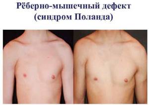
Poland syndrome
Poland syndrome is named after Albert Poland, who first described this type of chest deformity as a result of observations in school and refers to a spectrum of diseases that are associated with underdevelopment of the chest wall. This syndrome includes abnormalities of the pectoralis major, pectoralis minor, serratus anterior, ribs, and soft tissues. In addition, deformation of the arm and hand may occur.
The incidence of Poland syndrome is approximately 1 case per 32,000 children born. This syndrome is 3 times more common in boys than girls, and the right side is affected in 75% of patients. There are several theories regarding the etiology of this syndrome, which include abnormal migration of fetal tissue, subclavian artery hypoplasia, or intrauterine trauma. However, none of these theories have proven their validity. Poland syndrome is rarely associated with other diseases. Some patients with Poland syndrome have leukemia. There is a definite association of this syndrome with Moebius syndrome (unilateral or bilateral palsy of the facial nerve, abducens ophthalmic nerve).
Symptoms of Poland syndrome depend on the degree of the defect and in most cases they are cosmetic complaints. In patients with significant bone defects, the lung may bulge, especially when coughing or crying. Functional and respiratory problems may occur in some patients. The lungs themselves are not affected by this syndrome. In patients with significant muscle and soft tissue defects, decreased exercise capacity may become apparent.
Diagnostics
With a barrel chest, it is necessary to conduct a comprehensive examination of the patient. A pulmonologist prescribes the following types of diagnostics:
- spirometry;
- bronchoscopy;
- chest x-ray;
- ECG;
- sputum analysis for bacterial culture.

If osteoarthritis is suspected, a detailed X-ray examination of the ribs and spinal column is performed.
How can parents find out if their child has a congenital chest pathology?
Ryzhikov Dmitry Vladimirovich
Ryzhikov Dmitry Vladimirovich (Head of the Department of General Bone Pathology of the Federal State Budgetary Institution "National Medical Research Center for Pediatric Traumatology and Orthopedics named after G.I. Turner, Candidate of Medical Sciences, doctor of the highest qualification category, traumatologist-orthopedist)Most often, parents learn about a child’s disease in preschool or school age by accidentally noticing asymmetry or an unusual shape of the chest. This situation is noticed more quickly in families where a similar pathology was previously identified in other family members.
The disease progresses while the skeleton is growing, that is, on average until 15-17 years of age. (more about the causes - types and types of deformations)
Our center actively provides telemedicine consultations in Doctor/Doctor mode. For additional information about the work of the Telemedicine Center NMIC DTO named after. G.I. You can contact Turner by writing to us at [email protected]
For patients from regions of the Russian Federation, there is the possibility of correspondence consultation.
Treatment methods
Barrel-shaped breast deformation is just one of the symptoms of various diseases. You can get rid of such a defect only after treating the underlying pathology.
For chronic obstructive respiratory diseases and bronchial asthma, patients are shown the following bronchodilators:
- "Foradil."
- "Serevent."
- "Atrovent N".
- "Salbutamol."
These medications come in the form of inhalers. They relieve bronchospasm and make breathing easier.
For severe obstructive diseases and asthma, drugs with corticosteroid hormones are prescribed:
- "Prednisolone."
- "Dexamethasone."
Hormonal drugs are used both in oral and inhalation forms.
If it is difficult to discharge sputum, mucolytic medications are indicated:
- "Ambroxol".
- "ACC."
- "Carbocisteine".
These agents thin the mucus and facilitate easier removal of mucus from the bronchi.
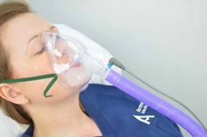
If the use of inhalers does not have the desired effect, then drug treatment is supplemented with oxygen therapy sessions. This helps to significantly improve the condition of patients.
Treatment for cystic fibrosis can only be symptomatic. Modern medicine cannot cure a gene mutation. However, the patient's condition can be significantly alleviated. Patients are prescribed bronchodilator and mucolytic drugs. These medications must be taken throughout your life. In case of severe blockage of the respiratory tract with mucus, the bronchi are washed with a solution of sodium chloride.
For osteoarthritis, chondroprotectors and intra-articular injections of drugs with hyaluronic acid are prescribed. In case of severe pain, taking non-steroidal anti-inflammatory drugs (Diclofenac, Naise, Ibuprofen) is indicated.
Patients are often interested in the possibility of plastic surgery for curvature of the chest wall. If the deformity is caused by serious lung diseases, then it cannot be eliminated through cosmetic surgery. After all, the volume of the chest in this case increases due to air retention in the respiratory organs. Usually, after remission is achieved, the shape of the chest returns to normal.
Congenital deformities
Congenital chest deformities include several types of pathologies. The cause of all disorders often lies in the malfunction of genes. An error in genes from the very beginning of fetal development predetermines a deviation in the development of breast bones. The deformity is expressed in the non-standard structure of the ribs, sternum bones or the absence of the necessary bone material. At the same time, muscles develop poorly.
Funnel deformity
This type of structural changes is diagnosed more often than other congenital disorders, mainly in the male half of society. The ribs, together with the sternum, are pressed inward, the size of the chest decreases, the spine changes its structure, and thoracic kyphosis develops.
The pathology is characterized by a genetic predisposition, as it tends to be inherited. Due to the disease, the respiratory process changes and the cardiovascular system suffers. In severe complications, the heart may become displaced.
There are several degrees of complexity of the disease:
Severity of pectus excavatum
- first degree - the depression is insignificant, about 30 mm, with all internal organs and the heart located in their places;
- second degree - the deepening of the funnel reaches 40 mm, the heart moves up to 30 mm;
- third degree - the funnel reaches a depth of more than 40 mm, and the heart goes to the side at a distance of more than 30 mm.
The patient feels the greatest discomfort at the moment of inhalation. At the age of about three years, the disease begins to manifest itself with a greater degree of severity - blood circulation is impaired, and deviations in the general development of the child are observed. The body's protective functions suffer, as a result of which the child often gets sick. As the size of the funnel increases, the number of concomitant diseases increases.
Funnel chest deformity
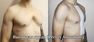
Carinatum deformity
Disturbances in the structure of the chest in this case develop due to excess cartilage in the ribs and sternum. The bones of the chest protrude noticeably forward and become keel-like. Every year the condition of the structure worsens. Despite such external changes, the lungs continue to work as usual. The heart suffers - its shape changes and it loses the ability to cope with serious loads. The patient feels a loss of strength, tachycardia and shortness of breath appear.
Plane strain
This pathology is characterized by a smaller cell volume, but there is no need for treatment. It belongs to the asthenic type and does not have a negative effect on the functioning or location of organs.
Deformity with cleft
A cleft, complete or incomplete, develops in a child during fetal development. As the child grows, the gap in the chest increases. The heart, blood vessels and lungs become more vulnerable. For children under the age of one year, the sternum bones are sewn together, and for older children, surgery is performed to fill the gap with a special implant.
Convex type deformation
The type of disorder is diagnosed extremely rarely. It is a protruding line at the top of the chest. It does not affect health in any way, but is a purely aesthetic defect.
Poland syndrome
It is a genetic disease and the child inherits it from his parents. A sign of the disease is the sinking of some areas in the sternum. The sternum, muscles, spine, ribs and cartilage may suffer from this. Treatment involves surgery.
In addition to congenital structural changes, acquired disorders are diagnosed. These include emphysematous change, paralytic, scaphoid, in which a scaphoid depression is formed in the chest, and kyphoscoliotic.
Prevention
How to prevent chest wall deformation? To do this, it is necessary to protect the respiratory system from harmful effects. Pulmonologists advise adhering to the following recommendations:
- completely stop smoking;
- avoid exposure to allergens, dust and toxic gases;
- when working in hazardous industries, undergo regular medical examinations;
- promptly cure inflammatory bronchopulmonary diseases.
If you have a systematic cough, wheezing in the chest and difficulty breathing, you should immediately consult a doctor. This will help avoid serious complications such as heart and pulmonary failure.