Non-stress CTG test (cardiotocography), also known as fetal heart rate monitoring, is a prenatal test that is done to check the health of the unborn baby. During the procedure, the fetal heart rate is monitored to see how it changes during physical activity.
Normally, your baby's heart rate should increase during activity later in pregnancy. However, in a condition such as fetal hypoxia (when it does not receive enough oxygen), this may be impaired, which is what this study reveals.
Typically, fetal CHT is recommended when the fetus is considered to be at increased risk of death. The test can be carried out after 26-28 weeks of pregnancy, since before this time the baby is not yet developed enough to respond to the test protocol. Some results may indicate that you and your baby need further monitoring, testing, or special care.
What is CTG?
Fetal cardiotocography is a non-invasive method that allows you to objectively assess the condition of the unborn child, based on observations of changes in heart rate in response to fetal movements or uterine contractions. Monitoring the fetal cardiac activity allows for the most objective assessment of indicators.
Would you like to make an appointment?
Request a call back
Fill out the form to make an appointment
Cardiotocography (CTG) is the most accessible and safe method of monitoring the functional state of the fetus.
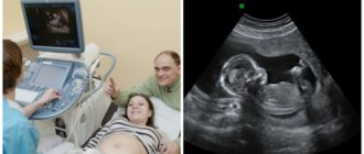
How is CTG (cardiotocography) performed during pregnancy? During the study, doctors use special devices - cardiotocographs, which are based on the Doppler effect. These fetal cardiac monitors record changes in intervals that occur between cycles of fetal cardiac activity, convert them into signals and provide information in the form of a graph. The presence of two sensors at once allows you to simultaneously assess the motor activity of the fetus and uterine contractions.
What is a cardiotocogram? This is a graph that represents 2 curves, one of which reflects the fetal heart rate (heart rate), and the other - fetal movements and uterine contractions (contractions).
Non-stress test for the unborn baby
Timely diagnosis of fetal pathology is an important task of perinatal medicine.
The deeper researchers penetrate into certain aspects of the narrow problem they are solving, the more obvious the limitations of our knowledge in the field of physiology and pathophysiology of the fetal period become. It is not always possible to draw the line between normality and pathology, to assess the severity of fetal damage, its reserves, ability to withstand birth stress, etc. But, on the other hand, with the help of objective methods it is possible to obtain reliable information about the vital activity of the fetus, which helps in choosing one or another tactic for its observation and treatment.
Cardiotocography is a method for assessing the condition of the fetus, based on the analysis of the variability of its heart rate at rest, movement, under conditions of uterine activity, as well as the influence of environmental factors.
A cardiotocogram is a simultaneous recording of the fetal heart rate and uterine contractions. An isolated recording of the fetal heart rate is called a cardiotachogram (tachogram), contractile activity of the uterus is called a tocogram (hysterogram), and fetal movements are called an actogram.
Currently, devices are being produced that, along with these capabilities, additionally measure oxygen tension in fetal tissues. In cardiotocography, fetal cardiac activity is usually recorded using ultrasound by the frequency shift of the wave reflected from the pulsating heart (Doppler effect).
In addition to the ultrasound method of obtaining information about the fetal cardiac activity, electrocardiography and phonocardiography can be used. Contractile activity of the uterus is recorded by determining the tension of the uterine wall or the amount of intrauterine pressure. Based on the method of obtaining information about the fetal heart rate and contractile activity of the uterus, indirect (external) and direct (internal) cardiotocography are distinguished.
The Alanda clinic uses external tocography with the BT-330 Bistos fetal monitor (Korea), in which fetal cardiac activity and the strength of uterine contractions are determined non-invasively, indirectly, through the anterior abdominal wall. Ultrasound is used to record the fetal heartbeat, and strain gauge sensors are used to record the tone of the uterus, placed on the belly of a pregnant woman.
To assess the condition of the fetus, both available recording channels must be used: cardiotachographic and tocographic. The duration of fetal heart rate recording if the curve is visually satisfactory should be 20 minutes. It may be shorter if the study reveals all signs of fetal well-being. If pathological or alarming rhythms are detected, CTG is performed within 40 minutes.
When the fetus is in a threatening condition, CTG usually reveals one or more pathological signs. During the physiological course of pregnancy, to screen the condition of the fetus, it is usually sufficient to take into account the presence of heart rate accelerations caused by fetal movements - non-stress test (NST).
The test is considered negative (reactive, normal) if, during 30 minutes of observation, at least 3 accelerations with an amplitude of at least 15 beats/min and a duration of at least 15 seconds are recorded on CTG. If 3 accelerations are recorded in a shorter period of time, the test is stopped, considering it reactive.
The test is considered positive (arreactive, pathological) if accelerations with an amplitude of less than 15 beats per minute are recorded on CTG or there are less than three of them within 30 minutes. Positive NCT is a reliable indicator of fetal well-being and prognosis for the newborn.
With areactive NCT, there is an increase in perinatal morbidity and mortality, as well as the frequency of development of a threatening condition of the fetus during labor and surgical delivery. When using a blood oxygen saturation monitor, it is possible to more accurately diagnose the threatening condition of the fetus when the fetal heart rate is unsatisfactory, which allows reducing the frequency of unjustified interventions during pregnancy and childbirth.
Natalya PAK, head maternity ward, Alanda clinic, Karaganda
Indications for CTG during pregnancy
When can a doctor recommend doing a CTG during pregnancy? This study is carried out as part of monitoring a normally developing pregnancy in the third trimester. In addition, cardiography is used to monitor the condition of the fetus in the following conditions:
- multiple pregnancy;
- oligohydramnios;
- increased risk of developing fetal hypoxia;
- high probability of complications in late pregnancy (hypertension, gestosis, etc.);
- anemia; · Also, a CTG of the fetus will need to be done in the intrapartum period (during childbirth).
Risks
This is a non-invasive test that does not pose any physical danger to you or your baby. The term "non-stressful" refers to the fact that there are no additional activities, such as artificially stimulating uterine contractions, to force the baby to move in the womb.
This test can either give you confidence in your child's health or be a cause for concern. A non-stress test can also give a false negative or false positive result. That is, failing to detect an existing problem or assuming that a problem exists when there is none, which may lead to the need for further examination.
It's also worth keeping in mind that while this test is recommended for women at increased risk of pregnancy loss, it is not always clear whether it will be useful.1
How is CTG done during pregnancy?
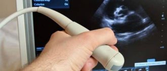
During pregnancy, indirect (external) CTG of the fetus is performed. To do this, one sensor with a special gel is placed on the mother’s abdomen, choosing the area where the fetal heart sounds can be heard best, and the second sensor is placed on the fundus of the uterus. The expectant mother, using a special device, records each episode of fetal movement by pressing a button. During the examination, the patient either lies on her side or is in a semi-sitting position. In order to obtain the most accurate information, the study should be carried out for at least 20-30 minutes.
How is it carried out?
Before the procedure
Your blood pressure will be measured immediately.
During the procedure
During the procedure, you will lie in a reclining chair and your blood pressure will likely be checked periodically.
Two monitor straps are placed on the abdomen. One will record your baby's heart rate and the other will record any uterine contractions that may occur. You may be asked to press a button when you feel your baby move, which will be noted in the results. The doctor will look to see if the fetus's heart beats faster during physical activity.
Typically, a non-stress test lasts 20 minutes. However, if your baby is inactive or sleeping, you may need to extend it for an additional 20 minutes - waiting for your baby to become active to ensure accurate results. The doctor may try to stimulate the baby manually or place a device that makes noise on the mother's stomach.
After the procedure
Once the test is completed, your doctor will likely discuss the results with you right away.
Fetal CTG – norm and deviations
- BHR is 120-160 beats per minute. When the values decrease, they speak of bradycardia, and when they increase, they speak of tachycardia.
- the amplitude of basal rhythm variability is 10-25 per minute. The presence of changes (variations) in the overall picture indicates normal interaction between the autonomic nervous system and the heart of the unborn child. A decrease in variability is normally detected during periods of fetal sleep. If such indicators are recorded during periods of wakefulness, the doctor may suspect chronic hypoxia (oxygen deficiency) in the fetus.
- the presence of two or more accelerations. On the CTG graph, accelerations are displayed as tall teeth.
- no decelerations. The graph appears as teeth pointing downwards.
- non-stress test (NST) indicators. By pressing the sensor button, the patient registers fetal movements. Normally, episodes of motor activity should correspond to acceleration (periods of increased heart rate).
Why is it carried out?
During CHT of the fetus, the fetal heart rate is monitored in response to physical activity. Normally, it will increase during the movement of the unborn child, and at rest it will decrease. The bottom line is that normal fetal activity and heart rate require sufficient oxygen. When its level is low, the fetus may not respond normally. Insufficient oxygen supply (hypoxia) can often be caused by problems with the placenta or umbilical cord.2
CTG may indicate the need for further monitoring, evaluation, treatment, or induction of labor to prevent fetal death.
Your doctor may recommend you take this test if you have:
- Multiple pregnancy with certain complications.
- A systemic disease such as type 1 diabetes, heart disease, or high blood pressure during pregnancy.
- Pregnancy that continues two weeks after the expected date of birth.
- History of complications in a previous pregnancy.
- Decreased fetal movements or possible problems with fetal growth.
- Rh sensitization (Rh conflict) is a potentially serious condition that can occur during a second or subsequent pregnancy when your blood is Rh negative and your baby's is Rh positive.
- Oligohydramnios (oligohydramnios).
Your doctor may recommend CHT once or twice a week, or sometimes daily, depending on your and your baby's health. For example, you may need regular testing if your doctor suspects your fetus is not getting enough oxygen.
Fetal CTG (cardiotocography)
Fetal cardiotocography (CTG) is a continuous recording of the fetal heart rate (HR), as well as uterine contractions, obtained using ultrasound transducers located on the mother’s abdomen; the signals are recorded on a tape in the form of a graph. The device for performing CTG is called a cardiotograph. The initial assessment takes 20 minutes. The position of the pregnant woman is lying on her side or reclining to avoid compression of the inferior vena cava. The procedure is painless and safe, easy to perform and decipher.
The result is assessed by a midwife or obstetrician-gynecologist.
Used to determine the well-being of the fetus in the 3rd trimester of pregnancy, since the heart rate depends on its functional state. Depending on the results obtained, further pregnancy management tactics and the amount of necessary medical interventions are planned.
Indications for CTG:
- Suspicion of intrauterine growth retardation;
- Deterioration of fetal movements noted by the pregnant woman;
- Preeclampsia (severe gestosis in the mother: high blood pressure, the appearance of protein in the urine, edema);
- Diabetes;
- Exacerbation of a chronic disease in a pregnant woman;
- Bleeding during pregnancy (threat of premature birth, placental abruption, etc.);
- Multiple pregnancy;
- Oligohydramnios or polyhydramnios;
- Pregnancy period is 41 weeks or more (signs of post-term pregnancy);
- Rhesus conflict;
- Antiphospholipid syndrome in the mother.
At rest, the fetal heart rate is 110-160 per minute; when the uterus contracts, the heart rate of a healthy fetus increases by 5-25 per minute, this is normal.
Acceleration of the heartbeat between contractions of the uterus (tachycardia) of the fetus is possible with:
- Fetal hypoxia;
- Chorioamnionitis (inflammation of the membranes of the fetus and infection of the amniotic fluid);
- Hyperthyroidism (increased production of thyroid hormones);
- Fetal or maternal anemia (decreased hemoglobin and blood iron levels);
- Fetal tachyarrhythmias (heart rhythm disturbances).
A slowing of the heartbeat between contractions of the uterus (bradycardia) of the fetus indicates severe hypoxia and is possible with:
- Prolonged compression of the umbilical cord;
- Umbilical cord prolapse;
- Epidural and spinal anesthesia;
- Eclampsia (severe gestosis, convulsions in the mother);
- Rapid descent of the fetus during labor.
It should be noted that heart rate is assessed not only between uterine contractions (basal rate, heart rate at rest), but also immediately after. All these data together give an idea of the condition of the fetus at the time of the examination.
In our clinic we perform CTG, as well as prevention and treatment of placental insufficiency, threatened miscarriage and premature birth in a day hospital.
Our highly qualified obstetricians-gynecologists will conduct a consultation, examination, answer all your questions and prescribe timely examination and treatment.