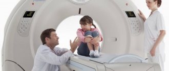
No branch of medicine can do without modern diagnostic techniques, which include CT, but patients are often concerned about how safe they are.
CT scans suggest radiation exposure. Therefore, it is especially important to understand what dose a person receives, what it depends on, and how often one can be examined without risk to health.
How much radiation does a person receive during a CT scan?
When receiving an excessive dose of radiation, various malfunctions begin in the human body, for example:
- changes in blood parameters (decrease in the number of leukocytes, platelets);
- internal bleeding;
- deterioration of the immune system;
- erythrocytopenia;
- oncological diseases;
- general deterioration of health.
It will be possible to avoid exceeding permissible standards if you know what the radiation exposure is for a particular type of computed tomography.
The radiation range is quite wide, for example, when examining the paranasal sinuses and skull, the dose is 0.6 mSv, and when scanning the extremities - 0.1 mSv.
The procedure has the greatest effect on:
- chest area – 7-10 mSv;
- pelvic organs – up to 9.5 mSv;
- gastrointestinal tract – 7-9 mSv;
- spine - depending on the department, the indicators can be 5-6 mSv (the greatest load when examining the condition of the lower back).
If the whole body is screened, a person will receive from 10 to 11 mSv. From a safety point of view, when checking 2-3 zones is required, it makes sense to undergo a comprehensive CT scan, because the total radiation exposure will be less than individually.
Currently, due to the coronavirus outbreak, the demand for computed tomography of the lungs has increased greatly. At LocalLab.ru you can find the diagnostic center closest to you and make an appointment online.
Patients should be aware that the law requires that when performing such diagnostics, you must document the radiation exposure to which you are exposed. According to the rules, the doctor must indicate this information in the expert report along with the parameters of the machine on which the CT scan was performed.
Keeping records of radiation dose is very important, especially if you have to undergo examination more than once a year.
Why can the radiation dose be different? What does it depend on?
The amount of radiation a person receives during a CT scan varies, and this is primarily due to the following factors:
- area of research (due to the fact that tissues of different densities “absorb” X-rays differently);
- irradiation area (there is a direct connection - the larger the area, the greater the load);
- a history of having recently completed a course of radiation therapy - the procedure has a stronger effect on the body of cancer patients, so they are often prescribed MRI as an alternative diagnostic method.
Additionally, the dose can be affected by the distance from the patient to the tube and the parameters of the computed tomograph. The latest generation models are more “gentle”, but at the same time accurate, so it is wiser to choose a clinic with modern equipment.
The most difficult thing is to calculate the radiation load when re-examining a particular area, because each body tolerates the procedure in its own way, plus a lot depends on how long ago the previous CT scan was done.
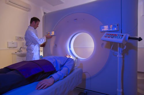
How is a CT scan of the spine performed?
Depending on the equipment used, linear and spiral CT are distinguished. The first is done on a tomograph, which simultaneously moves sensors and X-ray emitters in opposite directions. The area in the center of the scanning system is in focus. Structures located above or below the main axis are blurred. For linear CT scans of the spine, the minimum slice thickness is 3 cm.
Spiral computed tomography involves rotating the scanning system (tubes + detectors) around the table with the patient. At the same time, the conveyor moves in a horizontal plane inside the ring of the apparatus.
Modern tomographs take 2 or more (8, 16, 64) images per full revolution. The thickness of the scanned section is from 0.2 cm. This study of the spine is called multislice CT.
To improve the quality of photographs and prevent artifacts, the patient must remain motionless throughout the session. To do this, the body and limbs are fixed using special rollers and fasteners.
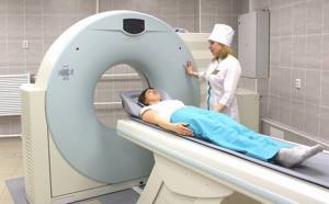
Computed tomography of the spine
After preparing the patient for the procedure, the doctor and laboratory assistant move to the next room where the monitors are located. Communication with the patient is maintained through a microphone.
The duration of the spine examination depends on how the CT scan is performed. The native procedure takes no more than 10 minutes. If it is necessary to administer a contrast solution, the scanning duration increases. After a series of routine scans, the patient is given an injection of an iodine-containing drug. The substance fills the vascular bed in the area of interest, after which the session continues. A CT scan with contrast takes up to 20 minutes.
Detectors record information about the density of spinal structures and transmit the data for processing by a computer program. The monitor receives layer-by-layer images in an axial projection. Based on the photographs obtained, the sagittal and frontal planes of the area under consideration are completed. If necessary, the 3D image is reconstructed.
What dose of radiation is considered safe for health?
Scientists cannot determine exactly how much radiation the body can receive without serious harm. Each person has their own reaction to radiation, and there are special standards for radiologists (no more than 20 mSv per year).
Russian legislation stipulates an indicator of 15 mSv - this is the maximum for an adult per year, while doctors strongly recommend not doing a computed tomography scan more often than 2 months, even if the dosage of the procedure is insignificant. For preventive examinations, a load of 1 mSv is considered safe. Exposure to radiation in excess of 3 Sv on the body has critical health consequences, but even with frequent CT scans a person will not receive such a dose.
It must be remembered that a CT scan is carried out exclusively as prescribed by a doctor (although a referral to medical centers is not always required), who determines:
- Do you need a CT scan or is it better to choose another diagnostic method;
- what specific area is to be examined;
- What power device is best for scanning;
- Is there any point in repeat screening?
Without the recommendation of a cardiologist, surgeon, orthopedist, gastroenterologist, etc. It's definitely not worth spending money and putting your health at risk. If the procedure is prescribed, then it is worth undergoing it as soon as possible, so as not to delay the start of treatment.
CT of the brain CT of the abdominal cavity CT of the teeth CT of the orbits CT of the jaw CT of the cerebral vessels CT of the abdominal aorta CT of the adrenal glands CT of the facial bones CT of the temporal bones CT of the lumbosacral spine CT of the thoracic spine CT of the elbow joint CT of the shoulder joint CT of the thoracic aorta CT of the cervical spine CT of the heart CT of the hip joint CT of the knee joint CT of the pelvis CT of the lungs CT of the kidneys CT of the sinuses CT of the larynx CT of the coronary vessels CT of the liver CT of the intestine CT of the vessels of the lower extremities CT of the hand CT of the pelvic bones CT of the foot CT of the chest CT of the vessels of the neck CT of the coccyx CT of the ankle CT bladder CT scan of the thyroid gland CT scan of the skull CT scan of the soft tissues of the neck CT scan of the wrist CT scan of the inner ear
X-ray diagnostics: discovering the “secrets” of our body
09.03.2016
The head of the X-ray diagnostic department of the REAVIZ multidisciplinary clinic, Candidate of Medical Sciences Anton Osadchiy answered questions from Komsomolskaya Pravda readers
— Hello, my name is Tatyana Mikhailovna. What is more accurate - CT or MRI? How often can a CT scan be done?
— Computed tomography (CT) and magnetic resonance imaging (MRI) are based on different principles of influence. CT uses X-ray radiation; with the help of CT, the physical and biological indicators of organs and tissues can be clarified. MRI shows the chemical and biological structure of organs. The use of these techniques depends on the pathology. CT is a priority for diseases of the chest organs (including the respiratory system), osteoarticular system, diseases of the endocrine system, diseases associated with the brain (including strokes), cardiovascular system, abdominal and pelvic organs. MRI is most effective for diagnosing inflammatory processes in the brain (encephalitis, meningitis, etc.), for pathologies of the spinal cord, joints (orthopedics), mammary glands, bile ducts, and oncology. As for the frequency of examinations, if there are indications, CT scans can be done no more than once every six months.
— Hello, my name is Yulia. The doctor told my father that he needed to have a CT scan before removing the kidney stones. What is it and why is it needed? Is any preparation needed for this procedure?
— The doctor is right: computed tomography is widely used in urology to determine the diameter and location of kidney stones before surgery to remove them, and also shows whether there are any disturbances in the outflow of urine. This is a painless procedure that takes 5-10 minutes. Within an hour after this, the doctor will describe and give you the results. CT scanning does not require special preparation and there are no contraindications.
— I am occasionally bothered by wheezing in my lungs, but fluorography is clear. Does it make sense for me to have an x-ray or CT scan?
— Fluorography is a screening method that allows you to identify major abnormalities. Therefore, if the results are clean, then most likely there are no serious pathologies. But to detail the condition of the lungs, computed tomography will be effective: it will allow you to visualize smaller details or foci of infection that are not visible on fluorography.
— Hello, this is Tamara Sergeevna. My son had a CT scan and received a radiation dose of 28-30 mSv. How dangerous is this exposure and what could be its consequences?
— 28 mSv is a completely non-critical radiation dose, which is eliminated independently without consequences for the body after a couple of months. The maximum permissible dose for a person is more than 90 mSv over six months - then there really is a danger of developing radiation sickness. By the way, the more sections are performed during a study on a computed tomograph, the less this load is. The REAVIZ multidisciplinary clinic uses a modern 64-slice Seimens Sensation 64 computed tomograph.
— Good afternoon, my name is Tatyana. I did an ultrasound of the cerebral vessels, which revealed that my blood flow was reduced, possibly due to osteochondrosis. Now the ophthalmologist says that decreased vision is also possible because of this. Is it worth doing a CT scan? What can this study find out?
— Tomography allows you to clearly visualize blood vessels and diagnose the causes of blood flow disorders that were identified on ultrasound. On CT, all narrowings, thromboses, atherosclerotic plaques, pathological bends from osteochondrosis and other pathological changes are clearly visible. Ultrasound and CT are two complementary diagnostic methods, the results of which allow the doctor to get an accurate picture and choose the right treatment.
— Good afternoon, this is Anastasia. Mom has a pacemaker, and we were told that an MRI cannot be done. Is CT possible?
— If a pacemaker is installed, MRI is indeed contraindicated, since magnetic radiation can cause malfunctions and even lead to the death of the patient. With CT, X-ray radiation does not affect the operation of the pacemaker in any way.
— Good afternoon, Olga Ivanovna is worried. Tell me, can an x-ray show a pinched sciatic nerve, or is it necessary to do an MRI?
— In your case, there is no need to do an MRI. Computed tomography (CT) is also effective in diagnosing root nerve entrapment. You can undergo an examination at the REAVIZ multidisciplinary clinic. If necessary, immediately after the examination you can invite a neurologist for consultation.
— Hello, my name is Lyudmila. I was scheduled for an MRI of the brain and was told that contrast would need to be administered. Is this necessary? After all, this is an additional burden on the body...
— An MRI is initially performed without contrast, but if the doctor has doubts, contrast may be needed. The substance that is used for this is harmless and is eliminated from the body in just a few minutes.
— Hello, my name is Vladimir. I had a CT scan of the abdomen three times with contrast injection, and I received a dose of about 26 mSv. Is it harmful?
— Triple CT scans are standard, you shouldn’t be scared. A radiation exposure of 26 mSv is not at all dangerous for the body. And the contrast is removed from the body within an hour.
— Hello, my name is Irina Vladimirovna. Please tell me how to check the performance of blood vessels? What type of diagnostics should I choose?
— If you have no complaints about your health, then most likely the blood vessels are working normally. If you are worried about arrhythmia or dizziness, then you should do an ECG, then an ultrasound with Doppler study to diagnose the state of blood flow. After this, you will need to do a CT angiography, which will allow you to evaluate the structure and location of the vessels, and, if present, see atherosclerotic plaques, stenoses, pathological tortuosity and other abnormalities.
— Hello, my name is Elena, I am 16 weeks pregnant, and my spine is bothering me. Can I have a CT scan at this time?
— You can’t do a CT scan during pregnancy. It is better for you to get an MRI, but only after consulting with your gynecologist.
— Good afternoon, this is Lyubov Viktorovna. How does MRI differ from CT? Which examination is better to choose for problems with the spine?
— Computed tomography (CT) and magnetic resonance imaging (MRI) are based on different principles of influence. CT uses X-ray radiation; with the help of CT, the physical and biological indicators of organs and tissues can be clarified. MRI shows the chemical and biological structure of organs. The use of these techniques depends on the pathology. For diagnosing spinal pathologies, hernias, protrusions, pinching, etc. CT is also used.
— Hello, my name is Svetlana. Frequent headaches bother me, the neurologist recommended an MRI. But it is very expensive... Is it possible to replace it with CT?
- Yes, you can undergo a CT scan instead of an MRI; it is no less informative than an MRI. A CT scan of the brain will help identify problems that may be causing dizziness and headaches and allow the neurologist to choose a treatment strategy.
Return to articles