This site was made by experts: toxicologists, narcologists, hepatologists. Strictly scientific. Tested experimentally.
Author of this article, expert: Gastroenterologist-hepatologist Ekaterina Kashukh
In short: The liver absorbs and half reflects ultrasound waves, which is why it appears uniformly gray on an ultrasound image. Tumors, calcifications, and areas of fatty degeneration do not transmit ultrasound so well and appear lighter in the image. To clarify the diagnosis, the doctor may prescribe you an additional examination, and then prescribe medications depending on the result.
- What does increased echogenicity of the liver mean?
- What does pathology look like on ultrasound?
- What causes increased echogenicity of the liver?
- What symptoms should you be wary of?
- How does a doctor identify the cause of such a picture on an ultrasound?
- How to be treated
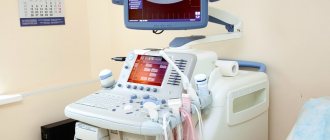
In modern approaches to the diagnosis of diseases of the parenchymal (dense) organs of the abdominal cavity, ultrasound plays an important role: ultrasound examination. Patients themselves also want to understand the terms that functional diagnostic doctors use in order to be able to assess their condition.
What does increased echogenicity of the liver mean?
Echogenicity is the ability of tissues and organs to absorb or transmit a signal (wave). An ultrasound machine is both a receiver and a transmitter at the same time. It sends out ultrasonic waves and then catches what is reflected back.
Echogenicity is the density of an organ during ultrasound, the ratio of absorption and reflection of a wave from an organ or tissue. The level of difference between the values of the sent and returned signal displays the picture on the screen. The degree of wave absorption depends on the density, elasticity, and humidity of the medium through which the ultrasonic waves pass.
Increased echogenicity of the liver on ultrasound can be due to various reasons. Contact your doctor to find out.
Ultrasound diagnostics allows you to see many indicators needed to assess the condition of the liver and surrounding organs:
- overall size of the organ
- ratio of the sizes of the right and left lobes,
- the condition of the gallbladder, the density of its walls,
- inclusions in the gallbladder,
- homogeneity of bile in the gallbladder,
- density of liver tissue - parenchyma,
- structure of large vessels of the liver,
- structure of large bile ducts,
- the presence of various formations in the liver.
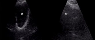
If space-occupying formations are detected in the liver tissue, then from the image one can guess their nature, structure, and state of the cells. It is in assessing the condition of the tissues, that is, the structure of the liver, that the assessment of echogenicity helps. In the picture, the echogenicity levels, when compared with the video image, represent a difference in contrast. The ultrasound picture is drawn with dark and light shadows. And the difference in colors means different echogenicity.
Any conclusion requires a standard. During the study, the standard of average echogenicity of the ultrasound machine is the renal cortex. The density (and, accordingly, echogenicity) of a healthy liver is close to the standard. The liver parenchyma is represented by hepatocytes; they provide an almost equal ratio between absorption and reflection of ultrasonic waves. In the ultrasound picture it looks like a smooth gray mass. But the organ has vessels, bile ducts, membranes of lobes and lobules - and they all differ in echogenicity from the liver parenchyma (tissue) in the picture.
For example: the bone reflects almost 100% of the signal that was sent. In liquids, on the contrary, the signal is completely absorbed. Parenchymal organs (that is, organs made of cellular tissue, including the liver) typically absorb and reflect 50% to 50% ultrasonic waves.
An expert on the website Pokhmelye.rf, gastroenterologist Daniela Purgina, warns against making a common mistake.
Do not self-medicate. It's ineffective. And sometimes, trying to treat yourself, the patient may waste precious time without achieving the desired result.
Nutrition plays an important role
It is not so difficult to limit your diet from meat, flour, sweets, smoked foods, foods with a flavor enhancer - glutamate, fatty foods, and especially stop reheating food prepared with a large amount of animal fats. Heated animal fats are the main evil.
Alcohol is not prohibited at all, but its naturalness and compliance with the dose are important. Rarely, but a lot is not good. Also, alcohol in the form of distillates (even if it is not moonshine, but slivovitz, grappa or rakia) and sweet dessert wines, liqueurs, and liqueurs hit the liver the hardest. Dry wine in a reasonable dose is acceptable.
What does pathology look like on ultrasound?
With pathological changes, the liver parenchyma becomes heterogeneous. As the echogenicity (hyperechogenicity) of the liver increases, the picture becomes lighter. Having seen in the ultrasound report a phrase about the hyperechogenicity of the liver tissue, the doctor can make an indirect conclusion about the presence of pathology.
A change in the picture in the image means a change in the tissue of the organ. Hepatocytes (working liver cells that make up the bulk of the organ) can be replaced by only two types of tissue:
- Scars (fibrosis) are rough collagen or elastic fibers that begin to “entangle” damaged (or dead) hepatocytes. This is a picture of cirrhosis.
- Fat cells (adipocytes) - fatty degeneration, or fatty degeneration of liver tissue. Dying cells are replaced by adipose tissue.
In either case, the hue in the ultrasound image will differ from the normal state, that is, from the average echogenicity of the liver tissue. In any case, the cause of the suffering of liver cells is always some processes that occur in the body. Let's tell you more about them.
What causes increased echogenicity of the liver?
Reasons for which a diffuse (spread) increase in the echogenicity of the liver tissue is detected:
- Acute hepatitis of various nature (viral, toxic and others).
- Metabolic liver diseases (eg fatty liver disease).
Advice to a sick person from gastroenterologist Daniela Purgina, an expert on the website Pokhmelye.rf.
If you have been diagnosed with fatty hepatosis (and this is one of the most common causes of diffuse changes in the liver), then the main treatment is weight loss. This is the simplest and most difficult at the same time. Alas, no drugs will remove fat from the liver, so you will have to make efforts for the sake of your own health.
The increase in echogenicity can be either diffuse, diffuse, covering the tissue completely, or local, that is, limited. Moreover, limited areas (individual formations of increased or decreased echogenicity) can have smooth or uneven contours. What does it mean?
Hyperechoic (that is, denser) areas can mask:
- benign tumors (usually hemangioma, vascular tumor);
- malignant tumors;
- metastases from other organs;
- stones;
- calcifications;
- areas of fatty degeneration of liver tissue.
Hypoechoic areas (with reduced density) are usually:
- cysts with liquid contents,
- hydatid cysts,
- abscesses.
When the doctor sees a separate area with a changed shade in the image, then we are talking about a lesion. During ultrasound diagnostics, there are a lot of subtleties that suggest the nature of the formation, for example:
- Benign tumors most often have smooth, clear edges.
- In a malignant tumor, growth most often occurs expansively: in all directions and uncontrollably, so that the contours are uneven and they grow without a clear boundary.
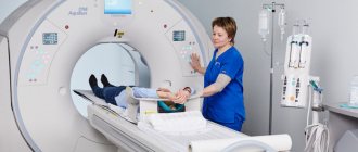
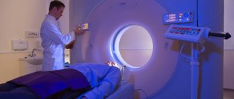
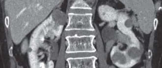
This division is conditional. There are features characteristic of each volumetric formation that a specialist looks at and describes. To clarify the diagnosis, the doctor may prescribe a computed tomography or MRI. In this case, layer-by-layer images with the introduction of contrast into the organ are very helpful in clarifying the nature of the formation. For information about what areas of abnormal density look like on CT and MRI in various diseases, read the article about hypodensity formations in the liver.
The liver is a very patient organ. If there are no changes in the biliary system, then it may not manifest itself clinically for a long time. However, over time, pathological processes in which the echogenicity of the liver is increased can lead to cirrhosis of the liver. Therefore, it is imperative to know exactly how the organ speaks about its malaise, so as not to miss dangerous symptoms.
Bibliography
- S. V. Shakhidzhanova, T. S. Pustovitova, “Some aspects of the diagnosis of focal liver pathology,” journal “Clinical Imaging” N19, December 2001.
- A.Z. Guseinov, T.A. Guseinov, “Modern diagnosis of liver tumors”, Bulletin of new medical technologies, electronic journal – 2016 – N 4, Tula.
- Zh.N. Kyzhyrov, B.B. Baimakhanov, M.M. Sakhipov, A.T. Chormanov, N.N. Birzhanbekov, E. Serikul, “Diagnostics of focal liver diseases”, Bulletin No. 1, 2021, Kazan.
- S.N. Berdnikov, V.N. Sholokhov, Yu.I. Patyutko, M.S. Makhotina, E.S. Chuchuev, K.E. Abirov, “Complex elastography and elastometry in the differential diagnosis of hyperechoic liver formations,” Journal: SonoAce Ultrasound No. 25, Moscow.
- O.M. Kurzantseva, “Some aspects of the diagnosis of focal nodular hyperplasia of the liver (fibronodular hyperplasia)”, Journal: SonoAce Ultrasound No. 30, Kemerovo.
What symptoms should you be wary of?
When you still feel well, but the following signs appear, do not ignore them. By contacting a doctor right now and starting treatment on time, you can avoid a lot of problems. Here are the most common early symptoms of liver damage:
- Pain or heaviness in the right hypochondrium is the most common first sign of liver problems.
- Itchy skin.
- Decreased appetite.
Next, consider the symptoms that develop when the liver is already severely damaged:
- jaundice;
- an increase in abdominal volume with a decrease in muscles;
- gynecomastia, or an increase in the volume of the mammary glands in men;
- menstrual irregularities, infertility;
- red palms. In the medical community they are called “liver”;
- the presence of dilated veins on the anterior abdominal wall;
- bleeding gums, bruising on the skin;
- change of size. The doctor can detect hepatomegaly even during the initial examination, during palpation (feeling with hands);
- the presence of a large number of spider veins - telangiectasias.
A case from the practice of gastroenterologist Daniela Purgina, an expert on the website Pokhmelye.rf.
There is nothing special in this case, but I remember it precisely because of the reaction of the patient herself. A 54-year-old woman came to the appointment and cried incessantly. As it turned out, the day before she went to have an ultrasound about dull pain in her right side. The ultrasound revealed diffuse changes in the liver, and the ultrasound diagnostic doctor told her that these were signs of liver cirrhosis. In fact, the woman did not have any liver cirrhosis: all her blood counts were normal, viral hepatitis markers were normal, and she had no signs of cirrhosis. The only thing the woman had was excess body weight and increased cholesterol levels. And diffuse changes in the liver were nothing more than fatty liver disease. The patient was recommended to lose weight and take medications to normalize cholesterol levels.
How does a doctor identify the cause of such a picture on an ultrasound?
If in the early stages the symptoms of liver damage are minimal, then deviations in biochemical tests usually appear much earlier. In a biochemical blood test, transaminases increase: AST and ALT, bilirubin. Sometimes the only sign of liver disease is an elevated level of gamma-glutamyl transpeptidase over a long period of time. With stagnation in the hepatic ducts, the amount of alkaline phosphatase increases.
If there is pathology in the tests and increased echogenicity of the liver on ultrasound, the doctor should suspect and look for a process that contributes to the destruction of organ tissue. If clarification of the diagnosis is required, the patient will be referred to a CT or MRI of the liver with the introduction of contrast.
Also in Russia, such a diagnostic procedure as liver elastometry using the Fibroscan device is becoming increasingly available. This method allows you to clarify the density of the liver and give a prognosis of the disease, but has its limitations. Thus, if transaminases increase by 3 times or more, it can give unreliable results.
In rare cases, a test such as a biopsy is required. It is invasive, that is, it requires breaking the integrity of the liver tissue for examination, therefore it carries some risks and is performed strictly according to indications.
How often does it occur?
Chronic liver diseases, according to WHO, affect 30% of the population, which is 2 billion people, 60% of them are people of working age. Professor T.D. Zvyagintseva provides data that the second place in the structure of this pathology is occupied by long-term exogenous intoxications (alcohol, nicotine, drugs, xenobiotics), second only to viral damage to hepatocytes. Fatty infiltration of the liver, according to WHO, occurs in every fourth resident of developed European countries, and the main reasons for the diffuse accumulation of fatty molecules in hepatocytes are alcohol consumption, obesity and diseases associated with impaired carbohydrate metabolism.