Have you wondered what the differences are between analog and digital mammography machines? Or about the main advantages and disadvantages of both types?
If yes, we believe that the information below will help you with this.
But first, a quick overview - what is mammography?
Mammography is a special type of breast imaging that uses low-dose X-rays to detect cancer. In addition, mammography machines continue to be a leading measure in the prevention and early detection of breast cancer. Mammography can detect changes in breast tissue up to two years before abnormalities can be palpated.
Mammography machines have changed a lot in recent years.
Analog mammography machines have been the standard for many years. However, in 2000, the FDA approved the first full-scale digital mammography machine, which greatly improved image quality. Then, in 2011, the first 3D mammography was introduced. This type of mammography machine added quality to the images.
However, 3D mammography systems are not very common on the market, so next we will talk about analogue and digital systems, since they are the most popular.
Differences between analog and digital mammography machines
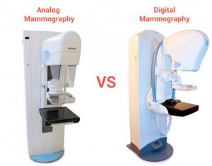
Analog mammography uses low-dose radiation that produces high-quality X-ray images and can detect tissue changes as small as 1-2 mm. This mammograph uses film cassettes and the result is a film image showing the breast from different angles and can be viewed on a special viewing board.
In contrast, digital mammography captures X-rays on a digital detector. This detector then converts the X-rays into electronic signals that are sent to a computer. As a result, computer images are available for viewing on a specialized high-resolution monitor.
Digital images can be further analyzed by radiologists using console/workstation options and tools. For example, magnification, masking light, inverting (negative images) and comparing them with previously obtained mammograms.
Fluoroscopic
1405 7.354 10.333 Computed tomography 468 9.959 4.661 Other 825 11.733 9.680 TOTAL: 105854 0.473 50.085 2013Is digital x-ray harmful?
X-ray examination is one of the most commonly used diagnostic methods. It is used to determine various diseases. Each of us has repeatedly faced the need to take an x-ray in our lives, but despite the fact that the procedure is well known to everyone, patients have questions about whether x-ray radiation is dangerous to health. In this article we will try to provide answers to the most frequently asked questions.
The peculiarity of X-rays is that they are able to reflect the internal structure of a person onto photographic film. This is used in various branches of medicine from dentistry to oncology and allows you to identify diseases in the early stages.
Is it dangerous to take digital x-rays?
The danger from x-rays only appears if the patient receives a large dose of radiation. The maximum permissible dose per year for humans is 150 mSv (millisievert). The procedures that the average person performs during the year do not exceed 20 mSv. In addition, modern digital X-ray machines are very different from their predecessors, both in the quality of the data received from them and in the radiation dose, which is several times lower in modern devices. If you try to compare the radiation dose that a patient receives from analogue and digital radiography, you will find out that for a digital X-ray machine it is 9-10 times less.
Of course, it is impossible to say that x-rays have a positive effect on human health. But it is important to understand that a doctor will never prescribe this procedure to a patient unless necessary.
Are X-rays contraindicated?
There are practically no contraindications to the procedure. X-ray diagnostics are not recommended for obesity, pregnancy, or for children under 16 years of age. However, if absolutely necessary, the procedure can be performed.
Is there protection against x-ray radiation?
The shorter the amount of time and the further you are exposed to radiation, the lower the radiation dose you will receive. The protective function in the X-ray room is performed by special protective aprons, caps and collars, which have a layer of lead inside.
Despite the fact that the radiation dose to the patient is carefully calculated, it is not recommended to conduct several X-ray examinations in one day - it is better to separate them and conduct them several days apart.
In conclusion, we will say that x-rays, like any other method of diagnosing diseases, have their advantages and disadvantages. It is important to remember that x-rays are performed only if there are certain indications in order to make an accurate diagnosis and draw up an individual treatment plan for the patient. An inaccurate or incorrect diagnosis can harm a person much more than an x-ray.
Follow doctors' recommendations and be healthy!
Fluoroscopic
Computed tomography 2257 5,782 13,049 Other TOTAL: 122396 0.266 32.586 2014Advantages and disadvantages of analog mammography machines
Advantages of analogue mammography machines:
- Analog mammography machines are much more affordable than digital mammography machines.
- Analog images can be converted to digital images using CR and saved as DICOM.
- These machines do not have a digital detector, which is both a fragile and expensive part, so repairs are usually less expensive.
- It is easy to find a company that will service analog mammography machines because they are easier to maintain.
These are good arguments for purchasing analog mammography systems, such as the Hologic Lorad M-IV.
Disadvantages of analog mammography machines:
- Analog images are less detailed than their digital counterparts.
- Analog mammography uses film. After receiving the image, you will need a CR cassette reader to convert the image to digital. As a result, it takes longer to acquire an image.
- Because two systems (mammographer and digitizer) work together, if one breaks down, the whole process is disrupted.
- It is more difficult to archive images produced by analog mammography machines if you do not have a CR.
- Sales of analog mammography machines are falling. Therefore, if necessary, it may be a little more difficult to find parts for repairs.
X-ray is an accessible diagnostic method
An X-ray image has long become synonymous with a generally recognized diagnostic method - accessible, informative, accurate, and inexpensive. Many modern research methods have been created based on X-ray radiation - computed tomography, angiography, densitometry, mammography, digital radiography. And even a new branch of medicine - x-ray surgical methods of diagnosis and treatment. What is the basis of all of the above?
A little history:
An article by Wilhelm Conrad Röntgen (as his last name is correctly spelled), in which he described the discovery of rays that would later be named after him, was published in 1896. X-rays can penetrate many opaque materials; however, it is not reflected or refracted. The transparency of substances in relation to the studied rays depended not only on the thickness of the layer, but also on the composition of the substance. Although the eye does not react to radiation, it illuminates photographic plates; he took the first photographs using X-rays. The discovery of the German scientist greatly influenced the development of science. After a short period of time, X-ray tubes found application in medicine and various fields of technology. For this discovery he was awarded the Nobel Prize in Physics in 1901.
X-ray diagnostics are widely used in medicine. Let's recall some of the terms that you may have heard in medical institutions.
Radiography is an image of the internal structure of the object under study, created by x-rays, on film or paper.
Fluoroscopy is an image of the internal structure of the object under study, created by X-rays, displayed on a luminous screen.
Digital radiography is the recording of studies obtained using X-rays on a digital medium, which makes remote X-ray diagnostics possible.
Computed tomography is a modern diagnostic method that allows you to obtain 3D images of organs and tissues.
General X-ray - for example, an overview X-ray of the chest organs. Allows you to assess the condition of a significant part of the body as a whole.
A targeted photograph is a photograph of a specific organ or its area. As a rule, special styling is performed for a targeted shot to achieve maximum visualization.
An image with an X-ray contrast agent - for a more accurate diagnosis, a drug with X-ray contrast properties is injected into organs or vessels. You must tell your doctor if you have previously had an allergic reaction to iodine or barium.
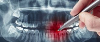
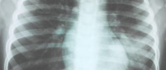
What can a doctor see in the image? Using X-rays, injuries are successfully diagnosed, the lungs are examined, various formations (stones, tumors), and areas of obstruction can be identified. With a contrast study of blood vessels, the doctor sees aneurysms, areas of blood vessels affected by atherosclerosis, etc. Different tissues transmit X-rays differently: bone tissue absorbs them almost completely, soft tissue partially retains them, and air allows them to pass through completely. Depending on this, shadows of different intensities are obtained on the film: in place of bones there are white areas, in place of soft tissues they are gray, layers of air appear black on an x-ray. X-rays are essentially negatives, so the lighter areas on them are called “shades.” For example, healthy lungs filled with air appear black on x-rays. The area of pneumonia (pneumonia) is a lighter spot, which doctors will call a shadow (see photo).
Using X-rays, you can make an accurate diagnosis for various injuries. For example, a fracture is visible as a darker “break” in a lighter “field” of the bone. Inflammation is usually visualized as a lighter area. Intestinal obstruction can be judged by the presence of gases in the organ or by changes in the shape of intestinal loops. Stones in organs look like light formations with clear boundaries and contours. If an X-ray is taken with a contrast agent and the image shows uneven filling of the organ, the doctor may assume the presence of a benign or malignant tumor. When examining vessels with a contrast agent, dilations are clearly visible - ruptures (aneurysms) are possible in this place.
How is an X-ray taken? Before the procedure, the patient must take off his jewelry, belt, remove all metal objects, phone, etc. from his pockets. In some cases, such as when examining the chest or spine, the doctor may ask you to undress to the waist. X-rays of the extremities can be performed with clothes on. Those parts of the body that are not examined are covered with special protective lead aprons. The doctor also puts on a protective suit and goes into the next room. Pictures are taken in different positions - mostly lying down or standing. Depending on the projection in which the image is needed, the doctor may ask you to change the position. Studies with contrast agents usually require some preparation. Your doctor will provide instructions for preparing for the study.
Modern X-ray machines (including those used in our clinics) provide minimal radiation exposure. For example, a general X-ray of the chest organs is no more than 0.15-0.3 mSv. We employ experienced x-ray technicians who minimize the impact of ionizing radiation on the patient’s body. X-ray examinations are performed in the left bank emergency room of the TERVE clinic on Partizana Zheleznyaka, 21A and the right bank emergency room of the TERVE clinic on Krasnoyarsky Rabochiy Ave., 150 p. 48 .
Prices for tests are listed on the pages of emergency rooms.
What is the research procedure like?
When performing radiography, there are no special features in preparation for the study. The digital method is organized in the same way as the analogue one.
First you need to understand where you can get tested. The installations for performing this diagnostic method can be located in a clinic, in a hospital, or in a tuberculosis dispensary. In order to sign up and then undergo the procedure, you must first consult with your doctor. He will explain whether there are indications, what shortcomings this examination has, and will suggest what specific options will be necessary for a more accurate and rational use of the results.
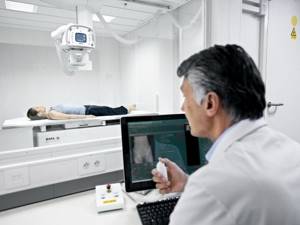
X-ray technician conducting a study
The procedure itself has no special requirements. In order to get the most out of the test, you need to listen carefully to the medical staff. If necessary, all jewelry, metal bracelets, and earrings must be removed, especially when performing a hand scan when the bones are small.
The result is assessed without the participation of the patient. A board-certified imaging physician or radiologist carefully reads the image at his desk. Digital radiography allows you to do this even with remote access, remotely. You can see the results the very next day. It makes no sense to draw any conclusions from the image yourself. Except, of course, when the patient has a medical education.
The study results do not contain a specific diagnosis.
They only reflect the specialist’s vision of a shadow picture of a particular area of the body. The diagnosis and treatment tactics must be determined by a clinician: therapist, pulmonologist, gastroenterologist, urologist, surgeon, traumatologist.

The final diagnosis is made by the attending physician, analyzing images and clinical data
Capabilities of mobile X-ray machines
A convenient and reliable mobile digital X-ray machine is increasingly used in medical practice. It is used to diagnose patients with limited movement in hospitals.
Such devices are equipped with:
- flat panel detector providing high resolution and fast image processing;
- touch display providing quick access to patient information;
- a movable tripod with a rotation angle of up to 270°, allowing you to quickly find the optimal shooting position.
- system for maneuvering in any, even cramped, spaces.
Digital technology in radiation diagnostics has not yet exhausted all its possibilities and continues to develop.