Ultrasound of the mammary glands and regional lymph nodes is prescribed to identify pathological changes in the tissues of the organs being examined. The prevalence of the ultrasound diagnostic method is explained by its safety, non-invasiveness, high information content and the almost complete absence of contraindications. The procedure can be prescribed independently or be part of a comprehensive diagnosis, including mammography, Dopplerography, radiography with contrast, etc. At the Hemostasis International Clinic, you can sign up for an ultrasound scan of the mammary glands and regional lymph nodes any day and undergo diagnostics at a time convenient for you. The study is carried out by diagnosticians of the highest category. A preliminary diagnosis can be obtained directly during the procedure. If necessary, our doctors will give detailed advice on the diagnostic results and prescribe effective treatment.
What are regional lymph nodes
Regional lymph nodes are a peripheral organ of the lymphatic system. It is the lymph nodes that perform the function of a kind of filter that cleanses the lymphatic fluid coming from organs and parts of the body. There are many groups of lymph nodes in the human body. They are called regional. Each group collects biological fluid from a specific organ. The regional group of mammary gland nodes includes the following types of lymph nodes:
- axillary;
- subclavian;
- parasternal.
If there is an increase in regional lymph nodes, this may be a sign of the development of breast pathology. Therefore, if there are suspicious symptoms, it is necessary to visit a doctor as soon as possible and do an ultrasound of the breast with regional lymph nodes at the initial stage of the disease. The earlier the pathology is diagnosed, the easier and faster it will be to get rid of it.
Why is inflammation of the lymph nodes in the lungs dangerous?
Lymph nodes play an important role in pneumonia. During the inflammatory process, a reaction of the lymph node to pathogenic microorganisms occurs - as a result of spasm and closure of the lymphatic vessel, inflammatory edema begins to develop. This reaction stops pathogenic microorganisms at the site of the inflammatory process and prevents the penetration of microbes into the bloodstream of the circulatory system. With pneumonia, the development of perilymphatic foci is observed, which are located along the lymph nodes.
Such changes are also observed in lymphogenous carcinomatosis (tumor cells spread through the lymphatic vessels) and sarcoidosis (a systemic disease characterized by damage to organs and systems of the body, damage to the lymph nodes). The lymphatic vessel may close, the drainage and cleansing function of the lymph nodes may be disrupted, and the infectious process will progress. Enlargement and pain of the lymph nodes of the chest cavity are observed with tuberculosis; enlargement of cervical, intra-abdominal, intrathoracic, axillary lymph nodes is typical for patients with HIV infection.
What can an ultrasound scan of lymph nodes show?
An ultrasound of the mammary glands and lymph nodes will show common changes in the mammary gland:
- Mastopathy. A non-inflammatory disease of the mammary gland, characterized by changes in the ratio in the structure of the organ of connective and epithelial tissue. The progression of pathology leads to the formation of cysts and fibrous tumors. The danger of mastopathy lies in the fact that at any time, under the influence of negative internal and external factors, the pathology can degenerate into a cancerous tumor.
- Intraductal papilloma of the mammary gland. This is a papillary growth formed from cells of the gland ducts. Papillomas are benign in nature and are diagnosed in the fair sex of any age, even in teenage girls. Multiple papillomas formed in the ducts of the organ can degenerate into a malignant neoplasm.
- Cystic changes. A common pathology that is asymptomatic in the initial stages of development. A cyst is a benign cavity neoplasm, the capsule of which is filled with serous fluid. However, with a combination of unfavorable factors, a cystic tumor can degenerate into cancer.
- Non-lactation mastitis. A disease characterized by the development of inflammatory complications in the mammary gland. Non-lactation mastitis is not associated with breastfeeding. The main cause of the pathology is a pathogenic infection that is activated as a result of immune dysfunction.
- Lactostasis. A common complication during breastfeeding. The reason for its development is a disruption in the process of milk outflow from any part of the mammary gland, caused by blockage or obstruction of the milk ducts. Lack of timely treatment of lactostasis leads to the development of mastopathy.
- Fibroadenoma. It is often formed against the background of hormonal disorders, the cause of which can be any gynecological disease. Fibroadenoma is an intermediate step between benign and malignant neoplasms, and therefore requires timely diagnosis and surgical treatment.
- Malignant tumors. Breast cancer is a common disease diagnosed in both young and old women. Malignant degeneration is a consequence of mutations in glandular tissue cells. Cancer requires early diagnosis and treatment. This is the only way to stop the progression of the disease and the development of irreversible consequences.
- Anomalies in the development of the mammary glands. The most common anomalies include the following: polymastia, polythelia, hypoplasia, complete absence of mammary glands, and nipple inversion.
Ultrasound of the mammary glands of regional lymph nodes if any pathological changes are suspected is one of the most effective and informative diagnostic methods that allows diagnosing the disease at an early stage of development.
Indications and contraindications for ultrasound of regional lymph nodes
Echography of the mammary glands and regional lymph nodes is prescribed for patients who contact a mammologist with the following complaints:
- discomfort, pain, feeling of fullness that does not go away for a long time;
- asymmetry of the mammary glands, when there is an increase or decrease in the organ, loss of shape;
- compaction, swelling, hyperemia;
- change in the shape and color of the areola;
- pain and inflammation of the axillary lymph nodes;
- pathological discharge from the nipple;
- menstrual irregularities.
Ultrasound examination is prescribed for women during pregnancy and after lactation. The procedure is indispensable during the period of monitoring the condition of breast prostheses, preparing for surgical treatment, and planning treatment tactics for previously identified pathology.
It is advisable to undergo an ultrasound of regional lymph nodes and mammary glands at least once a year for every woman after 30 years of age. Preventive diagnostics will help to identify any pathological change in the early stages, begin treatment in a timely manner and prevent the development of complications.
The procedure has no absolute contraindications. Diagnostics will have to be postponed if wounds, ulcers, or erosions have formed in the area under study. Also, the study will have to wait if a woman suffers from acute respiratory infections or acute respiratory viral infections, which are accompanied by a general feeling of poor health. After your condition returns to normal, you can sign up for the study by choosing a convenient date and time.
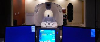
Advantages of X-ray of the axillary area at the CELT clinic
The diagnostic center of the multidisciplinary clinic CELT has a wide range of modern equipment that allows us to conduct comprehensive studies in various areas. Our diagnosticians have more than fifteen years of experience - each of them is a professional with a capital P, who is well acquainted with all radiology techniques and knows how to carry out diagnostics in such a way as to obtain all the necessary information. You can view our prices in the “Services and Prices” section or by calling the number.
Make an appointment through the application or by calling +7 +7 We work every day:
- Monday—Friday: 8.00—20.00
- Saturday: 8.00–18.00
- Sunday is a day off
The nearest metro and MCC stations to the clinic:
- Highway of Enthusiasts or Perovo
- Partisan
- Enthusiast Highway
Driving directions
Rules for preparing ultrasound of the mammary glands with regional lymph nodes
The procedure does not require special preparation. However, there are rules regarding the days of the cycle on which the study will be as informative as possible. At the beginning of the cycle, glandular structures branch and develop. After the end of menstruation, the ducts are restored. After ovulation, the gland begins to prepare for conception, so it increases in size and swelling. It is more advisable to carry out ultrasound scanning before the onset of ovulation. During this period, the mammary glands are not swollen, and the tissues and structure are clearly visible.
Optimal time for ultrasound diagnostics:
- 5 – 12 days with a 28-day menstrual cycle;
- 7 – 14 days in a cycle lasting more than 28 days.
In emergency situations, when diagnosis cannot be delayed, echography is performed regardless of the day of the cycle.
Interpretation of diagnostic results
Examination of regional lymph nodes and mammary glands takes 25–30 minutes. Directly during the examination, the doctor superficially informs the patient about the condition of the organs, the presence or absence of pathological changes. Ultrasound data is recorded in a protocol, which must be shown to the doctor who gave the referral for diagnosis. The specialist will decipher the results, notify the patient of the final diagnosis, and then determine a further action plan regarding the treatment of the identified pathology.
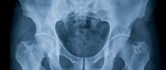
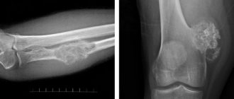
What does an x-ray of the axillary region show?
In the process of deciphering the results obtained, the doctor compares them with normal parameters, which makes it possible to determine soft tissue pathologies on x-rays. The pictures clearly show:
- axillary region;
- skin and subcutaneous fat;
- parts of the mammary glands.
As for the lymph nodes, the procedure allows you to determine them:
- dimensions;
- condition of glandular tissue;
- localization;
- contours;
- quantity.
When examining the mammary glands, an x-ray of the axillary region is carried out in combination with a number of other studies: mammography, pneumocystography and targeted puncture.
Benefits of breast ultrasound
Ultrasound examination has many advantages:
- non-invasive, painless, informative;
- almost complete absence of contraindications;
- can be used an unlimited number of times;
- does not cause radiation exposure to the body, so it can be prescribed to patients of any age;
- does not take much time;
- results can be obtained immediately after the examination;
- allows you to control and adjust the treatment regimen at any stage of treatment;
- affordable price.
Normal echo pattern
The structure of the breast changes with age. It includes the following types of fabrics:
- connecting;
- glandular;
- fatty.
Glandular tissue actively develops during pregnancy and breastfeeding. On ultrasound it is visualized as a homogeneous mesh with medium to large cells, covered with a hyperechoic capsule. After 60 years, fibro-fatty transformation is more pronounced. Lymph nodes are round in shape, hemogenic in structure, up to 10 mm in diameter. The capsule is smooth, continuous, hyperechoic.
Pathological changes
With pathological changes, the structure of the mammary gland becomes heterogeneous. If a hypoechoic neoplasm is visualized on the monitor screen, such a clinical picture may indicate the development of diseases:
- cystic tumor;
- galactocele;
- abscess;
- malignant tumor.
Hyperechoic neoplasms may indicate the progression of such pathologies:
- fibroadenoma;
- papillomas.
Enlarged lymph nodes and a hypoechoic structure indicate an inflammatory process. Changes in the membrane, thickening, and irregularities can be caused by a progressive purulent complication. A dense hyperechoic structure indicates a metastatic lesion, but more detailed diagnostics are prescribed to confirm the diagnosis.
How to treat inflammation of the lymph nodes of the lungs
Treatment of the pathological process in the lymph nodes of the lungs depends on the disease, the consequence of which was inflammation of the lymph node. If it is pneumonia, the doctor prescribes antibacterial therapy aimed at suppressing the causative agent of the disease. Inflammation of the lymph nodes is accompanied by fever, pain, and weakness. The doctor prescribes antipyretics and painkillers. Treatment of the inflammatory process in the lymphatic system begins with treatment of the underlying disease.
Inflammation of the lymph nodes can be a consequence of various diseases. At the Yusupov Hospital, the patient is sent for blood and urine tests, takes tests for antibodies to determine the causative agent of the inflammatory process in the lungs, the patient receives help from other specialists - an infectious disease specialist, an oncologist, a pulmonologist. You can make an appointment with a doctor by calling the clinic.