Ultrasound examination (ultrasound) is the most popular and informative method for diagnosing pancreatic diseases. During its implementation, specialists pay special attention to echo signs indicating the development of pathological processes in the organ. For example, an increase in the echogenicity of the pancreas, as well as a decrease in it, indicates the occurrence of various changes in the parenchyma of the pancreas, which may result from the development of serious diseases. And what does increased or decreased echogenicity mean during ultrasound examination, you will now find out.
What it is?
The term “echogenicity” refers to the ability of internal organs to reflect various radiations and impulses. It is characteristic of all tissues of the body, including fat. When performing an ultrasound of the pancreas, it is subjected to numerous ultrasound pulses, which are reflected from the gland tissue back to the device and create a kind of pattern reflected on the computer monitor. From this drawing, a specialist can determine the condition of the organ, the presence of neoplasms in it, as well as the occurrence of compactions in certain areas of the gland.
In this case, echogenicity is presented in the form of a gray scale displayed on the monitor, which has several shades. Its color directly depends on the ability of the gland to reflect ultrasonic impulses. However, in this matter, the settings of the diagnostic device play an important role, as well as some interference that may arise as a result of the mobile device being turned on, the patient having a heart device, etc.
It should immediately be noted that the degree of echogenicity is not determined “by eye”. Before recording it, an ultrasound examination of a healthy human liver is performed, during which the echogenicity of the organ is also determined. And only after that it is compared with the echogenicity of the pancreas.
As already mentioned, this indicator is displayed on the computer screen in the form of a gray scale, which can have different shades. So, for example, if a person has increased echogenicity of the pancreas, then on the monitor it looks like a bright spot, and hyperechogenicity appears as a white spot. If there are hypoechoic areas (low echogenicity) on the pancreas, they appear as dark spots, and moderate (average) echogenicity appears as a light gray area.
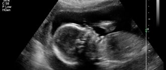
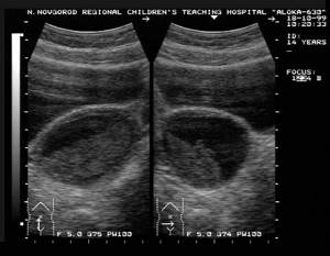
Ultrasound image of the pancreas
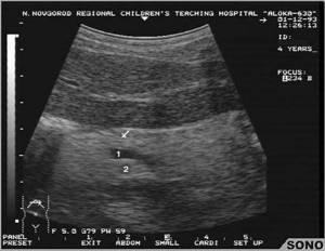
Sizes and features of the thyroid gland
The result of an ultrasound scan of the thyroid gland should reflect as accurately as possible the size of the organ, its structure and shape. Each person’s gland has individual sizes, which can change throughout life, so the norms for a particular patient are set directly by an endocrinologist or a specialist conducting diagnostics.
Normally, the size of the gland’s lobes should be as follows:
- length 2.5-4 cm;
- width – 1.5–2 cm;
- thickness from 1 to 2 cm.
With a good ultrasound result, the thyroid gland has a homogeneous structure, smooth edges and is characterized by the absence of compaction and swelling. The thickness of the isthmus of the gland normally ranges from 4-5 mm. The volume of the endocrine organ in men should not exceed 25 ml, in females - 18 ml.
Factors provoking changes in echogenicity
If the echogenicity of the pancreatic parenchyma changes, this may be due to various processes in the tissues of the organ, its ducts or the vessels with which it is penetrated. And if we talk about pathologies in which such pathologies are observed, we can highlight the following:
- the presence of calcium deposits in the ducts of the gland;
- tumor formation;
- development of inflammatory processes in gland tissues (for example, pancreatitis);
- the occurrence of necrotic processes in the organ (that is, the death of its tissues);
- the development of lipomatosis, in which healthy gland cells are replaced by fatty ones.
With the development of all these pathological conditions, the echo density of the gland, as a rule, increases or decreases. However, the presence of such deviations is not yet a 100% sign of the development of diseases. To make an accurate diagnosis, you will need to undergo a more detailed examination.
What is echogenicity?
Ultrasound research works on the well-known principle of echolocation. Since ultrasound is used for such diagnostics, different tissues of the body reflect it in their own way. The specialist sees a black and white image of the organs being examined on his computer monitor.
Each organ reflects ultrasound differently. Actually, what the doctor sees on the screen depends on this. The more fluid a particular organ contains, the darker it appears on the monitor, and vice versa.
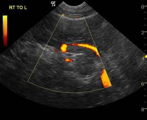
An example of increased echogenicity using the example of the pancreas. Pancreatic cancer.
The liquid is visible in black. And dense objects, accordingly, are visible in white. Actually, the property of human body tissue to reflect ultrasonic waves is called echogenicity.
This leads to another convention - the concept of “norm” regarding echogenicity is conditional. Again, this is due to the fact that each organ has its own density and echogenicity. The specialist knows what degree of echogenicity a particular organ should have and compares the norm with what he sees on the monitor. So he notices deviations in echogenicity in one direction or another, and based on this he makes a diagnosis.
Does a change in echogenicity always indicate the development of pathologies?
After a person undergoes an ultrasound, for the sake of his own interest, he begins to independently re-read the specialist’s report. And it often says that the echogenicity of the pancreas is increased, but only a few know what it is. Some, seeing such a conclusion, immediately begin to think that they have serious pathologies that require urgent treatment. But that's not true.
As already mentioned, diagnosis cannot be made solely on the basis of these indicators. If the echogenicity of the pancreas is decreased or increased, this indicates that the patient will need to undergo a more in-depth examination. After an ultrasound has been performed and changes in the ability of the gland to reflect ultrasound impulses have been identified, the diagnosis is made only taking into account the following indicators:
- symptoms that bother the patient;
- presence of abnormalities in stool and urine tests;
- changes in blood parameters (often with the development of gland pathologies, an increase in the level of certain enzymes produced by the pancreas is observed);
- size of the gland (also determined by ultrasound or CT), etc.
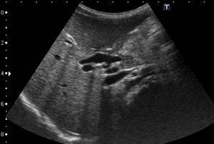
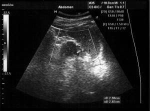
The image shows an enlarged pancreas
Only after receiving all the results of a detailed examination of the patient, the doctor can make an accurate diagnosis and prescribe the necessary treatment. If additional examination does not show any deviations from the norm, then this indicates normal functioning of the gland and the absence of pathologies, even if the echogenicity of the pancreas is increased.
Answers to the most frequently asked questions in case of liver diseases or pathologies
Liver screening is performed for patients with hepatitis B and C, cirrhosis of the liver, and residents of those regions that have high rates of liver cancer. Screening for autoimmune liver disease is necessary to detect autoimmune hepatitis and primary biliary cirrhosis. It is a blood draw from a vein, on the basis of which the presence of antibodies is detected:
- To the mitochondria.
- To smooth muscles.
- To microsomes of the kidneys and liver.
- To the parietal cells of the stomach.
What pathologies may increased echogenicity indicate?
Hyperechoic inclusions in the pancreas indicate the presence of dense areas in the organ that arise against the background of focal accumulations of fat cells. This phenomenon is medically called lipomatosis. Its treatment consists of maintaining a low-calorie diet and surgery.
With increased echogenicity of the pancreas, they also talk about the development of inflammatory processes in it, which appear when diseases such as acute or chronic pancreatitis occur. This indicator also means gluing (sclerosing) of the ducts of the organ, which is a frequent accompaniment of age-related changes in the body.
With increased echogenicity, we can also talk about the occurrence of fibrotic changes in the parenchyma of the gland, when a predominance of connective tissue is noted. As a rule, the development of such pathology occurs against the background of long-term chronic pancreatitis. At the same time, during an ultrasound examination, diffuse changes in the organ and its uneven contours are recorded.
Diffuse changes in pancreatic tissue
But it should be noted that if the pancreas has increased echo signs, then this may also signal that a person is leading an unhealthy lifestyle, namely eating large amounts of food, which puts a strong load on the organ, as well as a sedentary lifestyle. However, in this case, such deviations are temporary. If they are noted 1–2 weeks after the patient “goes on” a diet, then this indicates serious disturbances in the functioning of the gland and the development of the diseases described above.
What parameters does the doctor evaluate during an ultrasound examination?
First of all, the echogenicity parameter is important for an ultrasound specialist. Its normal parameter is called isoechoicity. In this case, healthy organs and tissues will be visible on the screen in gray.
Example of Hypoechogenicity
Hypoechogenicity is a decrease in echogenicity, and in this case the color becomes darker. In turn, increased echogenicity is called hyperechogenicity. Objects with the specified property are visible on the screen in white. With echo-negativity, objects will be visible in black. From this we can conclude: the lighter the object, the higher its echogenicity and vice versa. For example, kidney stones are hyperechoic: ultrasound does not pass through them. The doctor sees the upper part of such a formation and its shadow (it is acoustic).
Reduced echogenicity usually indicates that there is edema in the tissue or organ . And a full bladder will be visible on the monitor in black, and this will be the norm.
In addition, the following parameters are also assessed.
Structure.
Normally, it can only be homogeneous. If heterogeneity is noticeable, it will be described in detail. Based on such changes, one can judge the presence of pathological changes in the organ.
Contours.
Normally they are even. And the unevenness of the outline of the organ indicates an inflammatory process.
The unevenness of an object in an organ indicates that it is malignant.
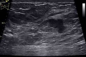
Diffuse and local changes in the gland
When performing an ultrasound examination of the pancreas and determining echogenicity indicators, specialists also pay great attention to diffuse and local changes in the parenchyma of the organ. These conditions may indicate various pathologies. For example, the diffuse structure of an organ may indicate significant changes in the tissues of the gland that occur:
- with lipomatosis (with this condition, a diffusely heterogeneous structure is revealed, but there are no pronounced symptoms);
- exacerbation of pancreatitis (in this case there are pronounced symptoms, which include severe pain, nausea, vomiting, etc.);
- the formation of neoplasms in the tissues of the gland (the clinical picture is increased gas formation, sudden weight loss, stool disturbances, etc.).
But it should be noted that diffuse changes in the pancreas can also be recorded due to poor diet, alcohol abuse, or even a common cold.
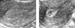
Differences between increased and decreased echogenicity
However, if diffuse changes are observed throughout the organ, then local changes are observed only in certain areas of the gland. And depending on the patient’s complaints and other ultrasound indicators (tissue density, organ size, etc.), the doctor can make a preliminary diagnosis. But it must also be confirmed using other diagnostic methods.
For example, local echo signs are recorded:
- during the formation of pseudocysts;
- deposition of fat cells;
- formation of stony deposits in the gland ducts;
- metastases (they are discovered during the development of stage 3–4 cancer).
It should be understood that regardless of what echogenicity (mixed, decreased or increased) was recorded by a specialist during the examination, these indicators should not be the main ones for making a diagnosis. In any case, differential diagnosis will be required.
What is high echogenicity?
The value of high echogenicity depends on the structure of the tissue. When this indicator increases in the glandular tissue, its normal cells are gradually replaced by scar or fatty tissue. It is also possible that calcium compounds may accumulate in this area.
Changes in tissue parenchyma are also possible. Let us remember that this is the main tissue of an organ that does not have a cavity. An increase in the echogenicity of the parenchyma indicates that the fluid content in it is reduced. This happens as a result:
- disturbances in the content of hormones in the body;
- disorders of metabolic processes (metabolism);
- unhealthy diet (especially the pancreas);
- presence of bad habits;
- parenchymal diseases;
- swelling due to inflammation or injury.
Treatment methods
If the echogenicity of the pancreas is reduced or decreased, the doctor will prescribe an additional examination, which may include:
- biochemical studies of blood and urine;
- CT;
- MRI;
- X-ray examination;
- stool analysis;
- gastroendoscopy, etc.
Only after receiving all the results of the examination, the doctor determines whether the person needs treatment or not. After all, as mentioned above, an increase in echogenicity can occur against the background of poor nutrition or a passive lifestyle. If, during a detailed examination, serious pathologies were identified, the doctor determines how to treat them. Treatment tactics depend on several factors - the type of disease, the patient's age and his general health. Therapy can be carried out either surgically or medically.
But in any case, when pathologies of the pancreas are detected, all patients, without exception, are prescribed a diet that completely excludes from the diet all foods that can negatively affect the functioning of the organ. Otherwise, treatment therapy is selected on an individual basis.
What does an increase in the degree of echogenicity of a particular organ mean?
Increased echogenicity of different organs is seen differently on ultrasound and has variable significance. Let's take a closer look at these changes.
Uterus
Hypoechoic area of the uterus with endometriosis
Normally, it has only a homogeneous structure. An increase in this indicator indicates that the patient has the following diseases:
- inflammation (diffuse echonegativity);
- uterine fibroid;
- fibroids (in this case, a light-colored object with sound amplification is visualized in the uterus);
- neoplasm (benign or malignant);
- endometriosis (resulting from hormonal imbalance or cancer). It is also characterized by blurred contours and heterogeneity of structure.
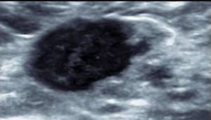
Ovaries
hypoechoic ovarian formation
The high-density area appears on the screen as a hypoechoic formation. Often these are the following objects:
- calcium deposits;
- benign and malignant formations.
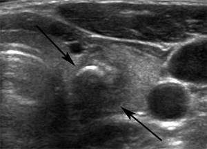
Pancreas
hypoechoic formation of the pancreas
An increase in the echo density of this organ indicates the development of acute or chronic inflammation in it. It can lead to the development of edema. Here are other reasons for the increase in ultrasound density of such an organ:
- flatulence;
- various tumor structures, including malignant ones;
- abnormal portal vein pressure;
- formation of calcifications;
- stones in the organ.
A diffuse increase in density indicates that healthy tissue in the pancreas is gradually being replaced by another. Scarring in this organ indicates that it is becoming smaller. This negatively affects the outcome of a particular disease. With fatty degeneration of an organ, its size does not increase. It occurs in diabetes and also in older people.
A transient increase in the ultrasonic density of the organ occurs with excessive consumption of fatty foods, irregular stools, or a lifestyle with a combination of alcohol. That is why, when the echogenic structure of the pancreas changes, a thorough diagnostic examination of the patient is required, in particular gastroenteroscopy.
Gallbladder
An area of high ultrasound density located in the gallbladder indicates that a stone has formed in the gallbladder.
A diffuse change in the ultrasonic permeability of the bladder towards an increase indicates that an inflammatory process is developing in it for a long time. In both cases, the doctor will see a white object.
Hyperechogenicity of the thyroid
Hypoechoic thyroid nodule
This phenomenon suggests that the amount of colloidal substance formed due to the effects of hormones is gradually decreasing. Often, hyperechogenicity in the thyroid gland is caused by the deposition of calcifications in its tissue. In all these cases, foreign formations in tissues are light in color, different from healthy tissues.
This condition occurs for the following reasons:
- insufficient amount of iodine in the body, which causes the phenomenon of endemic goiter;
- toxic goiter, which occurs as a result of damage to the thyroid gland by toxic substances;
- thyroiditis of an autoimmune nature;
- subacute thyroiditis.
An accurate diagnosis related to thyroid pathologies can be made not by the specialist performing the study, but by an endocrinologist. Often, ultrasound examination alone is not enough to make an accurate diagnosis. In addition, a hyperechoic object in the thyroid gland occurs due to cancer or sclerosis.
Mammary gland
hypoechoic formation of the mammary gland. Fibroadenoma.
In some cases, women have absolutely no need to panic about the increase in echogenicity of the mammary glands. In menopause and postmenopause, such a change is normal, as the amount of connective tissue in the tissue increases. But if the hyperechogenicity of the mammary gland in young women and girls indicates that there was inflammation in the organ, which affected the structure of the organ.
A high-density formation is visualized as a light-colored object. Analysis of the image may indicate that the gland is progressing:
- atypical cyst;
- calcified area;
- area with fibrous tissue.
The heterogeneity of the structure of the mammary glands also indicates that there are some foreign changes in it. The doctor can determine their nature and, accordingly, prescribe treatment.
Kidneys
Hyperechogenicity of the kidney is visualized on the monitor in different ways, depending on the pathology. In diabetic nephropathy, the size of the kidney is increased. However, the renal pyramids are characterized by reduced echogenicity. On the contrary, an increase in this indicator for the parenchyma is observed with glomerulonephritis, especially with a severe course.
Areas with increased density are also determined in the following pathologies: malignant kidney disease, especially hypernephroid cancer;
- myelomas;
- kidney infarction;
- accumulation of calcifications in the renal parenchyma.
Spleen
An increase in ultrasound density may also occur in the spleen. It directly depends on the patient’s age, but should not be greater than that of the liver. If the increase in ultrasound echogenicity of the kidney does not depend on age, then this may indicate the following pathologies:
- increased portal vein pressure;
- Konovalov-Wilson syndrome;
- amyloidosis;
- increase in blood iron.