Introduction
On December 28, 2012, the Government of the Russian Federation approved the Strategy for the Development of Medical Science in the Russian Federation for the period until 2025. “The goal of the strategy is the development of medical science aimed at creating high-tech innovative products that, based on the transfer of innovative technologies into practical healthcare, ensure the preservation and strengthening public health. The strategy is aimed at implementing state policy in the field of healthcare, improving the quality and accessibility of medical care to the population of the Russian Federation, including the development of innovative products, the development of critical technologies and the development of competencies...” (cited from [1]).
In the process of implementing the Strategy in 2021, “Introduction of innovative medical technologies, including a system of early diagnosis and remote monitoring of the health status of patients” is designated by the Russian Ministry of Health as one of the main areas of activity for established national medical research centers. The importance of solving the problem “Introduction of innovative medical technologies, including a system of early diagnosis and remote monitoring of the health status of patients” (clause 4b, 4th paragraph) in May 2021 was emphasized in his decree by the President of the Russian Federation V.V. Putin [2].
One of the directions for solving this problem could be monitoring of the microcirculation system. The oscillatory nature of tissue perfusion is a fundamental feature of the microcirculation system and is due to the simultaneous functioning of various regulatory mechanisms at the level of arterioles [3–5]. The oscillatory nature of microcirculatory blood flow in human skin is recorded by such non-invasive research methods as capillaroscopy [6], laser Doppler flowmetry [5, 7], thermometry [8, 9], infrared spectroscopy [10, 11], plethysmography [12, 13] and other research methods [14].
To analyze oscillatory processes of a physiological nature, which are mostly non-stationary (signal characteristics change over time), wavelet analysis is now increasingly used, which in a number of medical applications has become an alternative to the amplitude-frequency Fourier transform in the analysis of heart rate variability, encephalography, electrogastroenterography and other physiological research methods. With laser Doppler flowmetry, amplitude-frequency wavelet analysis of fluctuations in skin perfusion makes it possible to detail the functional state of resistive microvessels in a wide range of diseases - arterial hypertension, diabetes mellitus, myocardial infarction, chronic heart failure, occlusive-stenotic lesions of the vascular bed of the lower extremities, obesity, bronchial asthma, systemic connective tissue diseases and other pathological conditions [15].
The purpose of this work is to demonstrate a new method for studying microcirculation in humans, a preliminary analysis of its diagnostic capabilities and prospects for application.
Web capillaroscopy method
The method is based on the development of Russian scientists who use a conventional web camera to obtain physiological information (application for a Eurasian patent for an invention No. 201700326 dated 07/08/17 “System for telemetric monitoring of parameters of the patient’s vital functions”). The method is based on the analysis of changes in pixel contrast with fluctuations in skin perfusion.
The study is carried out as follows: the patient sits motionless in front of a web camera, which records video (30 frames per second) of the skin. In Fig. 1 per color The inset shows an example of recording from a laptop webcam. For ease of perception and further analysis of information, the object of study (in this case, a person) is divided into four sectors, each of which has a specific color. In Fig. 1 dotted rectangle highlights the “Filtered Cr” sector, in which a 10-second fragment clearly shows how each heartbeat is accompanied by a change in the signal - blood flow. It can be seen that the amplitude of pulse oscillations varies from cycle to cycle.
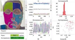
Rice. 1. Web capillaroscopy of the face using a laptop camera for 3 minutes.
To correctly interpret the data obtained, it is fundamentally important to understand from which part of the microvasculature (MCR) of the skin we receive information. The most detailed study of the structure and spatial organization of skin MCR was carried out by I. Braverman [16], who, based on a large array of biopsy material, using the technique of microtome sections 1 μm thick, electron microscopy and computer modeling, showed that 1 mm3 of the papillary layer of human skin in non-acral sections contains a typical microvascular module. The microvascular module of the skin includes one feeding arteriole with a diameter of less than 30 microns ascending from the depths of the dermis, which is divided into 5 precapillary arterioles, forming a network of capillaries. The network of capillaries passes into postcapillary venules, merging into 9 collecting venules, which flow into one draining venule with a diameter of less than 50 microns descending into the skin. The author draws attention to the fact that the structural organization of the MCR is identical in all preparations, with the exception of age-related differences in the number of metabolic microvessels [16]. The angioarchitecture of the MCR of the skin is such that only capillaries reach the very surface - the arterial section ascends from the depths, which at the very surface of the skin makes a 180° turn (transitional section), passing into the descending venous section of the capillary. The approximate depth of the transitional section of the capillaries is about 100-120 microns from the surface of the skin.
Thus, the most superficially located section of the MCR is the transitional section of the capillaries, and the nature of the blood flow during web capillaroscopy is recorded precisely in them. Anatomically, the transitional section of the capillaries is the “pole” of the systemic circulation opposite the heart, where the transition of the arterial system to the venous system also occurs. From a functional point of view, the transitional region is a zone of equilibrium between the processes of filtration and reabsorption, which underlie the bilateral transendothelial transport of water, water-soluble and low-molecular substances. The key parameters for the efficiency of the filtration-reabsorption mechanism of metabolism are the values of hydrostatic pressure at the “input” to the capillary and at the “exit”.
As was shown in works with video capillaroscopy in the area of the nail bed of the fingers, blood flow in the capillaries of the skin is oscillatory in nature, which is explained by the functioning of various regulatory mechanisms at the level of muscle microvessels - precapillary arterioles [6]. The oscillatory nature of capillary hemodynamics is due exclusively to the vasomotor activity of precapillary arterioles, since the capillary itself is a monolayer of endothelial cells, and its effect on the hemodynamic parameters of blood flow is indirect through the processes of filtration and reabsorption. Vasomotion (vasa - vessel, motion - movement) of precapillary arterioles is caused by the simultaneous action of 3 regulatory mechanisms - myogenic, neurogenic, endothelial. These mechanisms, based on the principle of positive and negative feedback, continuously regulate the tone and size of the lumen of precapillary arterioles, modulating the volume of arterial blood flowing to the exchange link to values that are optimal for transcapillary exchange.
In Fig. 2 per color The inset shows an analysis of signal changes during web capillaroscopy for 3 minutes. Data are shown for the left half of the forehead (blue sector in Fig. 1 ). When analyzing a video fragment over its entire length (3 minutes), the oscillatory nature of the signal is clearly visible, which is a characteristic sign of microcirculatory blood flow. Knowing the frequency of functioning of regulatory mechanisms, we can quite clearly distinguish the alternation of endothelial (1) and neurogenic (2) vasomotions, which differ from each other in both frequency and amplitude (Fig. 2) .
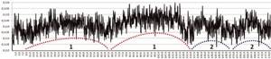
Rice. 2. The nature of perfusion for 3 minutes in the skin of the left half of the forehead (blue sector in Fig. 1).
Dashed lines: 1 - endothelial vasomotions; 2 - neurogenic vasomotions.
The data presented allow us to conclude that the web capillaroscopy method registers changes in blood flow in the transitional sections of the skin capillaries, making it possible not only to obtain digital information of a physiological nature, but also, regardless of the patient’s location, to transmit it through various communication channels to a medical institution for further analysis .
Any areas of the patient’s skin are available for web capillaroscopy, but one of the most promising areas is the skin of the face, which is of particular interest for several reasons. The first includes regional features of the innervation of skin microvessels, which are represented mainly by somatic sensitive (afferent) and autonomic sympathetic (efferent) regulatory systems. The direct participation of the parasympathetic nervous system in the regulation of microvessels is considered proven only for facial skin [17, 18]. Thus, the study of microcirculatory blood flow specifically in the facial area makes it possible to study both sympathetic and parasympathetic mechanisms of neurogenic control of the vasomotor activity of resistive microvessels at the “pole” of the systemic circulation opposite the heart.
The second important aspect is the characteristics of the blood supply. It is known that the scalp receives nutrition from the external carotid artery system and only the skin of the central part of the forehead is supplied with blood by the supratrochlearis (a. supratrochlearis) and supraorbital (a. supraorbitalis) arteries, which are the terminal branches of the ophthalmic arteries included in the internal carotid artery system (Fig. 3 on color insert) . The long-standing interest of researchers specifically in the ophthalmic artery basin is due to the fact that disturbances in the microcirculatory blood flow in the eye area (the fundus and bulbar conjunctiva) are associated with various types of cerebral circulatory disorders [19-21], and the microcirculatory bed of the eyes is considered as a “window” into the cerebral system. microcirculation.
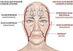
Rice. 3. Blood supply system to the skin of the facial part of the skull.
The red color indicates the arteries from the external carotid artery system, the black color indicates the terminal branches of the ophthalmic arteries that are part of the internal carotid artery system. Dotted squares highlight areas of the skin to analyze the nature of cerebral microblood flow in the basins of the right and left internal carotid arteries.
In the available literature, the only pilot study was found in which, using laser Doppler flowmetry with wavelet analysis, it was shown that the nature of microcirculatory blood flow in the skin of the right and left half of the forehead differs significantly both in the level of tissue perfusion and in the vasomotor activity of microvessels depending on the side and volume of vascular lesions in the brain [22]. The authors also note significant regional differences in the nature of microcirculation of the forehead skin in the first 3-7 days of restoration of cerebral blood flow after thrombolytic therapy. The results obtained suggest that analysis of the nature of perfusion in limited areas of the forehead skin during web capillaroscopy (dotted squares in Fig . 3 ) may be a useful tool for assessing cerebral blood flow in isolation in the territories of the right and left internal carotid arteries.
Capillaroscopy is a non-invasive study of capillaries, capillary blood flow and aggregates of blood cells. The work is based on the method of studying biological objects using a special microscope equipped with a digital camera. The study is carried out on a device developed by JSC. Since 2011, CAB has been a resident of the Innovation Center.
The study is carried out non-invasively, painlessly, in real time.
The structure of the capillary itself, the zones around the capillary, the blood flow in it, and the presence of aggregates of blood cells are examined.
The scope of application is very wide: cardiology, neurology, phlebology, endocrinology, rheumatology, sports medicine , etc.
Capillaroscopy makes it possible to determine signs characteristic of changes in the vascular bed during arterial hypertension and to quantify the presence of hidden edema.
This method is of great help in correcting drug treatment in patients with cardiac pathology that involves a high risk of thrombosis - with atrial fibrillation, after heart valve replacement, coronary artery stenting, with thrombophlebitis.
This method allows you to evaluate the effectiveness of treatment by the aggregation state of the blood and the state of blood rheology in hematological practice. Capillaroscopy is an indispensable diagnostic method for diabetic angiopathy, Raynaud's syndrome, systemic lupus erythematosus, and scleroderma. In sports medicine, the method is applicable to assess the training regimen and recovery process.
The object of study is the cuticle of the fingers or toes. The capillaries of the nail bed are examined using an optical system (powerful microscope) at a magnification of 200x, 400x, 800x , during which all key parameters of microcirculation are calculated using a computer program.
The study is carried out on an empty stomach or several hours after a light meal. Before the study, do not drink strong tea, coffee, alcohol, or drink excessive amounts of liquid. Smoking is prohibited before the examination.
Rational care of the nail bed is necessary: do not expose the skin of your fingers to gasoline, washing powder, soda, acetone, varnish, etc., and do not cut off the skin near the nail bed.
Restrictions on capillaroscopy:
- fungal infections of the tissues of the nail phalanges;
- burns of the hands and/or feet;
- traumatic lesions of the hands and/or feet;
- mechanical damage to the cuticle of the fingers;
- deterioration of light scattering of the skin of the fingers due to exposure to aggressive chemical compounds.
| Name of service by nomenclature | Price |
| Functional diagnostics | |
| Code: A03.13.002 Capillaroscopy | 1000.00 |