Timely identification of the problem is the key to successful treatment of vascular diseases. To ensure effective diagnosis, specialists develop and use various techniques, one of which is duplex scanning of the main arteries of the head. Duplex scanning is an alternative to Doppler ultrasound. The main difference between duplex scanning is that the doctor receives information not only about the state of blood flow, but can also assess the condition of the walls of blood vessels and determine the parameters of blood flow. Duplex scanning of the main arteries of the head is a modern instrumental diagnostic method used to obtain information about the direction and speed of blood flow, and the condition of the walls of the great vessels.
During duplex MAG, the main arteries of the head and neck are examined using an ultrasound scanner. In the neurology department of the Yusupov Hospital, doctors are attentive to the wishes of patients, so the study can be performed at the most convenient time. Comfortable rooms are equipped with modern equipment; with their help, neurologists receive accurate information about the condition of blood vessels and blood flow.
Description of the study
Duplex examination of the great arteries is painless and safe for the patient. Before the scan, the doctor talks with the patient, studies his complaints, symptoms, and the results of other studies in order to have the most complete clinical picture.
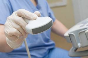
MAG duplex scanning lasts from 15 to 30 minutes. The doctor applies a special gel to the required area of the skin, which promotes better transmission of the sound signal, after which the specialist begins to move the sensor and continuously monitor the image on the monitor on which the sections are reflected. After the examination, a conclusion about the scan results is immediately issued.
The mechanism of duplex scanning of the arteries of the head and neck is based on the principle of echolocation, in which the vessel is imaged in two planes (across and along). When performing a duplex study, the area in question is displayed in color. The doctor gets the opportunity to study the walls of blood vessels, surrounding tissues, lumen, speed and direction of blood movement.
A little theory
The most commonly used is B-mode. It allows you to form an image on the device’s monitor, most often it is a picture in shades of gray. Ultrasound, penetrating into tissue, is reflected differently from different surfaces, goes back to the sensor, and through a series of transformations, a visualization of the organs being studied is formed on the screen.
Next comes M-mode. Its task is to inform about the movement of structures, allowing it to be studied in time. It is most often needed for research into heart disease.
Doppler ultrasound was founded . This method takes as its basis the property of ultrasonic waves. The reflection of a wave from moving objects causes a shift - a changed frequency. This change in frequency depends on the speed of movement of the structures being studied. For example, movement is directed towards the sensor - an increase in frequency, directed from the sensor - a decrease.
At the moment, there are two types of Doppler ultrasound (USD):
- blind (included in functional diagnostics and does not apply to ultrasound),
- B-mode (modern).
In the first case, the scanning depth is set blindly, without a “visible” image of the vessel on the screen, which leads to an increase in the number of diagnostic errors.
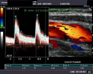
In Dopplerography, a spectral Doppler mode is distinguished, the direction and speed of blood flow in which is displayed graphically on the scanner screen. On the graph, the horizontal axis corresponds to the time value, and the vertical axis shows the speed. Above the level of the horizontal axis are signals towards the sensor, and signals below the axis are movement away from the sensor.
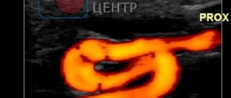
Color Doppler displays the direction of blood flow in color. This method of ultrasound imaging contains visualization of blood flow in the heart (also used for blood flow in relatively large vessels). Color designations are accepted: red for the direction to the sensor, blue for the opposite flow.
Indications for use
The main factor that has a negative impact on the treatment of vascular diseases is untimely contact with a neurologist and late diagnosis of the disease. The circulatory system is closed in a cycle, so disruption of the blood supply to tissues in one area inevitably causes problems in another. Main indications for duplex scanning of the great arteries:
- problems with vision, coordination, understanding, or speech production;
- headache and frequent dizziness;
- fainting or attacks of impaired consciousness;
- unsteadiness of gait;
- numbness of the limbs.
Duplex scanning of the main arteries of the head is also carried out if any pathologies of the vascular system are detected during other studies. Diagnostics are necessary so that the neurologist can assess the effect of the disease on the brain and prescribe adequate treatment.
There are two types of duplex scanning: two-dimensional mode and Doppler effect. The two-dimensional mode is used for visualization of blood vessels. The doctor receives information about the direction and speed of blood flow using the Doppler mode. These studies are indicated in the presence of the following pathologies or symptoms:
- fainting;
- sharp and frequent headaches;
- a feeling of coldness in the extremities or their swelling, cyanosis;
- development of trophic ulcers;
- sudden changes in blood pressure;
- tinnitus;
- weakness in the legs when walking.
The Neurology Clinic of the Yusupov Hospital employs neurologists with extensive experience who treat patients even with serious illnesses that other specialists have refused. An individual approach, modern treatment methods, high-precision and effective equipment allow us to promptly identify problems and develop ways to eliminate them.
Make an appointment
Methodology
No special preparation of the patient is required before the study. Taking medications should be carried out as usual, but you must inform your doctor about taking medications. Duplex scanning is performed in a comfortable and calm environment for both the patient and the staff. During duplex MAG, the main arteries of the head and neck are examined in the supine position.
Before scanning, the doctor applies an acoustic gel to the surface of the patient’s skin; it increases sound conductivity and makes it easier for the device to glide over the surface of the skin.
Then a special sensor is placed in place of the vessels being examined. When installing sensors, the doctor takes into account the location of the “ultrasonic windows” - areas of the skull with the thinnest bones or those with holes. Through these zones, the ultrasound beam easily enters the skull.
This diagnostic method does not cause discomfort in the patient. During the test, the doctor may ask the patient to take a certain position or hold his breath. The duration of the study does not exceed half an hour.
Experienced doctors include the main advantages of this method: the ability to obtain adequate characteristics, identify various vascular diseases in the early stages of development, the ability to detect the presence of a blood clot, and the low invasiveness of the method.
Identified changes
A number of diseases affect the vascular wall, which makes it possible to identify both specific and nonspecific pathologies of the vessel wall using ultrasound.
Among these diseases, ultrasound-related signs include aneurysms, vascular thrombosis, arteriovenous malformation and stenotic atherosclerosis.
Aneurysm of the internal carotid artery
The processes of non-stenotic atherosclerosis, as well as angiopathy and vasculitis processes do not have characteristic ultrasound signs.
Atherosclerosis
Non-stenotic atherosclerosis leads to disturbances in the IMC (intima-media complex) of large arteries, a uniform change in echogenicity, the vascular walls are pathologically evenly (unevenly) thickened. At the same time, the increase in the overall narrowing of the lumen in the vessel due to the listed disorders does not exceed 20%. The thickness of the vascular wall does not relate to pathology up to 0.7 mm in the carotid arteries, up to 1.2 mm in the area of the brachiocephalic trunk and the mouth of the right subclavian artery.
Stenosing atherosclerosis causes atherosclerotic plaques, leading to an increase in the narrowing of the lumen of the vessel by approximately 20%. Each atherosclerotic plaque is assessed as a potential source of embolism. To do this, they study the structural features of plaques: length, echo density, the presence of hemorrhages and calcifications in the structure, surface ulcerations, and so on.
The ultrasound picture is not specific during vasculitis, as it depends on the development of the pathology: diffuse wall deviations, damage to differentiation into layers, heterogeneous echogenicity, etc.
Temporal arteritis
Homogeneous diffuse wall thickening appears during temporal arteritis in the area of the superficial temporal artery, while the branches often also exhibit low echogenicity. This leads to damage to the differentiation of the wall into layers; There may be small calcifications.
Diabetes
In diabetes mellitus, a characteristic feature is microcalcification of the walls of small arteries (the so-called Minkenberg calcific sclerosis).
Hypoplasia
Hypoplasia is the most common pathology of the vertebral artery. Reduction of arterial diameter to less than 2 mm with characteristic deviations in the flow curve. Ultrasound signs are revealed by the hemodynamic value and level of severity of hypoplasia. In this case, patients may be bothered by migraines and dizziness, which worsens when turning the head.
Arteriovenous malformation
Arteriovenous malformation refers to a differently diametrically pathological vascular network (usually an arteriovenous fistula). Blood is discharged into the veins directly from small arteries, bypassing the capillary bed. As a result, there is a threat of hypertrophy of the draining veins with manifestations of hyalinosis and calcification. Hemorrhages are also the result of thinning of the vascular wall. As a result, arteriovenous shunting develops, which entails “intracerebral theft” and damage to cerebral hemodynamics. Various deviations are often observed at the vertebral arterial entry into the canal, where the transverse processes of the cervical vertebrae are located. Often the vertebral artery enters the canal at the level of C4 (fourth cervical vertebra) - a high entry. Often, hemodynamics do not change due to this deviation. With ultrasound, there are cases when the vertebral arteries pass completely outside this canal.
MAG research methods
Ultrasound of cerebral vessels can be performed in several modes, each of which allows one to obtain certain information. Often, a patient who has been referred by a neurologist for diagnostics does not fully understand the features of these techniques and the results obtained.
Doppler ultrasound of the main arteries of the head is performed using a sensor operating on the basis of the Doppler effect. In this study, the reflection of sound waves from moving objects is measured, the result is an image of the vessels in 2D mode, which shows problem areas. Doppler sonography is used to determine obstructions and blood flow parameters.
Duplex scanning of the main arteries of the head allows the doctor to obtain information about the anatomy of the vessels, their condition, direction and speed of blood flow. This method is highly informative; it allows you to identify even minor disorders before the appearance of serious symptoms. Triplex scanning of the main arteries of the head is a duplex scanning mode, when using which the doctor receives a color image. The scanning device operates in three modes.
The neurology clinic performs Doppler sonography of the main arteries of the head and other studies. The Yusupov Hospital is equipped with modern equipment, so the research results are as accurate as possible, which helps to successfully verify the diagnosis.
DS MAG - study of the vessels of the head
In our clinic you can undergo a modern non-invasive examination of the vessels of the head using duplex ultrasound scanning.
The essence of the method
The duplex scanning method is based on the ability of ultrasonic waves to be reflected from the tissues of the human body and the boundaries between them, and in this study the doctor receives two sections of the vessels at once - longitudinal and transverse. It is important that the image is constructed by an ultrasound machine in real time and provides maximum useful information about the walls of blood vessels, adjacent tissues and narrowing of the lumen. Doppler ultrasound performed during the study provides data on blood flow speed and other parameters.
The lifestyle of a modern person is characterized by inactivity, constant stress and consumption of large amounts of calories. With age, all this leads to the fact that the walls of blood vessels are damaged on the inside, and plaques begin to be deposited in places of microdamage. A number of other processes also occur that threaten a person with serious consequences, including stroke and death. The most effective way to prevent problems or begin to treat them at the earliest possible stage is timely diagnosis. Among diagnostic tools, duplex scanning of the main arteries of the head is one of the most informative and effective.
Indications for use
A doctor may prescribe DS MAG if one or more of the following symptoms are present:
Periodic occurrence of tinnitus; Loss of consciousness, fainting, seizures; Poor coordination and balance, falls, dizziness, especially associated with changes in body position; Headache; Visual impairment (blindness in one eye, spots in front of the eyes); Tremor; Head injury; High intracranial pressure, etc.
Advantages of the method
DS MAG has a number of advantages, such as:
Information content (during the examination, the doctor receives a lot of data on the basis of which he makes a general conclusion); Non-invasive and painless (all measurements are taken without incisions or punctures); No side effects or contraindications; The ability to repeat the study after any period of time and any number of times, which allows you to regularly monitor the progress of treatment; No need for special training; The duration of the procedure is short, no more than 30 minutes.
Progress of the examination
During DS MAG, the patient lies on the couch. The doctor applies a gel that conducts ultrasound waves to the skin and places an ultrasound probe on the skin. By moving and tilting the sensor, the doctor receives the clearest and most informative image and makes an interpretation based on it.
Make an appointment
In our clinic you will find everything you need to conduct a high-quality examination and absolutely reliable results. Our equipment was produced by the world's leading manufacturers of medical equipment and meets international standards for products in this area. All duplex scanning specialists have many years of experience, which allows them to notice even a problem that does not yet affect the functioning of the brain, but will inevitably have one in the near future. Thanks to this, our clients have the opportunity to prevent the development of negative manifestations.
To make an appointment, you just need to contact us by phone or go to the reception. The clinic staff will agree with you on the day and time of your appointment. A diagnostic specialist will see you exactly at the appointed time.
Duplex MAG: scanning of the main arteries of the head and neck
At the neurology clinic, part of the Yusupov Hospital, experienced doctors diagnose various diseases using modern equipment. Duplex scanning of the main arteries of the head and neck allows you to identify circulatory disorders. The results of the study allow the neurologist to make a diagnosis and choose effective treatment tactics.
Experienced doctors at the clinic are proficient in the technique of duplex ultrasound scanning, which is used to identify various forms of cerebral ischemia, monitor the condition of blood vessels after strokes and in cases of hypertension and cerebral atherosclerosis.
The Yusupov Hospital not only diagnoses diseases, but also treats them, and works with patients who require rehabilitation after illnesses.
Each patient of the neurology clinic can receive the necessary information about research, treatment of diseases, and rehabilitation by calling the Yusupov Hospital.
Triplex scanning of the great arteries: what research shows
If there is a need to undergo an ultrasound of the main arteries, then this can be done at the Yusupov Hospital almost in the center of Moscow and receive specialist advice with detailed diagnostic results. And all this at an affordable price.
DS of the main arteries of the head allows you to obtain information:
- about the structure of the vessel wall, its tortuosity. This information is important not only when making a diagnosis, but also before performing surgery;
- about the speed and direction of blood flow;
- about the presence of a blood clot or narrowing of a vein, the image is displayed in color mode.
Color duplex scanning of the main arteries of the head, in comparison with other methods, allows doctors to obtain the most complete information, since the ultrasound machine is configured to operate in three modes. This method is an alternative to x-ray examination with a contrast agent, which was previously used in medicine.
Neurologists at the Yusupov Hospital use ultrasound scanning of the main arteries of the head, the price of which is optimal, to diagnose a number of serious diseases. By contacting a neurology clinic, you entrust your health to experienced specialists who work even with those patients who were rejected by other clinics.
Make an appointment
What do the vessels show?
First, the B-mode examines the location of the vessels, the presence of hemodynamically significant deformations in their course, the structure of the vessel wall, the presence of ASP (atherosclerotic plaques) or blood clots in the lumen. The thickness of the IMT is determined, otherwise the intima-media complex, which corresponds to the inner and middle layers of the arteries, and the degree of differentiation of the IMT into layers or the absence thereof is described. If plaques are present, their height, extent, echo structure, surface contour, and indicator of narrowing of the vessel lumen are indicated.
When a thrombus is detected, the location of attachment of its base, size, echo structure, and the presence of signs of flotation, namely the mobility of the free section of the thrombus, are described, which sharply increases the threat of fragmentation - thrombus separation, blockage of the distal branches of blood vessels and ischemia of the organs supplied by them.
Next, using the color Doppler mode, the nature and direction of blood flow in the studied arteries and veins is assessed. Normally, blood moves through the carotid and vertebral arteries in the direction from the heart (chest) to the brain, and through the jugular and vertebral veins in the opposite direction.
Spectral Doppler also allows you to assess the nature and direction of blood flow (laminar, turbulent; anterograde, retrograde), and in addition, determine a number of its exact characteristics: speed (maximum, average, minimum; time averaged, etc.), indices of peripheral resistance (pulsative, resistive), pressure gradients, etc. This way, objective information about the blood flow in the vessel being studied is obtained.
Doppler ultrasound of the main arteries of the head: indications for performing
An ultrasound examination is prescribed by a neurologist for patients who have factors contributing to the development of diseases of the cardiovascular system, as well as during the treatment phase. Ultrasound scanning of the main arteries of the head, the price of which in the neurology clinic is available for patients with various financial capabilities, is performed in the following cases:
- for early diagnosis of thrombophlebitis;
- for thrombosis;
- for varicose veins, diagnosis is especially important in the severe stage of the disease;
- when preparing the patient for surgery to determine the location of the lesion;
- for pathologies of deep veins;
- when a patient is diagnosed with atherosclerosis of the main arteries of the head.
Neurologists at the Yusupov Hospital are attentive to the condition of each patient, therefore, before making a diagnosis, a comprehensive diagnosis and collection of information are carried out. When referring a patient for examination, the specialist explains what a triplex scan of the main arteries of the head shows and what results can be expected.
Duplex and triplex
With Doppler ultrasound, an ultrasound scanner performs Dopplerography in duplex or triplex. First, we find the vessel in B-mode, then to obtain information about the blood flow, we turn on the color Doppler mode, then in its lumen we set the area of the required scanning depth (control volume mode for data measurement) and obtain the flow spectrum. Duplex scanning is a combination of any two scanning modes (B + spectral or B + color Doppler), triplex scanning is the simultaneous use of all three modes (B-mode + spectral + color).
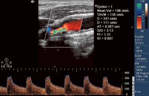
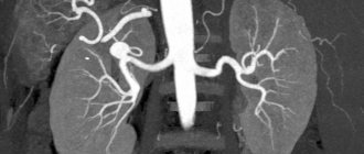
Dopplerography picture of cerebral vessels using triplex mode
As we see, ultrasound and Doppler Dopplerography of the main arteries (vessels) of the head are carried out primarily to detect anomalies in the circulatory system, measure its pathogenetics and hemodynamics. This helps in monitoring the effectiveness of treatment, angiography and the involvement of a vascular surgeon when considering surgical treatment.
Triplex scanning MAG in Moscow
A person who is scheduled for diagnosis seeks to find a clinic in close proximity to his home or place of work. If triplex scanning of the main arteries of the head is required, then the Nagornaya metro station is one of the most accessible and convenient for travel.
Near the Nagornaya metro station there is the Yusupov Hospital, which has several clinics, including a neurology clinic. Experienced doctors treat patients with various diseases of the nervous system. To diagnose violations, specialists use modern high-precision equipment. In the neurology clinic, the main arteries of the head are examined using ultrasound machines.
Patients of the clinic can undergo various tests at affordable prices. For the convenience of clients, various options for making an appointment are provided; in addition, you can visit a specialist at a convenient time without a long wait in line. In order to make an appointment with a neurologist and undergo an ultrasound scan of the main arteries of the head, please contact the clinic staff by phone.

Duplex MAG: service price
People diagnosed with stenosing atherosclerosis of the main arteries of the head and other vascular diseases strive to find a clinic that will provide medical care at an affordable price. At the Neurology Clinic of the Yusupov Hospital, the price for diagnostic and therapeutic methods is optimal. In addition, the hospital provides rehabilitation programs for patients who have suffered strokes, suffer from dementia and other diseases.
A beneficial option for the rehabilitation of patients after Alzheimer's disease, Parkinson's disease, stroke, multiple sclerosis are comprehensive programs, including examination by experienced doctors, development of a rehabilitation program and other activities. If necessary, duplex scanning of the main arteries of the head and neck and other studies are performed.
When contacting the Yusupov Hospital, you can be confident in the accuracy of the diagnosis, the quality of treatment, and the friendly attitude of the staff. A personal quality manager helps patients resolve any issues. For the convenience of patients and their relatives, the clinic has created the necessary conditions: the ability to receive medical care 24 hours a day, healthy and tasty food, comfortable rooms with satellite TV and wireless Internet. If you need a triplex scan of the main arteries of the head or a consultation with a neurologist, please contact the clinic staff to make an appointment.
Duplex scanning MAG in Moscow
At the neurology clinic of the Yusupov Hospital you can undergo a MAG duplex scan. Diagnostics are carried out using highly informative ultrasound equipment that has sensitive sensors, due to which a specialist can objectively assess the parameters being studied and make a diagnosis based on the information received.
The experience and professionalism of the medical staff of the Yusupov Hospital allows us to successfully diagnose even minor problems and quickly choose tactics for eliminating them, using innovative techniques during therapy and recovery. Doctors at the Neurology Clinic, together with other specialists, provide detailed consultations to patients and their families in an accessible manner. Thus, the patient has information about the state of health, the course of the disease, and risk factors.
MAG duplex scanning in the neurology clinic is performed in accordance with current standards and legislation. The Yusupov Hospital offers affordable prices in the segment of highly informative ultrasound examinations. You can find out the details of the procedure, sign up for a consultation and diagnostics through a special form on the website, after which a clinic employee will contact you to clarify the details or by phone.
Make an appointment