The cervical spine consists of seven vertebrae, which are responsible for performing the most important motor and conductive functions. Displacement or pinching of these vertebrae can cause serious illness. To identify these diseases and make a diagnosis, it is necessary to do an MRI of the cervical spine.
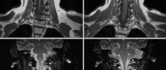
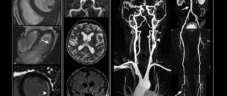
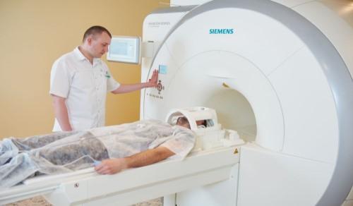
MRI of the cervical spine (tomography of the neck) is a modern study that allows the doctor to identify not only the pathology of bone tissue, damage to the spinal column and spinal cord, but also to diagnose disorders in the soft tissues surrounding the intervertebral discs. Neck tomography has important advantages over alternative diagnostic methods. For example, CT and X-rays of the cervical spine are used to study bone structures, ultrasound is used to scan soft tissue, and Doppler sonography is used to study blood flow. MRI of the cervical spine allows for examination of soft tissues, the bone skeleton, and the vascular system. The advantages of tomography include non-invasiveness and the absence of radiation exposure. MRI of the neck is safe for human health and can be done several times in a short time (for example, to track the dynamics of treatment). At the Kutuzovsky Children's Center you can do an MRI of the neck using a Philips Achieva tomograph with a power of 1.5 Tesla. Such a tomograph is considered optimal, as it provides expert quality with minimal distortion of information. As a result, a clear three-dimensional image (if necessary, layer-by-layer) of the area under study is formed on the monitor. MRI of the neck is a series of layer-by-layer images, with the help of which doctors detect even initial changes in this area.
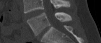
The Philips Achieva tomograph allows you to obtain thin enough layers that eliminate projection overlaps caused by other structures that are observed during X-rays of the spine. Therefore, magnetic resonance imaging of the cervical spine is a highly informative diagnostic tool. Thanks to the additional functions and capabilities of the tomograph, doctors can set various parameters, conducting a more detailed examination of the pathological area. If it is necessary to diagnose oncological diseases (these include bone and soft tissue tumors, metastases), an MRI of the cervical spine is done with the introduction of a contrast agent. MRI of the cervical spine has no radiation effect on the body and is a completely safe diagnostic method for human health. If necessary, MRI of the neck can be performed repeatedly and at any frequency.
So, MRI of the cervical spine is a modern diagnostic method that helps identify various diseases and injuries of the spinal cord, soft tissues and vertebrae located at the level of the craniovertebral junction and to the lower part of the neck. This diagnostic method allows you to recognize various neoplasms, identify circulatory disorders, pathologies and anomalies in the intervertebral discs, etc.
What does an MRI of the neck show?
During a magnetic resonance imaging study, layer-by-layer images are created that allow one to determine all the anatomical features of the patient’s neck. A tomography examination helps to detect even minor abnormalities in areas that are poorly visualized with other examination methods.
What diseases can be diagnosed:
- vasoconstriction;
- damage to nerve roots;
- spinal deformity;
- vertebral damage;
- change in shape, thinning of the vertebrae or intervertebral discs;
- vertebral displacement;
- protrusion;
- intervertebral hernia;
- osteophytes;
- neoplasms.
The examination helps to identify pathological changes in bone tissue, cartilage and soft tissue, nerve endings and blood vessels. MRI with contrast, which is slightly more expensive, is required to detect tumors and areas of myelin loss, including in the early stages.
Magnetic resonance imaging of the cervical spine
If a patient complains of dizziness or pain, numbness of the back, then using an MRI of the cervical spine, the correct diagnosis can be quickly established.
No special training is required. General recommendations include the absence of metal jewelry and clothing items, and decorative cosmetics with metal particles. The last meal is taken 2 hours before the MRI with contrast.
During the entire procedure, the patient lies on a comfortable couch, which is located in the internal cavity of the tomograph tube. During the examination, you can ask the specialist questions, but you cannot move. The duration of a simple study is 20-30 minutes, and the contrast method lasts 45-90 minutes.
To carry out the procedure, the patient's body is located inside the equipment, where a strong field is created. The resulting images are stored in computer memory. A contrast agent may sometimes be used to obtain a clearer image. The specialist injects the drug into a vein before the study, after which it is removed without harm to the body.
Advantages of MRI diagnostics at the Central Clinical Hospital of the Russian Academy of Sciences
- Safe, no radiation exposure, multiple repeat examinations possible
- High diagnostic efficiency
- Experienced doctors guarantee the accuracy of the diagnosis and the quality of the completed protocol
Magnetic resonance imaging is used to diagnose:
- neoplasms;
- osteochondrosis;
- arthrosis;
- listhesis;
- arthritis;
- neuritis;
- deformations of the spinal column, individual vertebrae and intervertebral discs;
- vasospasms;
- blood clots
Possible symptoms and indications for MRI
A doctor may order a tomography to examine the following diseases:
- spinal cord compression;
- volumetric neoplasms or metastases in the spinal cord, spine;
- anomalies of congenital dysplasia and vascular diseases;
- osteochondrosis, osteomelitis, tuberculous spondylitis, etc.;
- with abnormal development of the spinal column;
- for fractures, displacement or dislocation of the vertebrae;
- in metabolic-dystrophic conditions (rickets, osteoporosis);
- for disc pathologies (protrusions and hernias);
- in preparation for surgery and further monitoring of the patient’s recovery.
The advantage of performing an MRI of the cervical spine is that instrumental intervention is not required for a correct diagnosis. The examination is painless and safe (no stress). It is also highly efficient; many photographs are formed in three perpendicular planes.
Indications for the study
Unfortunately, diseases of the cervical spine often give vague symptoms, which makes it difficult for the doctor to make the correct diagnosis and prescribe the correct course of treatment. Magnetic resonance imaging is an effective way to differentiate pathologies of the spine and surrounding tissues.
The most common indications for magnetic resonance imaging:
- frequent pain in the neck;
- muscle spasms;
- numbness of the face or back of the head.
A neck examination should be performed if examination of the brain and blood vessels does not produce results, and the patient has:
- weakness;
- headache;
- nausea;
- tremor;
- feeling of “current” in the body;
- numbness of the limbs, certain areas of the body;
- pressure surges;
- irregular confusion or speech;
- periodic coordination disorders.
These symptoms may indicate pathological changes in the cervical spine. Research is required even if anxiety symptoms tend to spontaneously appear and disappear spontaneously.
Regular examination is required for previously diagnosed diseases in order to monitor the dynamics of the patient’s condition. Observations require pathology of an autoimmune, degenerative-dystrophic, deformational, inflammatory nature. An annual examination allows you to identify new pathological changes, adjust the course of treatment, prevent the progression of pathology and the development of secondary diseases. Urgent investigation is required for injuries. MRI is often prescribed as a preoperative examination.
How much does an MRI of the spine cost in Moscow?
Prices for magnetic resonance imaging examinations at the multidisciplinary CELT clinic are among the most affordable in Moscow. The cost of MRI of the lumbosacral region and other parts of the spine is approximately the same, since the time and labor intensity of the studies are practically the same. You can sign up for an MRI examination of the spine at CELT by receiving a referral from your attending physician with a preliminary diagnosis and indicating the examination area, by telephone or from the administrator of the radiology department directly at the clinic.
Contraindications:
- excess weight (if it is not possible to place the patient inside the tomograph);
- the presence of non-removable metal objects in the body (braces, pacemaker, etc.), tattoos made with metal-containing ink;
- I trimester of pregnancy, lactation.
If you have kidney disease, a study with a contrast agent may be contraindicated, so you should inform your doctor about the presence of such diseases in advance. The procedure itself lasts 30–40 minutes. The patient removes all metal items and lies down on a sliding table, which is placed in the tomograph. It is important to lie still during the examination, otherwise the pictures may not be clear.
Get specialist advice or make an appointment
+7 (499) 400-47-33
Restrictions to the procedure
An MRI scan part may be rejected for the following reasons:
- presence of metal in the body;
- pregnancy in the early stages;
- gadolinium allergy;
- failure of important organs.
Screening is not carried out when the patient arrives at the research center unconscious and in severe pain. The procedure is postponed until a later date, when the person’s well-being is normalized.
What pathologies can be identified
The main purpose of the examination is to identify pathologies of the structures of the spine and spinal cord. Using screening techniques, the following diseases or conditions are detected:
- disc protrusion;
- degenerative deviations of cervical segments;
- sclerotic formations (cholesterol plaques);
- pinched nerve endings;
- osteochondrosis, osteoporosis, spondylosis, spondyloarthrosis;
- cause and location of Frank's nerve pinching;
- dynamics of inflammatory processes in soft tissues and roots;
- spinal canal stenosis;
- spinal cord meningitis;
- metastases of malignant tumors from other organs;
- malformations of blood vessels in the spinal canal, vertebral bodies, spinal cord (hemangiomas, malformations);
- localization and size of the infectious focus;
- consequences of injuries;
- anatomical features of the spine.
If the cervical vertebrae are regularly displaced, it is necessary to undergo diagnostics at the first discomfort.
The cervical spine should be examined if the following symptoms occur:
- noise and ringing in the ears;
- surges in blood or intracranial pressure;
- occasional nausea;
- numbness of the upper limbs, neck;
- enlarged lymph nodes;
- goosebumps along the spine;
- painful shooting in the upper limbs, jaws;
- repeated fainting.
MRI of the sacroiliac joints and coccygeal area of the spine (MRI of the coccyx)
Today, MRI of the sacroiliac joints and coccyx is the only research method that allows diagnosing diseases specific to these particular parts of the spine with a high level of accuracy. The popularity of this study is justified by its safety and non-invasiveness.
Indications for the use of MRI of the coccyx:
- pain syndrome of unknown etiology,
- traumatic damage to bone structures.
Indications for examination of the sacroiliac joints:
- traumatic changes
- inflammatory changes in this area (characteristic of Ankylosing spondylitis, etc.),
- tumors.
If magnetic resonance imaging of the spine (or any other organ) has been performed previously, doctors recommend bringing these results with you. Then specialists will be able to guarantee a more reliable result, as well as tracking the dynamics of the process (disease).
Magnetic resonance imaging of the spine does not require special preliminary preparation.
About the presence of contraindications, and they are almost the same for all types of MRI studies, be sure to consult a specialist!
Among the main contraindications:
- the presence of a pacemaker, various foreign metal bodies, implants, metal pins, plates and structures for osteosynthesis;
- claustrophobia, inadequate mental behavior;
- severe chronic diseases in the stage of severe decompensation.
Currently, magnetic resonance imaging (MRI) has taken first place in the diagnosis of most diseases of the spinal cord and spine, pushing into the background methods such as myelography and computed tomography (CT) of the spine.
MRI of the cervical spine, what the procedure will show
Outpatient medicine uses two types of magnetic resonance imaging scanners for diagnostics: open and closed. The 1.5 Tesla Philips Intera tomograph used in the DiMagnit MC is of the closed type. The device is optimal for most MRI screenings.
The duration of the procedure depends on the number of tasks set by the doctors. On average, 15 minutes is enough to study the cervical spine. A more extensive examination with contrast injection may take up to 30 minutes.
Classic version (native MRI)
After making sure that the subject has no metal objects, the center’s specialists proceed directly to MRI scanning. The event is carried out in stages:
- the person lies on the horizontal surface of the tomograph on his back;
- the surface is pulled into the medical equipment;
- the diagnostician goes into the next room to control the MRI system via a computer;
- During the diagnostic procedure, the patient hears sounds produced by the tomograph (pops, clicks);
- upon completion of the manipulations, the patient receives a disc with the results in his hands (for an additional fee, it is possible to copy the recording onto film or a flash drive).
During the examination, there is an audio connection between the doctor and the patient.
To obtain high-quality images, you must remain completely still while the magnetic resonance equipment is operating.
Option with contrast agent
MRI diagnostics of the cervical region with simultaneous administration of a contrast agent is often prescribed if the presence of a neoplasm or demyelinating disease is suspected. The contrast agent has the ability to accumulate in the area of the tumor or area with an active blood supply.
Peculiarities:
- if screening is recommended to be carried out on an empty stomach, it is necessary to provide the body with a supply of fluid several hours before the start of the event;
- Preliminary measures are taken to prevent an allergic reaction to contrast;
- the contrast agent is given intravenously (injection).
The cost of the contrast agent is calculated based on the weight of the subject.
Indications for MRI of the lumbar and other parts of the spine
Patients who complain of the following are examined:
- frequent headaches of unknown origin;
- recurring dizziness of unknown origin;
- visual impairment;
- hearing loss, tinnitus;
- sensory disturbances in the hands;
- impaired mobility of the spinal column;
- spinal injuries;
- violations of motor functions of the limbs;
- dysfunction of the pelvic and abdominal organs;
- spasms of the muscles of the back and buttocks;
- nagging pain in the lower back and sacrum, etc.
Main services of Dr. Zavalishin’s clinic:
- consultation with a neurosurgeon
- treatment of spinal hernia
- brain surgery
- spine surgery
Why choose us?
The equipment of the MRI Center in Moscow allows you to work with a section 0.05 mm thick. The diagnostic center is equipped with the latest tomographs, allowing scanning 4-16 times faster, with the ability to 4D visualization, and correction of data acquisition bias. The price of tomography depends on the area of the area and the need to use a contrast agent. We have a flexible system of discounts, including for preferential categories of citizens. The price list for the services of branches in Moscow includes prices for MRI, options for obtaining results, and consultations.
To make an appointment at the clinic, leave your contact information or call back at one of the telephone numbers indicated next to the branch address. The final cost of diagnostics includes additional services that are provided as needed.
Decoding images
After completing the manipulations, diagnostic doctors begin to analyze the results. At DiMagnit MC the patient receives a disk with images and a transcript.
If any deviations from the norm are detected, the specialist doctor will convey the information to the client in an accessible form and give recommendations on further actions.
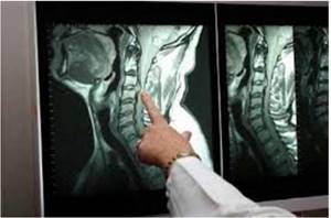
An MRI specialist identifies pathology in the area of the cervical vertebrae