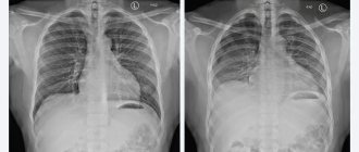
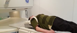
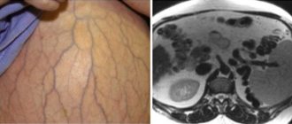
A CT scan of the pancreas is a non-invasive way to examine this abdominal organ. It is most often carried out in medical centers during computed tomography of the abdominal cavity and retroperitoneal space. The procedure is performed using a multislice computed tomograph, whose slicing capacity can vary from 12 to 640 slices per scan revolution. The quality and cost of the examination will largely depend on the slicing capabilities of the tomograph. Yes, for adult patients, this diagnosis does not require a doctor’s referral. For young children, this scan is carried out only on the recommendation of a doctor, if there are significant medical indications. This is due to the fact that spiral computed tomography is associated with radiation exposure to the body. On average, the training dose is 3-5 mSv per diagnostic session.
Free diagnostic consultation
If in doubt, sign up for a free consultation. Or consult by phone
+7
All about diagnostics
What is a CT scan of the pancreas?
The computed tomography method is based on the specific property of X-ray radiation to be absorbed by body tissues. During scanning, a beam of X-rays is applied to the area under study. These rays pass through the body. High-precision computer equipment processes data on the residual radiation force, and based on it, three-dimensional images are created.
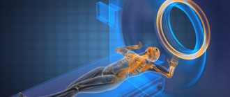
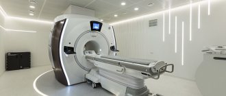
The device itself can be a computed tomograph and a multislice computed tomograph. A CT scanner has detectors and emitters located around the perimeter of the Gantry ring. At the entrance of the examination, this ring rotates around the patient's body, and the rays pass through the tissue from different angles. In a multislice computed tomograph, the number of detectors and emitters increases, and not only the ring itself moves, but also the tomographic table. Thus, MSCT is capable of making a larger number of slices per scanning revolution.
Contraindications to the procedure
Diagnosis of pancreatic pathologies using a computed tomograph is contraindicated:
- women carrying a child. X-rays have a teratogenic effect.
- patients of the children's department (up to 14 years old);
- for psychopathological disorders;
- people with kidney pathologies in the decompensated stage;
- patients with oncohematological diagnosis – myeloma.
Relative (relative) contraindications are the inability to immobility (pain, increased excitability), the constant need to monitor vital signs (blood pressure, heart rate, breathing).
The examination is not possible if the patient’s weight category exceeds 130 kg. The tomography machine is not designed for heavier weights. If the patient underwent an X-ray of the stomach with a barium suspension, the time interval between examinations should be 10 days.
Indications
The following symptoms may make patients think about the need to do a CT scan of the pancreas:
- pain in the upper abdomen (the area below the breastbone);
- weight loss;
- jaundice;
- vomit;
- chronic pancreatitis;
- obesity;
- development of diabetes;
- bulky, unusually smelly, light-colored feces;
- disturbances in the intestinal tract, digestive problems;
- suspicion of the presence of tumor formations;
- control over the condition after operations and monitoring the progress of treatment.
Features of surgical treatment:
- Carrying out preoperative biliary decompression. The use of a plastic or metal stent minimizes the risks of complications and mortality after surgery.
- Portal and mesenteric vein infiltration no longer affects resectability.
- The choice of surgical method is influenced by the location and spread of the tumor.
For conditionally resectable cancer, neoadjuvant therapy is prescribed to downstage the disease and to allow tumor removal. The assessment of possibilities depends on the experience of surgeons, radiologists and gastroenterologists.
The prognosis for operable pancreatic tumors is extremely unfavorable. Complete cure is rare. In advanced cases, most patients die within 5 years after surgery. This is why timely consultation with a doctor is so important.
Prices for treatment of pancreatic cancer
- Consultation with an oncologist— RUB 5,100.
- Consultation with a chemotherapist— RUB 6,900.
- Gastropancreaticoduodenal resection - RUB 318,100.
- Distal resection of the pancreas - 168,800 rubles.
- Pancreaticoenteroanastomosis - RUB 114,900.
- Total pancreatectomy - RUB 284,600.
- Carrying out intrapleural chemotherapy (infusion, without the cost of drugs) - 19,100 rubles.
- Immunotherapy (without the cost of medications) - RUB 15,000.
Make an appointment with an oncologist
Book a consultation 24 hours a day
+7+7+78
CT pancreas with contrast
If the doctor suspects the presence of a tumor in the pancreas, he will order a computed tomography scan of the pancreas with contrast. Sometimes a radiologist may suggest a contrast enhancement procedure. This happens in situations where, during a native examination, signs of a tumor are revealed, and in order to carry out a differential diagnosis, it is necessary to administer contrast.
The contrast agent for computed tomography is an iodine-based substance. It is injected intravenously into the patient’s elbow area; the composition spreads throughout the body through the circulatory system and stains the tissues, increasing their tissue contrast in the photographs. In this way, visualization is improved, and the doctor can see smaller tumors, for example, metastases, determine the clear location of the voluminous neoplasm and, based on the type of accumulation and release of the contrast agent, judge whether the tumor is benign or malignant.
Permissible frequency of CT scanning
A CT scan poses a certain risk of radiation – ionizing radiation. In modern devices, the radiation exposure received by the patient slightly exceeds the single permissible dose. The norm is 12 μR/h (micro-Rengen/hour), which is equivalent to 0.12 μS/h (microsievert/h).
For CT scans of the gastrointestinal tract, the dose of X-rays ranges from 5 to 14 microroentgen per hour. Frequent administration of a contrast agent can cause excess iodine in the body. An excess of a microelement leads to the development of intoxication, disruption of cardiac activity and thyroid function.
Based on this, the recommended frequency of tomographic procedures is once a year. For severe pathologies that require regular monitoring, three examinations per year are allowed. The interval between examinations should not be shorter than six weeks.
How is tomography performed?
Before starting the examination, the patient must take a lying position inside the tomograph. Then the subject is injected with a contrast agent if the scanning is carried out using a contrast protocol. In some cases, during the contrast procedure, you may experience a feeling of nausea, vomiting, malaise or weakness. You should immediately report these symptoms to your doctor, who will promptly take measures to stop the allergic reaction to the contrast agent.
The scanning procedure itself takes an average of 3-7 minutes, during which the subject must lie as still as possible and follow the operator’s commands to hold his breath. The radiologist will need another 30-40 minutes to decipher the diagnostic data. The result of the CT examination will be a written conclusion from the doctor and recording of the tomograms on electronic or film media.
Contraindications and restrictions
Native CT of the pancreas, performed without contrast, is considered safe for most patients, but it is not prescribed to pregnant women because of the harmful radiation exposure to the fetus.
Children under 3 years of age should be approached with caution when performing a CT scan. Radiation of a child at such an early age is undesirable, so parents are better off choosing a safe MRI of the pancreas or ultrasound. MSCT for children under 3 years of age should be done only in extreme cases for serious medical reasons.
The administration of contrast is contraindicated in patients with an allergy to iodine, as well as in patients with renal failure. It is possible to determine whether nephropatients can undergo a CT scan of the pancreas with contrast by taking a blood test for creatinine.
CT in a comprehensive examination of the gland
The pancreas (P) is a unique organ that simultaneously performs two functions: exocrine (exocrine) and intrasecretory (endocrine). To diagnose pancreatic diseases, laboratory and instrumental methods are used. The first include:
- CCA (general clinical analysis) of blood;
- biochemical blood test;
- blood test for alpha-amylase;
- OAM (general urinalysis);
- coprogram (stool analysis).
Hardware techniques include:
- Ultrasound;
- retrograde cholangiopancreatography;
- magnetic resonance imaging (MRI);
- computed tomography.
For differentiated diagnosis of oncological pathologies, a biopsy is performed - tissue sampling for histological examination. The prerogative aspects of a computer examination are:
- The ability to detect neoplasms and identify pathological lesions of internal organs at the initial stage of development. This significantly increases the patient's chances of a full recovery.
- Information content. The use of a contrast agent during the examination allows us to assess the problem as objectively as possible.
- Non-invasive. The examination is carried out without damaging external tissues.
- Short procedure interval. A CT scan takes a quarter of an hour, with contrast – half an hour.
- Painless. During the procedure, the patient does not experience discomfort.
The disadvantages of CT include:
- X-ray radiation. Despite the minimal radiation exposure, the examination is not completely harmless.
- Lower information content compared to MRI in determining cystadenocarcinoma and adenocarcinoma.
Reference! Cystadenocarcinoma of the pancreas is a benign cystic neoplasm that has degenerated into cancer; adenocarcinoma of the pancreas is a malignant tumor of glandular cells and ducts.
The question of which procedure (CT or MRI) is better is decided by the attending physician. The diagnostic method is selected depending on the expected pathology and individual characteristics of the patient.
Preparation
For a high-quality diagnosis, the examinee must provide the doctor with all available medical information related to the pathology:
- excerpts from case histories;
- ultrasound, x-ray, tests for tumor markers;
- direction for scanning.
If a multislice CT scan is performed with contrast, patients with kidney disease will need to have a blood test for creatinine. It will help specialists decide whether to perform a CT scan with contrast.
An allergy test is a very important aspect of preparation for contrast MSCT of the pancreas. It is carried out to avoid anaphylactic reactions to iodine-containing contrast material directly in the medical clinic before the examination.
You need to prepare for a CT scan of any organs of the abdominal cavity and retroperitoneal space. Three to two days before the tomography, you should start following a diet that helps reduce gas formation. The patient should give up fresh vegetables and fruits, cabbage, legumes, baked goods, baked goods and other high-fiber foods. The evening before the study, no later than 21:00, a light dinner is allowed, after which food intake should be stopped. After dinner, you can take two tablets of espumisan or activated carbon to reduce gas formation. Tomography of the pancreas is performed on an empty stomach. The minimum fasting period should be at least 6 hours. On the day of the study, 30-40 minutes before the start of MSCT, you need to take two No-Spa tablets.
The issue of preparing for a CT scan of the pancreas should be approached responsibly. If diet requirements are not followed, CT scan results may be incorrect.
Decoding
A radiologist interprets the computed tomography data. He evaluates the obtained tomograms to ensure that the organ’s indicators correspond to the norm. If the diagnostician finds anatomical abnormalities, he describes them in detail in terms of structure, size, extent and degree of invasion. If a contrast examination was performed, the doctor additionally notes the behavior of the tissues in terms of accumulation and release of the contrast agent. He draws up all his findings in the form of a written report and accompanies them with CT images recorded on electronic media or printed on film. This entire package of documents is transferred to the patient with recommendations on further steps.
Study Details
- Doctor's referral: Required for CT scans for children
- Preparation: Yes in case of CT angiography, CT OMT, CT OBP
- Application of contrast: As prescribed by a doctor and with CT scan of blood vessels
- Time: 5-7 minutes
- Contraindications and restrictions: Pregnancy, allergy to iodine on CT with contrast, renal failure
- Results delivery time: 30-40 minutes
| List of studies | Price | Stock |
| CT scan of the urinary system (kidneys, adrenal glands, bladder, urinary tract) | from 2500 rub. | At night |
| CT scan of the abdominal cavity (part of the esophagus, stomach, liver, gallbladder, pancreas, spleen, small intestine) | from 2500 rub. | At night |
| CT scan of the retroperitoneum (kidneys and adrenal glands, ureters, soft tissues and lymph nodes) | from 2500 rub. | At night |
| CT virtual colonoscopy (large intestine) | from 4100 rub. | At night |
Diagnostics
- Ultrasound examination (ultrasound) of the abdominal organs makes it possible to detect the presence of a tumor, determine its size and location in the structure of the organ.
- Transesophageal ultrasound. A thin flexible endoscope equipped with an ultrasound sensor is inserted through the mouth into the esophageal cavity. This method is more informative than classical (transabdominal) ultrasound for the purpose of assessing the condition of the initial parts of the digestive tract (stomach, pancreas, duodenum, etc.).
- Endoscopic retrograde cholangiopancreatography (ERCP). A diagnostic method that, unlike transesophageal ultrasound, has the advantage of advanced diagnostics. During the examination of the duodenum, a radiopaque substance is injected into the lumen of the common bile and main pancreatic duct (which open into its cavity). From the image transmitted to the monitor, it is possible to determine the general patency and condition of the biliary tract (biliary system), areas of narrowing, and obstruction.
- Diagnostic laparoscopy. An endoscope equipped with an optical device is inserted directly into the abdominal cavity. This imaging method also allows you to record a tumor formation, determine its size, location and extent of the process.
- Tumor markers. An increase in the level of CA19-9 and CEA in the blood of the subject is also one of the diagnostic criteria for the development of a malignant process in the body.
- In order to clarify the volume, localization of tumor lesions, as well as identify metastatic lesions, diagnostics are carried out using CT, MRI, PET-CT.
Diagnostic methods 3 and 4 also allow you to take a biopsy (tissue fragment) of the pathological formation. Histological examination is the most reliable diagnostic option, allowing one to determine the presence/absence of cancer cells and establish the type of tumor, which is extremely important when selecting therapy, as well as for prognostic reasons.


