Indications for CT scan of the lungs
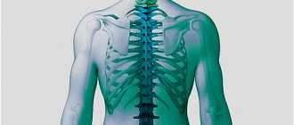
Computed tomography is a universal diagnostic method that allows one to assess the condition of the lungs, bronchi, trachea, lymph nodes and even chest bones in one procedure. CT allows one to detect pathologies in all structures of the lungs, both diffuse and focal. The specialist sees in the resulting images the location, nature, prevalence of the pathology, and the degree of damage to organs. Multislice computed tomography of the lungs, performed on a modern Toshiba Aquilion RXL tomograph at the Miracle Doctor clinic in Moscow, allows you to diagnose acquired and congenital lung anomalies:
- malignant and benign formations,
- pulmonary tuberculosis,
- cystic fibrosis,
- inflammation of the pleura,
- pneumonia of various origins, including the consequences of a previous coronavirus infection,
- structural changes in the bronchi, etc.
The test helps diagnose the cause of cough, shortness of breath, chest pain, prolonged fever and sputum production, and other symptoms.
A computed tomography scan of the lungs is appropriate not only for the indicated symptoms and suspicions of the listed diseases. The study is carried out for preventive purposes for the following categories of people:
- persons over 55 years of age;
- smokers;
- workers in hazardous industries (metallurgists, miners, etc.);
- people with chronic obstructive airway diseases.
Advantages of lung CT over other diagnostic methods
- Painless and safe. The study is carried out on a modern device that allows you to reduce the radiation dose without losing the quality of the images to the minimum required. During the procedure, the patient does not experience any discomfort.
- Highly informative. The sensitivity of the lung tomography method tends to 100%, since the tomograph’s ability to obtain 32-slice images allows you to visualize minimal deviations in the structure of tissues, identifying signs of pathology at the earliest stages.
- Speed and accessibility of results. The procedure is carried out approximately within half an hour, including the period of patient preparation. The specialist deciphers the results and writes a conclusion within an hour. The results of the study are available for viewing on a computer monitor, recording on DVD, and upon request, sent by e-mail.
How is a CT scan of the lungs performed during the COVID-19 pandemic?
During the coronavirus epidemic, experts recommend limiting visits to public places, including clinics. However, there are situations when a person’s life and health depend on the speed of the examination. In this case, going to the doctor cannot be postponed. This is especially true for diagnosis of progressive symptoms of COVID-19.
Our medical center is fully prepared to safely examine patients. All diagnostic rooms in the clinic are regularly sanitized. Our employees fully comply with the instructions of epidemiological services. If necessary, all patients who need examination will be able to undergo a CT scan of the lungs and quickly obtain an accurate result.
The diagnostic procedure is as comfortable as possible for the person. It does not cause pain, does not require special preparation and does not involve subsequent rehabilitation.
The procedure will last 10-15 minutes. The time the patient spends in a public place is minimized.
If you follow all the instructions, regularly use personal protective equipment and maintain social distance, you can reduce the risk of infection several times.
Contraindications to computed tomography
CT scan of the lungs is a simple and safe procedure at a reasonable price, while being very effective and informative. However, there are a number of contraindications to its implementation, including:
- During pregnancy and breastfeeding;
- obesity (patient's body weight exceeds 200 kg);
- the patient has symptoms of ARVI or bronchitis;
- the patient is in serious condition.
In addition, CT scans with contrast should be avoided in patients diagnosed with renal failure, severe forms of diabetes mellitus, or individual intolerance to the components of the contrast agent.
Moreover, the list of contraindications for performing a CT scan of the lungs with contrast is somewhat broader than with the usual method of performing this study:
- age under 14 and over 70 years;
- bronchial asthma;
- tendency to allergies;
- thyroid cancer and hyperthyroidism;
- long-term therapy with NSAIDs, diuretics or beta-blockers.
Computed tomography of the chest
Computed tomography of the chest (another name is MSCT examination of the chest) is today the best method for visualizing the state of the respiratory organs (lungs, pleura, trachea, bronchi) and mediastinal organs (pericardium with the heart located in it, blood vessels, including aorta, esophagus, lymph nodes located in this area).
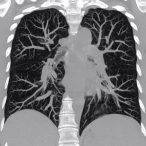
Computed tomography of the chest organs allows us to identify such lung diseases as alveolitis, sarcoidosis, pulmonary emphysema, pneumonia, tuberculosis and some others, dilation of the bronchi and alveoli, cicatricial narrowing of the bronchi, cysts, foreign bodies in the bronchi, pulmonary circulation disorders, tumors of the lung tissue, pleura , mediastinum.
Most often, computed tomography is performed as a clarifying study based on the results of chest x-ray. No special preparation is required for the study. If necessary, the study can be performed with a bolus injection of contrast agent.
The examination time is up to 20-30 minutes.
Preparation and performance of CT scans of the lungs and bronchi
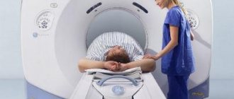
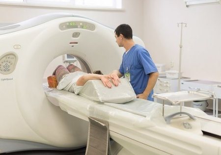
The study does not require any special preliminary preparation. It is better to come in comfortable, wrinkle-resistant clothing, as the procedure is performed in a lying position. Before a CT scan, you will need to remove metal objects, including jewelry, piercings, glasses, dentures, hearing aids, and hair clips, which can affect the quality of the image. Women will be asked to remove bras containing metal wires or clasps.
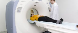
The patient is placed on a special table in a supine position. Straps and pillows may be used to help the patient maintain proper positioning and remain still during a CT scan. Random movements can blur images and degrade the quality of the examination.
Scanning occurs in several stages:
- the table with the patient quickly passes through the scanner, determining the correct starting position for scanning using projected light lines;
- the actual scanning occurs as the table moves slowly through the scanner ring. During the procedure, you may hear a slight buzzing and clicking sound - this is the internal parts of the CT scanner working in the process of taking images;
- Once the scan is complete, the patient may be asked to wait while the radiologist checks that the images are of sufficient quality for accurate interpretation.
The actual CT scan takes less than 30 seconds, and the entire process, including preparation, usually takes about 30 minutes.
Although the scan is painless, the patient may experience some discomfort from having to remain still and alone while the radiologist in the next room makes the necessary adjustments.
Interpretation of results - doctor's opinion
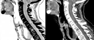
Within an hour after the examination, the radiologist analyzes the results obtained and transfers the conclusion and recording on the selected medium to the patient and sends it by e-mail. When interpreting the results of a CT scan of the lungs and bronchi, indicators of location, size limits of the structures under study, and the presence of anomalies are taken into account. Normal indicators imply the absence of pathological changes, the preservation of the anatomical structure, and the absence of free fluid in the pleural cavity. There are also no changes in the chest wall, thrombotic masses in the lumen of the aorta, pulmonary arteries, or trunks. Pathologies on CT scans of the lungs and bronchi have different classifications and features. The result directly depends on the nature of the disease; not only pathological changes are taken into account, but also accompanying symptoms.
How does CT work?
In many ways, a CT scan works like other x-ray tests. Different parts of the body absorb x-rays in different amounts. This property allows organs to be distinguished from each other on an X-ray or computer image.
In a typical x-ray, a small amount of radiation is directed through the part of the body being examined. A special electronic image recording plate captures the image. On an x-ray the bones appear white. Soft tissues such as the heart or liver are displayed in shades of gray. The air looks black.
With a CT scan, electronic X-ray detectors rotate around the patient. They measure the amount of radiation absorbed. The examination table moves during scanning so that the X-ray beam follows a spiral path. A special computer program processes the data to create layer-by-layer cross-sectional images of the human body. Images are displayed on the monitor. Obtaining a CT image is sometimes compared to slicing a loaf of bread into thin slices. When image fragments are reassembled using computer software, the result is a highly detailed multi-dimensional visualization of the organ.
Modern CT scanners can scan large areas of the body in just a few minutes. This speed is especially important for those who find it difficult to remain motionless even for the short time required to obtain images—the elderly and the seriously ill.
Contraindications
- Pregnancy, because the study involves x-rays.
- When using contrast, contraindications are: severe renal and heart failure, thyrotoxicosis, individual intolerance to the contrast agent (iodine-containing contrast agent).
If you are looking for where to get a CT scan of the lungs or chest organs right now, contact Family Doctor JSC. CT examinations are carried out in Polyclinics No. 5 (Barrikadnaya metro station) and No. 12 (Teply Stan metro station) - during clinic hours, as well as in the Hospital Center - around the clock. Prices for all types of research are listed below. You can undergo a chest CT scan without a doctor's referral by simply signing up for the test at any of the specified clinics.
Multislice computed tomography (MSCT)
MSCT is the most modern method for visual diagnosis of structural changes in internal tissues and organs and functional systems of the body.
Using this method, the following are examined: the brain, ENT organs, chest organs (heart, coronary arteries, trachea, esophagus, lungs), abdominal organs (intestines, gall bladder, stomach, pancreas, kidneys, liver), small organs pelvis (ovaries, uterus, fallopian tubes, vagina, prostate gland, bladder), spine, joints.
Main tasks of MSCT:
- Identification of problem areas in ENT organs;
- Assessment of lung condition;
- Assess the condition of the coronary arteries and prevent coronary heart disease;
- Visualization of blood clots during thromboembolism;
- Identification of the extent of the oncological process;
- Difference between malignant and benign neoplasms.
Indications for MSCT:
- Malignant formations in the liver, pancreas, kidneys, bladder;
- Degenerative changes in the spine, with intervertebral hernias;
- Multiple injuries (especially in emergency situations);
- Pulmonary tuberculosis (to identify foci);
- Suspicion of extraorgan tumors of the abdominal cavity and retroperitoneal space;
- Diseases accompanied by severe pain.
Contraindications:
- Pregnancy;
- Claustrophobia (MSCT may be performed using sedatives);
- Lactation;
- Allergy to contrast (if the procedure is performed with contrast);
- Obesity (the tomograph is designed for weights up to 150 kg).
How to prepare for MSCT
Preparation for the procedure depends on the direction of the study. It is recommended to conduct the study on an empty stomach; it is necessary to stop consuming food and water at least 4 hours in advance. If you need to take medicine, you can take it with a small amount of water.
Special preparation before the session is needed when examining the abdominal cavity, pelvic organs and kidneys. It consists of carrying out activities aimed at cleansing the intestines: this is a diet for several days before CT, taking enzyme preparations and activated charcoal, and a cleansing enema.
How is MSCT performed?
On average, the procedure takes from 30 to 60 minutes, but in some cases it can reach 2 hours (depending on the scope of the study). The procedure is painless, but the only inconvenience is the need to lie motionless for several minutes to half an hour.
There are no contraindications after undergoing the MSCT procedure, and there are no restrictions either. The patient calmly returns to his normal life and awaits the diagnostic result.