PET/CT refers to a high-tech method of nuclear medicine, which is used to study the functions, structural and morphological features of organs and tissues.
The method is based on the sequential use of computed tomography and positron emission tomography, combined into one device.
To visualize pathological foci and obtain images, radioactive isotopes are used, which have the ability to accumulate in affected cells.
Description of technology
The PET (two-photon emission tomography) technique is based on the registration of gamma quanta of equal energy that arise during the decay of a radionuclide from an administered radiopharmaceutical.
A positron formed by the positron emission of an isotope interacts with an electron, followed by annihilation.
The radiation source is captured by detectors and transmitted to the recording system of the tomograph, which determines the exact coordinates of the signal.
With the help of a complex of converters, the glow from the interaction of gamma quanta with detectors is transformed into an electromagnetic pulse.
Next, the received pulses are recorded in the form of a graph, or sinogram. Computer processing of the sinogram is completed by performing a three-dimensional reconstruction of the isotope distribution in the study area.

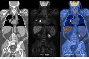
Computed tomography allows you to obtain an image of the organs being examined and identify the localization of the pathological focus.
Combined PET/CT machines perform sequential scans, then the software creates an anatomical picture of the organs with an image of metabolic processes superimposed on them.
The abbreviation PET/CT represents the combination of two X-ray tomography techniques to obtain diagnostic information.
Benefits of PET/CT
PET CT replaces several diagnostics at once. Also positron emission tomography:
- is distinguished by high accuracy (almost 100 percent) of the obtained data;
- helps identify emerging diseases;
- painless;
- no side effects;
- with methionine is not contraindicated during brain examination;
- one scan covers all organs at once;
- safe for health;
- can be performed more than once without causing harm to health;
- does not cause discomfort;
- excludes unnecessary or ineffective surgical and drug therapies.
PET CT is a new technique that allows you to evaluate the functioning of organs. The main task of diagnostics is not a simple snapshot, but a high-quality color image that allows you to see the internal processes taking place in the body. With cancerous tumors they change, as can be seen from the colors and their intensity.
Advantages and disadvantages of the method
PET has many advantages over other methods of ionizing radiation. For clinicians, the most valuable of these are:
- 100% reliability of results;
- ease of use due to clear protocols and an automated system;
- high throughput due to simultaneous data collection and processing, image reconstruction, and results analysis;
- visual diagnostic results;
- absence of pain and discomfort during diagnostics;
- no need to use additional diagnostic methods, including invasive ones;
- short terms for assessing the effectiveness of the therapy (1-2 weeks).
Disadvantages include high cost, the need to locate the Cyclotron near the diagnostic center, and the length of preparation and procedure.
Failure to comply with diagnostic deadlines after administration of a radionuclide increases the risk of false results.
What changes does PET scan show?
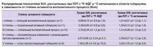
PET images are graded on a 4-level isotope accumulation intensity scale. When interpreting data, physiological foci of radionuclide accumulation are taken into account.
Level scale:
- levels 1-2 indicate foci of an inflammatory nature;
- level 3 - the probable nature of pathological changes includes tumors or metastatic lesions;
- level 4 - serves as a sign of a primary tumor, metastases.
The method shows the presence of atypical foci in organs and tissues, detects degenerative, dystrophic and post-traumatic changes.
PET with contrast reveals pathological activity of lesions based on accelerated metabolism. In the brain, PET scans trace the distribution of blood flow, which affects all aspects of brain activity.
Decoding the results
After a PET scan, the results are usually interpreted within two days. If necessary, a conclusion can be made in a few hours. If the patient has previously taken photographs or records of MRI, CT, X-ray, etc., then these materials must be provided to the doctor in advance. They are important for comparison and final conclusion. The interpretation is carried out by a radiologist or nuclear physicist.
Based on the resulting images, tissues that have responded to irradiation are analyzed and when they differ from the norm. PET CT data are assessed using a special scale that has several colors:
- Levels 1 and 2 indicate inflammation in specific organs.
- 3rd – for primary neoplasm.
- 4th – for the appearance of metastases.
After a PET scan, decoding may take varying amounts of time. This depends directly on the organ being examined and the pathology detected. The longest wait for results is after a brain scan.
PET/CT in neurology
Positron emission tomography helps detect:
- Brain pathologies that lead to senile dementia (for example, Alzheimer's disease). Emission tomography helps to detect even minor changes when there are no symptoms of pathology yet. It also allows you to distinguish Alzheimer's from Parkinson's and Huntington's.
- Areas causing epilepsy.
- Processes leading to Parkinson's pathology.
- Compare brain activity. In depressed patients it decreases, in healthy people it remains normal.
- Other brain pathologies.
Positron emission tomography is done for seizures that occur due to epilepsy, as well as to evaluate brain activity after a stroke. Sometimes the diagnosis is made for schizophrenia.
PET/CT in oncology: cancer diagnosis
Positron emission tomography is the most informative way to diagnose and monitor cancer treatment. Using PET-CT:
- Oncology is detected at the first stage, before the pathology progresses.
- Areas of tissue affected by cancer cells are determined. They absorb more radioactive glucose than healthy areas. The boundaries and dimensions of the malignant neoplasm are clearly visible in the picture.
- Tumors smaller than one centimeter are detected.
- It is possible to find metastases that are not detected by other methods. PET-CT examination makes it possible to assess the size of lesions, their spread, germination into the tissue of neighboring organs, and identify pathological foci that are still forming.
- The cause of the neoplasm is determined.
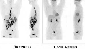
After chemotherapy, emission tomography makes it possible to evaluate the effectiveness of therapy. Due to the reliability (95 percent) of the data obtained, surgical intervention can be avoided. Instead, a specific therapeutic regimen is prescribed.
When assessing neoplasia of the lymphatic system, positron emission tomography replaces a whole range of diagnostics - MRI, X-ray, etc. Diagnostics makes it possible to distinguish oncology from inflammation, draw up a surgical plan, and determine the stage of cancer. The affected lymph node can be detected even before it enlarges.
PET in cardiology: diagnosis of heart diseases
Emission tomography shows areas of the heart where the blood supply is disrupted. The technique is used for incipient ischemia and to detect past infarctions. Using positron emission tomography, you can distinguish between living and dead areas of the organ and determine the condition of the heart muscle.
This is mainly necessary before surgery. If the tissues are still alive, then balloon dilatation or bypass surgery is sufficient. If dead areas are found, this may be an indication for a heart transplant.
Incorrect results
Unreliable results are due to low resolution on equipment monitors, improper preparation, or due to movement during sensor operation. Also, the results are affected by high sugar levels, the remaining effect after chemotherapy or radiation therapy. Therefore, after them you need to wait a certain time.
PET in the diagnosis of malignant neoplasms
The first use of PET in oncology dates back to 1991. Today, 70% of all PET procedures performed in the world are aimed at identifying malignant tumors.
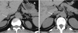
Central cancer of the right lung
The method allows you to determine the exact location of the tumor, identify relapse, stage of the process, differentiate malignant from benign tumors, and find regional and distant metastases.
Standard PET diagnostics determine the metabolic activity of the lesion based on the absorption of the injected isotope.
The method is capable of identifying pathological foci of a tumor nature with a size of at least 7 mm. In cases where the tumor intensively absorbs the radiopharmaceutical, formations ranging in size from 3 mm are detected.
Based on PET, the course of the disease is predicted, a radiation therapy plan is drawn up, the effectiveness of the treatment, and the radicality of tumor removal during surgical interventions are assessed.
Types of radioactive drugs for PET
Radiopharmaceuticals make it possible to study biological processes occurring in the human body.
Based on the data obtained, the metabolism of glucose, oxygen, secondary active transport of amino acids, and protein biosynthesis are assessed. Tissue hypoxia is detected and the rate of growth of tumor cells is determined.
Types of isotopes for tomography:
- 18F-fluorodeoxyglucose - used to diagnose malignant neoplasms, determine the stage and extent of the process. In cardiological practice, a drug with glucose is prescribed to patients to assess the viability of the myocardium before surgery. In patients with epilepsy, it reveals foci of hypometabolism in the brain;
- 11C-methionine - prescribed to detect brain cancer, multiple myeloma, tumors of the lymphatic system. Diagnoses pathology of the parathyroid glands: adenoma, carcinoma;
- 11C-choline – used for the diagnosis of prostate diseases: adenoma, cancer and its metastases. Detects hepatocellular cancer, differentiates from benign formations;
- 18F-sodium fluoride - used to detect pathology of the skeletal system in oncology. Prescribed for the diagnosis of benign and malignant bone formations, post-traumatic changes, inflammatory and degenerative processes.
When is a PET scan indicated?
The PET method combined with CT is used to search for foci of pathological accumulation of the isotope, indicating the development of the disease.
The procedure is carried out in the absence of clinical symptoms, to monitor the process over time and evaluate therapeutic measures.
PET CT indications for diagnostics:
- formations of the head and neck: diagnosis of the nature of the tumor;
- neoplasms of the thyroid gland: identification of differentiated and medullary carcinoma with the stage of the process;
- malignant tumors of the lung: large cell carcinoma, squamous cell carcinoma, adenocarcinoma;
- cancer of the upper gastrointestinal tract - damage to the esophagus and stomach;
- cancer of the lower gastrointestinal tract - damage to the colon;
- pancreas cancer;
- tumor diseases of lymphatic tissue: lymphogranulomatosis, non-Hodgkin lymphoma;
- malignant neoplasm of the breast;
- malignant skin tumors – melanoma;
- formation of bones and soft tissues;
- neoplasms of the genitourinary tract;
- brain tumors;
- neurological diseases caused by the occurrence of pathological foci in the substance of the brain (temporal and extratemporal epilepsy);
- brain diseases associated with vascular damage and thrombosis: ischemic and hemorrhagic stroke, carotid artery stenosis;
- traumatic brain injuries: assessment of the volume and degree of brain damage in the early and late post-traumatic period;
- degenerative and dystrophic diseases of the brain: Alzheimer's, Huntington's, Parkinson's diseases.
Lymphoma
Indications and prohibitions for PET/CT
PET CT is done when a more thorough examination is necessary, if other methods do not provide a complete picture. Positron tomography scanning is indicated for:
- pancreatic pathologies;
- study of already detected foci of oncology to assess the presence and growth of metastases;
- prostate cancer;
- vascular pathologies of the brain;
- failures of the musculoskeletal and motor systems, if the diseases appeared due to impaired bone density or tissue deformations;
- strokes (and after them);
- suspected sarcoma;
- lymphomas and melanomas;
- brain examination after surgery (especially if previous computed tomography or MRI interpretations are questionable);
- suspicion that a malignant neoplasm has been detected;
- epilepsy attacks (emission tomography shows the location of the source of the disease).
PET CT also has contraindications:
- Seriously ill patients who are unconscious and unable to independently control their movements.
- PET-CT cannot be done on people who have just had a biopsy or laparoscopy. Otherwise, there is a risk of damage to soft tissue at the surgical site.
- PET-CT is prohibited to be used in conjunction with radiation therapy. Otherwise, complications after it will only intensify. A 3-month interval is maintained before positron tomography scanning.
- Also, PET-CT is prohibited during chemotherapy. This contributes to the emergence of irreversible processes in the brain. After chemotherapy, a month's break is also necessary before performing PET-CT.
- Pregnancy, lactation period.
It is not recommended to do PET-CT for infectious patients or for any inflammatory processes. This may affect the reliability of the results.
Contraindications for the study
When referred for diagnostics, with the help of anamnesis and additional clinical studies, patient conditions are identified that serve as an absolute or relative contraindication to tomography.
The following persons are not allowed to take a PET scan:
- children under 18 years of age;
- pregnant women, regardless of gestational age;
- patients suffering from diabetes mellitus in subcompensated or decompensated form;
- with severe pain that makes it difficult to remain still during the procedure;
- obese patients whose body weight exceeds the permissible technical standards of the tomograph;
- with pulmonary emphysema.
The question of the benefit and advisability of diagnosing patients in a severe, unconscious state is considered on an individual basis.
Persons with mental and movement disorders, with a phobia of limited space, undergo tomography after the administration of sedatives.
Lactating women abstain from breastfeeding for 3 days after the procedure. The child is transferred to adapted milk formulas, and the milk is expressed.
Diagnosis restrictions:
- Tomography is performed no earlier than 30-90 days after surgery. The exception is cases when detection of distant metastases from the primary tumor site is required;
- for patients prescribed chemotherapy, the procedure is carried out before treatment, or 30 days after completion of the course;
- if there are results of PET-CT performed before the start of chemotherapy, re-diagnosis to assess the effect of drugs on the tumor process is carried out 7 days after the end of the administration of chemotherapeutic agents;
- For patients who have undergone radiation therapy, the procedure is scheduled after 90 days.
Description of PET/CT
Positron emission tomography (PET) is a scan of the entire body or specific parts of the body using a radioactive substance. It makes the metabolic processes that occur in the body visible on the screen. Biological substances are taken to administer substances. They are labeled with radionuclides. The resulting substances are administered intravenously, in tablets or by inhalation.
CT (otherwise known as computed tomography) is a diagnostic that reveals foci of various pathologies due to structural changes in tissues or organs. In this case, it is possible to accurately determine their location.
Despite many advantages, PET has poor anatomical information, and computed tomography can detect a tumor only if it greatly increases in size. The combination of two methods allows you to see the most accurate clinical picture even at the initial stage. The image is displayed on the screen in real time.
PET principle
The diagnostic principle is based on the speed difference in metabolic processes in the body. As a result, radionuclides accumulate in certain areas in different quantities. This process also differs in the time it takes for contrast to accumulate. The highest level of metabolism is in places where cancerous tumors are located. Therefore, they are easy to distinguish from healthy areas.
A radioactive substance is infused into the bloodstream, which is produced in a cyclotron. The product lasts for 60 minutes, then begins to gradually disintegrate. The drug contains fluorine or iodine. The more the drug accumulates in specific areas, the brighter they become.
Such areas are called “hot”. Dull areas of the image are considered “cold”. In malignant neoplasms, the level of metabolic processes is much higher than in healthy areas. Therefore, cancerous tumors are highlighted very brightly.
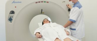
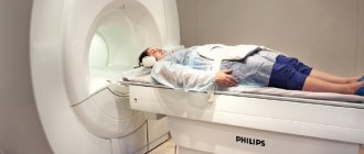
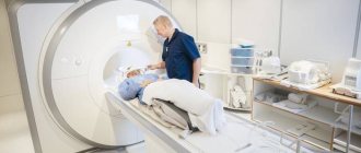
Mechanism of action of the PET/CT device
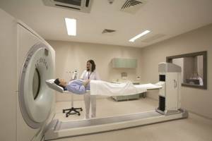
The equipment for PET CT is similar to a standard tomograph. This is a large ring with many sensors that respond to radiation. The device is equipped with a couch that slides inside. The operation of PET-CT equipment is based on positron emission. The dose of radiation received by a person is no more than that of radiography and computed tomography.
After the radiation drug is injected, it seeps into the tissue and begins to emit rays, which the scanner picks up. Through it they are transferred to a computer, where they are processed using a special program. The result is shown on the monitor in 2 or 3 dimensional graphic models.
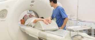
Preparing the patient for the procedure
The patient prepares for tomography with radiopharmaceuticals 6-12 hours before diagnosis. Avoid eating food, carbonated drinks, and sweet juices.
Medicines containing glucose are discontinued. The patient refuses physical activity, avoids overwork and stress factors.
Upon admission to the diagnostic center, the patient is allowed to rest for 15-20 minutes and drink 500 ml of clean water. After determining the level of glucose in the blood, which should not exceed 8-11 mmol/l, a catheter is installed in the vein of the elbow. The person being examined is helped to change into comfortable clothes that do not contain metal elements.
The patient is placed on the couch in a supine position. The radiopharmaceutical is administered using a syringe or an automatic dispenser.
Then 500 ml of 0.9% NaCl is infused by infusion; during brain tomography - 20 ml of 0.9% NaCl. After administration of the drug, the patient is left in a motionless position with his eyes closed in a darkened room for 30-60 minutes. During this time, the product is distributed and the concentration in organs and tissues is equalized.
It is prohibited to use a mobile phone, talk, chew gum, or strain muscles. Before the scan, the patient is asked to urinate or the bladder is catheterized.
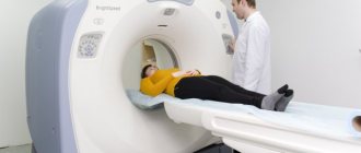
Passing diagnostics
The patient sits on a sliding table and is immersed inside the tomograph. Position – lying on your back with your hands behind your head.
At the first stage, a low-dose CT scan of the planned area or the whole body is performed. Then they begin PET diagnostics.
The pelvic area is scanned first to avoid artifacts from the bladder filling with urine.
The duration of the procedure ranges from 10 minutes for a brain diagnosis to 70 minutes for a full body examination.
When studying the brain, a repeated delayed diagnosis is carried out 180 minutes after the administration of the radiopharmaceutical.
At the end of the tomography, the patient is placed in a rest room for 40-60 minutes to reduce the radiation level.
After the procedure, the doctor gives recommendations on drinking fluid in the amount of 1500-2000 ml per day and observing radiation safety measures.
Complications and side effects
Adverse events that may occur during PET scanning are associated with the administration of a contrast radiopharmaceutical.
Occurs in 10-20% of cases. Patients with a history of allergic reactions are given premedication before tomography.
If consequences develop in the form of severe systemic complications, emergency intensive therapy is carried out.
| Local reactions: | Systemic reactions with organ damage: |
| · redness, itching, burning, urticaria at the injection site of the radioactive drug; · damage to the skin, vascular wall, hematoma; · inflammation of the nerve; · abscess, necrosis of underlying tissues; Pain when moving the elbow joint. | · anaphylaxis, Quincke's edema; collapse, loss of consciousness, cardiogenic shock; · convulsions, paresis; acute renal failure; pulmonary edema, status asthmaticus; · thyrotoxic crisis. |
Considering the radioactive radiation emanating from the subject, patients are advised to avoid pregnant and lactating women, children, and crowded places after the procedure. For two hours, it is prohibited to approach the specified category of persons more than 1 meter.
Risks associated with PET/CT
PET CT is absolutely safe for health, since the received radiation dose does not exceed the x-ray norm. Contrast agents used for examination are given or administered only after preliminary questioning for the presence of allergic reactions. The drugs are used in strict dosages. The above reduces to a minimum the possible risks of any complications.
They can appear only in the case of a slight excess of the administered radioactive drug. The body may react to this with itching or burning of the skin, nausea. Sometimes temporary claustrophobia appears. These side effects do not require treatment. They go away on their own within 2 hours. After the PET scan, you need to drink a lot of still water. This is required to accelerate the removal of radioactive substances from tissues.
Radiation exposure to the patient
The combination of radiation received from X-rays from a CT scan and from the administered radiopharmaceutical drug constitutes the amount of radiation exposure to the patient.
Without taking into account delayed scans, the radiation dose for a full body diagnosis does not exceed 10-13 mSv. This dosage is considered unsafe and harms the body.
| Effective radiation doses when administering 1 MBq of radiopharmaceutical (FDG) | |
| Children 0-12 months. | 0.13 mSv |
| Children 1-5 years old | 0.073 mSv |
| Children 5-10 years old | 0.047 mSv |
| Children 10-15 years old | 0.032 mSv |
| Adults | 0.027 mSv |
Note: For a complete diagnosis of the body, 370-400 MBq of radiopharmaceuticals are required.
Diagnostic price
The PET procedure combined with CT is an expensive diagnostic method. The patient pays the cost of the radioactive drug, CT scan, delayed PET scan, disc and imaging scans.
The average price for the procedure is 60-80 thousand rubles. Some clinics offer a 10-25% discount program for the second and subsequent diagnostic sessions.
The information content of PET, close to 100% and sensitivity of 97-98%, allows one to find out the nature of the disease in one procedure.
The correct approach to the technique determines the correct choice of patient management and treatment plan, on which the health and life of the patient depends.
The high cost of PET justifies the range and value of diagnostic information that cannot be obtained by any other known method.
Cost and price of PET/CT in Moscow: where to do it?
Equipment for PET CT is installed in large medical centers and is not available in many Russian regions. The cost of an examination in Moscow varies on average from 30,000 to 65,000 rubles. Some locations may offer discounts of up to 10 percent.
In Moscow, positron tomography scanning can be done at the clinic on Mayakovskaya (multidisciplinary center). The approximate cost of the examination is 65,500 rubles. Emission tomography is also carried out at the European Medical Center on Shchepkina Street, 35. The cost of the examination there reaches 87,000 rubles.
During a positron tomography scan, the internal organs of the entire body are examined. The examination shows the presence of neoplasms, their boundaries, malignancy or benignity. Emission tomography helps to recognize the onset of cancer when there are no obvious signs of the disease. This helps to prescribe timely treatment and prevent the occurrence of metastases.
Related articles:
- Computed tomography (CT) of the intestine
- How to check your colon without a colonoscopy
- Mammography or breast ultrasound: which is better?
- The test is positive, but the ultrasound does not show pregnancy
- Results and features of the spine MRI procedure