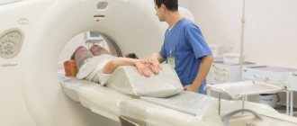
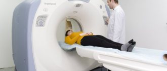
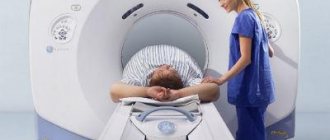
A method such as magnetic resonance imaging has long been widely used in medicine. Due to the fact that different tissues react differently to electromagnetic waves passing through them. A high-tech MRI device records the tissue response, which is expressed in a picture on the tomograph screen. It is worth saying that at present, magnetic resonance imaging today is an informative way to “look” into the internal organs, notice the disease in time and prescribe the right treatment. Magnetic resonance imaging of the head is a 100% safe diagnosis that does not require a penetrating method; the doctor has the opportunity to examine the organ in detail as it could be done in reality.
MRI is an informative way to examine the brain and intracranial space. Currently, the capabilities of modern tomographs are wide, especially for such a complex area as the brain. The doctor can conduct a detailed diagnosis of the circulatory system of the brain, which is necessary not only for making a diagnosis, but also for monitoring treatment.
Why do the eyes bleed and the baby that matured in... the ovary
There are many examples where magnetic resonance imaging (MRI) made it possible to identify hidden pathologies or, on the contrary, to refute a previously made diagnosis and avoid unnecessary surgery.
By the way, the latter is also very relevant, especially against the backdrop of increasing cases of surgical aggression towards patients. You can find many sites on the Internet that talk about MRI as an advanced and very effective diagnostic method. But we decided to start with examples that actually took place, which the gynecologist at the Medservice clinic told us about.
How to behave during scanning
For any medical procedure there is a list of instructions and rules of conduct. Magnetic resonance imaging is no exception. When going for screening, it is important to remember:
- before entering the room with equipment, remove all metal elements from the body, check pockets, clothing should also not have ferromagnetic fasteners or decorative components;
- You can set up music or watch a movie on the internal screen of the tomograph before the procedure begins, this applies to putting on earplugs and other preparatory issues;
- To obtain clear images, you need complete immobility; you cannot change the position of your body inside the device;
- you need to carefully listen to the instructions of the doctor who addresses the patient via the public address system, asks to hold the breath or completely fix the position.
Before sending for a scan, you need to talk with the specialist on duty, talk about the disturbing nuances in the tomography, possible contraindications, suspicions of pregnancy or permanent implants in the body. Only a doctor can determine whether a patient can undergo an MRI session.
MRI and pregnancy
Despite the fact that MRI is partially contraindicated during pregnancy, there are cases when it saves the lives of both mother and child. In one of the regions of Udmurtia, a woman who was 32-33 weeks pregnant came to the maternity hospital with complaints of abdominal pain. She was hospitalized due to risks of premature birth.
The results of the examination and ultrasound of the pelvic organs did not allow us to determine treatment tactics and make a decision - to allow childbirth or to prolong treatment. The lady was sent for an MRI, where it was revealed that the pregnancy was ectopic - ovarian. The lady was sent for surgery. Only a miracle and an MRI saved both mother and child.
Is it necessary to do an MRI of the brain with contrast for headaches?
For headaches, you need to start your diagnostic journey with a basic, non-contrast MRI of the brain. If the initial MRI examination reveals tumor abnormalities, the patient will be offered a contrast agent. During diagnosis, a contrast agent is injected to enhance the signal in the area of possible anomaly. With contrast, you can reliably determine the structure, size, boundaries and zones of pathological processes in the brain. MRI with contrast will give the doctor the opportunity to see the extent of the spread of the tumor abnormality to the surrounding structures, reliably assess dynamic changes and see the number of foci.
Previous Next
It is advisable to make MRI a habit
A lady consulted a gynecologist with complaints about the appearance of bloody, pink discharge from the genital tract. Her cheerfulness and youthful appearance beyond her years did not foretell any unpleasant surprises.
An ultrasound of the pelvic organs revealed a polyp in the uterine cavity. In order to clarify the diagnosis, the gynecologist referred the patient for an MRI, where complete destruction of the muscle tissue of the uterus was visualized.
Apparently, the process lasted two to three years; in fact, we are talking about the second or third stages of cancer. The patient was referred to an oncologist.
Neither ultrasound nor external examination made it possible to timely determine the presence of malignant tumors. MRI allows you to detect malignant and benign formations at the earliest stages, when their size does not exceed 0.5 mm. With ultrasound, the lower limit of accuracy is about 1 cm, but this is subject to the highly qualified doctor and the resolution of the equipment. It must be of an expert class. That is why it is very important to conduct MRI not only as prescribed by the attending physician, but also for preventive purposes. For example, during medical examinations.
What does a patient experience during an MRI?
When the patient is sent for examination, he is given a brief instruction. It is not difficult to follow the prescription, since you just need to lie in a relaxed state and listen to the doctor’s instructions via speakerphone. In most cases, a person does not feel anything, some manage to rest and even take a nap. Depending on the area of scanning, a session takes on average 30-40 minutes. Particularly sensitive people describe the following sensations:
- 1. The noise from the equipment is too loud. During operation, the gradient coils in the device produce mechanical noise in the form of crackling, humming, and clicking. The more powerful the equipment, the louder the sounds seem. This is considered normal, most people don’t pay attention. But if the patient is sensitive to noise and comes for screening with headaches, this can cause discomfort.
- 2. Slight heating of the study area. If this is a slight warming in an area of the body, there is nothing to worry about. If sensitive heating or burning begins, it is better to immediately inform the specialist on duty or press the “panic button” inside the tomograph.
- 3. Fatigue from prolonged immobility. For most patients, a fixed body position does not cause discomfort, but if there is tension in the body or pain in the spine or limbs, a long lying position can lead to increased pain, numbness and other unpleasant sensations.
- 4. Anxiety, fear of an unknown procedure. This applies to emotionally excitable people, as well as young children who are afraid to be alone in an unfamiliar environment. In this case, you need to understand that you are being monitored by doctors and other employees who are in the next room behind a glass partition. In special cases, loved ones are also allowed to be in the room during the session.
- 5. Claustrophobia. A panic attack can occur suddenly, even if the patient has not previously experienced such attacks. If the study takes place in a device with closed walls, it seems that it is difficult to get out of the equipment on your own. In fact, one side is always open, there is air conditioning and lighting inside the installation, sometimes there is a screen for watching movies and listening to music. You can always stop the session by pressing the “panic button” or by communicating with the clinic staff over the loudspeaker.
Some types of MRI examinations require holding your breath for a while.
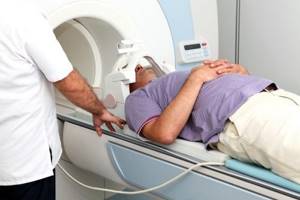
Patients with respiratory problems, as well as heavy smokers, note that even short breaths without exhaling cause a feeling of lack of air. It is difficult to hold your breath for several seconds in these cases, but you have to be patient to get good pictures of the chest or abdomen. The described situations are observed quite rarely in the total number of procedures performed: every 20th patient expresses minor complaints. Most often this is due to individual characteristics, which is not typical for the majority of subjects.
Advantages of MRI in gynecology
MRI allows you to obtain complete images of human organs and tissues at any level and in any given plane with an assessment of their functional integrity. If necessary, you can obtain layer-by-layer series of images with slice thicknesses from 4-5 mm to 0.8 mm. MRI with contrast provides a more accurate verification of the tissue identity of tumors, identifying small metastases at the earliest stages of development. In addition, tomography allows you to assess the structure and condition of bones, soft tissues, blood vessels, ligaments, and cartilage.
Feelings when contrasting
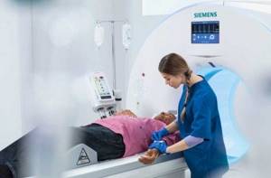
A special scanning mode involves preliminary administration of a contrast agent. It looks like a solution that is injected into the bloodstream through a syringe or a special injector. During contrast, a person feels:
- an injection that lasts a few seconds;
- coolness in the body or, conversely, a surge of heat (no more than 1-2 minutes);
- drowsiness;
- sudden desire to go to the toilet “in a small way”;
- slight dizziness or nausea;
- tingling at the injection or catheter site;
- metallic taste on the tongue.
If the contrast does not cause severe nausea, lightheadedness, itching all over the body, or difficulty breathing, these sensations are considered normal. If your health deteriorates seriously, be sure to notify your doctor so that rehabilitation measures can begin as soon as possible. After the contrast procedure, you can also do your usual activities. The use of intravenous dye does not affect mental activity or driving.
When should an MRI be performed?
MRI should be performed if the presence of benign or oncological formations is suspected. Tomography of the pelvic organs with contrast is applicable for cancer patients to determine the area of spread of metastases and damage to neighboring organs.
MRI of the pelvis is indicated when identifying the causes of infertility in men and women (obstruction of the fallopian tubes), in case of injury, the presence of anomalies and structural pathologies, and inflammatory processes in the organs of this part of the body.
Tomography of the pelvic organs in women for use in the diagnosis of endometriosis. The endometrium is the hormone-sensitive tissue lining the uterine cavity that can implant in a variety of tissues, including the ovaries, fallopian tubes, bladder, peritoneum, rectum, and other distant organs. These fragments, in accordance with the phases of the menstrual cycle, undergo the same cyclic cycles as the endometrium in the uterus, which is accompanied by pain, an increase in the affected organ in volume, and bleeding. Endometriosis can lead to infertility. Diagnosing endometriosis and localizing distant metastases is quite difficult, but MRI provides such an opportunity.
When planning untimeliness, MRI allows you to assess the condition of the uterine scar in mothers who gave birth by cesarean section. Images in the axial and frontal directions make it possible to identify problem areas and assess the risks of scar failure, which is essentially an assessment of the risk of maternal death.
MRI of cerebral vessels
Conditions such as frequent fainting and dizziness may be the result of a malfunction of the vascular system of the brain. In this case, the attending physician refers the patient to a survey tomography. Using the angiographic mode, doctors have a unique opportunity to track the process of blood flow in real time. In this case, the angiologist can assess the state of all functional indicators, note the presence of spasms or see the cause of poor blood flow. Thus, it is possible to identify blood clots, aneurysms and other pathologies of the vascular system.
Contraindications
Despite the fact that MRI is considered one of the safest diagnostic methods, there are some contraindications to its use. We are mainly talking about the presence in the patient’s body of foreign bodies that contain metal particles, including implants, dentures, shunts, dental crowns, insulin pumps, pacemakers, and vascular clips. It is impossible to carry out the procedure if the patient’s weight exceeds 130 kilograms, since the tomograph chambers are quite narrow. MRI is not recommended in the first weeks of pregnancy. But for serious indications, the study is possible.
As for claustrophobia, a good solution is to study with open-type tomographs, one of which is installed at the Medservice clinic.

