Computed tomography procedure
In modern medicine, diagnostic methods that provide a large amount of information in a short period of time are highly valued. These characteristics acquire particular significance when studying the state of the urinary system, the pathology of which does not always manifest itself in a clear clinical picture. Computed tomography of the kidneys and ureters allows one to assess the structure and degree of pathological changes in organs in one session, indirectly analyzing their functions.
What does a CT scan of the urinary system show?
Computed tomography is based on the principle of X-rays, as in conventional x-rays. Unlike the latter, CT is a more advanced method. Narrowly directed beams enter the human body at different angles, which are recorded at the exit by special sensors. Since these rays are retained in the body by dense, radiopaque structures, their resulting strength varies. The computer processes the information received and produces an image - a tomogram, which shows “slices” of the human body at all levels and displays formations that differ in their structure.
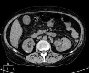
Kidney CT scan
A regular X-ray does not visually distinguish between muscle and fat tissue; it only shows bone and other hard structures. A computer tomogram, thanks to special processing, allows you to differentiate the density of all organs included in the image.
A CT scan of the urinary system evaluates the condition of several organs at once:
- adrenal glands;
- kidneys: parenchyma and pyelocaliceal system;
- ureters;
- Bladder;
- vessels of the renal pedicle;
- regional lymph nodes.
In addition to the urinary system itself, the tomogram reveals the lumbar
vertebrae, intestinal loops. Computed tomography allows you to assess the size of organs, their location, the presence of injuries, tumors, and stones. Inflammation is determined by indirect signs and its presence can be unequivocally stated only after passing a urine test and undergoing an ultrasound examination of the retroperitoneal organs.
Diagnostic results
When choosing a center for performing a CT scan of the kidneys in St. Petersburg, pay attention to what you get as a result of the study. It is better to give preference to those centers where the result is issued immediately after the diagnosis, and not the next day. This means that the doctor is present on the shift, and does not describe the images remotely. This option is better, since in different clinical situations it may be necessary to adjust the tomograph settings to suit the needs of a particular patient.
Preparing a report takes some time, from 30 minutes to 2-3 hours, since the scan results in a huge number of images, up to several thousand, and the doctor needs time to correctly evaluate and interpret them. If the patient does not have the opportunity to wait to receive the conclusion, some centers provide the ability to send it to the patient’s email.
The next point that you need to pay attention to when choosing a center is the format for displaying images. It is preferable to issue results on electronic media - a CD or flash card, because All images obtained during the scanning process can be recorded on them. If a film is issued with a printout of the images, it usually only contains about 20-30 images, which does not always reflect all the detected changes. In such centers, patients are often asked to pay additionally for a disc with all the footage.
CT scan of the urinary system with contrast
In some cases, there is a need to obtain more detailed information about the condition of individual organs, their function, and blood supply. Thanks to the contrast, a CT scan of the urinary system becomes brighter and clearer, sometimes even formations that were previously invisible are displayed. It is often possible to identify X-ray negative urinary stones, narrowings, deformations of the ureter, and the collecting apparatus.
Since the genitourinary system is difficult to access for examination, computed tomography in this area is almost always performed using contrast agents. In addition to significant diagnostic benefits, this technique can cause a number of problems. The contrast agent is administered intravenously, and the kidneys filter it from the blood, thereby “highlighting” the entire urinary system on the tomogram.
Contrast is an iodine-containing compound and can cause a number of complications, most often allergic reactions. The indicator creates additional stress on the kidneys, which can be dangerous if their function decreases.
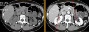
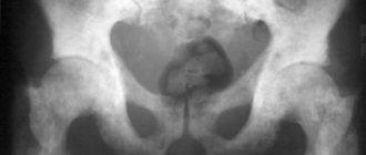
CT scan of the kidneys without contrast (left) and with contrast (right): arrows indicate vessels in the renal hilum
Why contrast is needed
Thanks to the introduction of contrast, it turns out to be as detailed as possible. As a coloring agent, specialists usually use non-ionic preparations based on barium or iodine: Omnipaque, Optirey, Ultravist, Xenetics. They are introduced into the bloodstream intravenously or by drip through a special catheter. The contrast quickly spreads across the body, making the image bright, clear and detailed. It is especially relevant for detecting areas with impaired blood flow. If the blood supply is excessively intense, the presence of a tumor can be assumed, but if no blood flows into the area under study, there may be an area with necrotic tissue.
Iodine-containing preparations are safe and are usually well tolerated by patients. Adverse reactions may include a salty, metallic taste in the mouth. Sometimes, headache. The medicine is completely eliminated from the body within two days. It can cause allergies, the symptoms of which are burning in the stomach, skin rash, itching, swelling of the mucous membranes, difficulty breathing. In this case, it is necessary to take antihistamines. Therefore, if during the scanning process a person feels unwell, it is necessary to inform the diagnostician about this via a two-way communication device.
Kidney CT with contrast is prescribed because it allows you to clearly determine the size and boundaries of the organ, vessels and neoplasms in the early stages.
What is included in a urinary system examination?
A CT scan of the urinary system includes examination of the condition of several organs at once. The greatest diagnostic weight is occupied by information about the condition of the kidneys and bladder, as the largest and most often “sick” organs. Data is collected on the condition of the ureters, adrenal glands, and lymph nodes. The vertebral bodies and intestinal loops that fall within the tomography area are studied last and often do not provide any necessary information related to the disease of the urinary system.
When examining the kidneys on a computed tomogram, their size, location, and shape are assessed. Often an organ has congenital developmental anomalies, which may not manifest themselves in any way and are diagnosed by chance. Any kidney disease leads to a change in its structure, and tumors are completely visible as separate formations. The adrenal glands are more often affected by tumors. The collecting system and ureters are susceptible to urolithiasis and injury, which is also clearly visualized in the image. The bladder may contain polyps or calculi and be damaged by external influences, including rupture of the organ. Assessment of the condition of regional lymph nodes is required to determine the stage of the oncological process.
Side effects
Because CT scans are performed using contrast, there may be side effects. Most are caused by hypersensitivity to iodine, the main component of the pharmacological drug. The type of symptoms determines the severity of the allergic reaction:
- in the mildest case, the allergy will manifest itself as dry mouth, a feeling of dizziness, nausea and headache. One-time vomiting is possible, as well as the appearance of an itchy rash on the skin (urticaria);

Urticaria - allergic skin rash - Repeated vomiting can lead to dehydration, so it is considered a moderate allergy;
- Severe forms include angioedema and anaphylactic shock. In the first case, swelling of the face, tongue and neck can cause significant difficulty breathing and oxygen deficiency in the body (hypoxia). The second case leads to a sharp disturbance in vital signs: pulse, blood pressure.

Quincke's edema is a form of allergic reaction to contrast
Quincke's edema - video
The likelihood of allergic symptoms occurring can be anticipated. The risk increases sharply in the following situations:
- if there have been similar reactions to contrast in the past;
- with a general tendency to various kinds of allergic reactions: urticaria, food allergies, etc.;
- with symptoms of exacerbation of chronic diseases.
In this case, you can take certain measures in advance and introduce antihistamines (Suprastin, Tavegil) and steroid hormonal drugs (Prednisolone, Dexamethasone, Hydrocortisone). If symptoms of severe allergic reactions appear, hospitalization and intensive care in the intensive care unit are required.

Prednisolone is a powerful antiallergic drug
Indications and contraindications
CT scan of the urinary system has indications and contraindications. You should resort to the procedure when:
- suspected urolithiasis. CT allows you to determine the presence, size and location of stones, and indirectly judge their composition. More information can be obtained by using contrast;
- inflammatory processes in the kidneys, pyelocaliceal system, ureters, bladder. According to the study data, one can judge the extent of the process’s prevalence, its acute or chronic nature;
- neoplasms: their location, size, involvement of surrounding organs and tissues are assessed. Based on indirect signs (spreading into neighboring structures, damage to lymph nodes), one can assume the benign or malignant nature of the tumor. Contrast agents also provide greater tumor imaging;
- congenital anomalies of the urinary system. From 10 to 30% of people have these features. In the vast majority of cases, the anomalies do not manifest themselves clinically. Sometimes they contribute to the development of infectious and inflammatory processes and the formation of stones;
- injuries and their consequences.
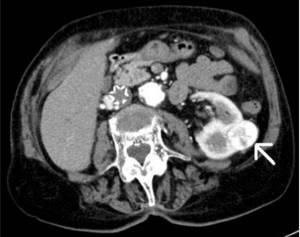
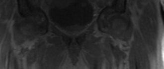
A tumor of the left kidney identified on a CT scan of the urinary system with contrast (indicated by an arrow)
Contraindications to computed tomography:
- pregnancy;
- body weight more than 150 kg.
When using contrast enhancement, the range of restrictions expands:
- intolerance to the drug itself and iodine-containing substances;
- increased thyroid function;
- renal failure;
- taking oral hypoglycemic drugs (metformin).
What are the dangers of kidney pathologies?
Few people know that kidney pathologies can cause heart attacks and strokes. When they are damaged, a disturbance in the metabolism of calcium and phosphorus occurs. Calcium is deposited in the walls of blood vessels, causing thrombosis.
There are other dangers:
- Nephroptosis or displacement of the kidney. If left untreated, it leads to chronic inflammation, hypertension and kidney failure, which threatens organ loss.
- Pyelonephritis refers to inflammatory diseases. It is often asymptomatic or “masked” as an acute respiratory infection. In severe cases, a purulent focus appears in the body, which can lead to sepsis or removal of the kidney.
- Kidney stones are a common pathology that often requires emergency medical care and surgical intervention. In a kidney blocked by a stone, acute inflammation occurs, threatening urosepsis.
This is not the entire list of diseases with serious consequences. This is why it is so important not to self-medicate, but to consult a doctor and regularly take blood and urine tests.
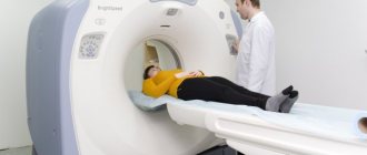
How to do a CT scan of the urinary system
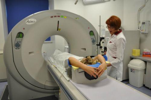
Carrying out any computed tomography is an absolutely painless procedure. A CT scan of the urinary system takes a maximum of 15 minutes. If contrast is administered, the duration may increase by another 5 to 10 minutes.
Throughout the procedure, the patient lies on his back on the tomograph table. A contrast agent, if necessary, is administered intravenously. The device's sensors pass over the lumbar and pelvic area without affecting all other areas of the body. The procedure is completely painless and does not cause any discomfort.
All received data is processed by a special program into graphic images and displayed on different media: on an image and on a disk. The results of a computed tomography scan of the kidneys and ureters are assessed by a doctor who describes the image obtained and makes a conclusion.
What is contrast agent used for?
Contrast is introduced into the patient's body so that the resulting image quality is improved. This allows you to more accurately diagnose pathological processes or monitor the effectiveness of therapy.
Non-ionic preparations that contain iodine are used as a contrast agent. Thanks to iodine, all kidney structures are visible in great detail, so the doctor can more accurately examine the tomogram and make a diagnosis. The kidneys are parenchymal organs, so if CT is necessary, the use of contrast is recommended (in the absence of contraindications).
How to prepare for a CT scan of the urinary system
Proper preparation for any diagnostic procedure is the key to its quality and effectiveness. A CT scan of the urinary system is performed on a full bladder - this is necessary for a reliable assessment of the condition of the urinary tract. To achieve tight filling, it is enough to drink a glass of water 1 – 2 hours before the test and not go to the toilet. Another important point is to fast for at least 4 hours before the tomography.
In cases where contrast administration is required, renal function should first be assessed - a blood test for creatinine. The result must be fresh, made no more than 10 days ago. Creatinine numbers allow you to calculate the glomerular filtration rate - the main indicator of kidney function and the presence of renal failure.
Carrying out the procedure
A CT examination begins with the patient laying on the machine table and attaching a system for automatically injecting contrast into a vein. The drug is administered through a regular plastic cannula installed into the lumen of the vessel. After the procedure is completed, it is removed from the vein without any problems. The following is the approved research algorithm:
For infants, the examination is performed under general anesthesia in the presence of an anesthesiologist. When the drug is administered into a vein, a feeling of fever and nausea may occur, which disappear without external intervention and are not considered side effects.
CT or MRI of the urinary system
Despite some similarities between computed tomography and magnetic resonance imaging, these diagnostic methods have significant differences. CT better visualizes dense, radiopaque structures: most stones, foreign bodies, bones. The contrast allows us to judge the condition of the urinary tract. MRI visualizes soft tissue well and is more suitable for diagnosing inflammatory processes, tumors, and is safe for pregnant women. MRI cannot be performed on people with any implants (artificial joints, pacemaker, insulin pumps, wires, plates, other foreign bodies), while CT has no such restrictions.
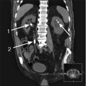
Stones in the pyelocaliceal system and ureter identified on CT (indicated by arrows)
Advantages of this method
The main advantage of computed tomography is the ability to create a three-dimensional image of the area or organ being examined, which is important when preparing for surgery. In terms of its effectiveness, CT is significantly superior to ultrasound diagnostics, excretory urography and conventional x-rays.
In terms of studying oncology at an early stage of development, the accuracy of a computed tomograph cannot be compared with any other diagnostic method. Only a tomograph using contrast can accurately determine the location of the tumor, its size and etiology.
CT scan of the urinary system at the MAGNIT center
In our center, computed tomography is performed on a modern German Siemens tomograph, which provides one of the lowest radiation loads on the patient’s body. The results are provided on the day of the examination, and the doctor who conducted the examination explains in detail to the patient the essence of the identified changes.
To make an appointment for a CT scan of the urinary system, you need to call the center at +7 (812) 407-32-31 - our employees will tell you about all the features of the procedure and help you choose the optimal time for diagnosis.